Association Between Neutrophil-To-Lymphocyte Ratio And Heart Failure In Patients With Acute St-Elevation Myocardial Infarction: A Case-Control Study.
- P S Puneeth Kumar , 3rd Year PG Resident, Department of General Medicine, Katihar Medical College, Bihar, India
- Professor P.K. Agrawal , Professor, Department of General Medicine, Katihar Medical College, Bihar, India
- Professor Mrityunjay Pratap Singh , Professor, Department of General Medicine, Katihar Medical College, Bihar, India
- Shaik Nayeem , 3rd Year PG Resident, Department of General Medicine, Katihar Medical College, Bihar, India
- Chinnala Sai Krishna , 3rd Year PG Resident, Department of General Medicine, Katihar Medical College, Bihar, India
- Nikhil Sharma , 3rd Year PG Resident, Department of General Medicine, Katihar Medical College, Bihar, India.
Article Information:
Abstract:
Background: Heart failure is an important complication of acute ST-elevation myocardial infarction (STEMI) and is associated with adverse clinical outcomes. Inflammation contributes to myocardial injury and ventricular dysfunction following acute myocardial infarction. The neutrophil-to-lymphocyte ratio (NLR), calculated from routinely available complete blood count parameters, is a simple and inexpensive marker of systemic inflammatory response. This study evaluated the association between NLR and heart failure among patients with acute STEMI. Methods: A hospital-based case-control study was conducted in the Departments of General Medicine and Cardiology at Katihar Medical College and Hospital, Katihar, Bihar, India, over 18 months from March 2024 to August 2025. A total of 100 patients with acute STEMI were included, comprising 50 patients with heart failure (cases) and 50 without heart failure (controls). Demographic and clinical characteristics, Killip classification, admission NLR, and left ventricular ejection fraction (LVEF) assessed by two-dimensional echocardiography were recorded. NLR was calculated as the absolute neutrophil count divided by the absolute lymphocyte count. Results: The mean age was 58.18 years among cases and 50.76 years among controls, while males constituted 72.0% of both groups. Mean BMI was 27.52 kg/m² among cases and 25.18 kg/m² among controls. Cases had higher mean systolic and diastolic blood pressure and heart rate than controls. Among cases, 18 (36.0%) were classified as Killip class II, 21 (42.0%) as class III, and 11 (22.0%) as class IV, whereas all controls were classified as Killip class I. Mean LVEF was substantially lower among cases than controls (34.78 ± 4.62% vs 56.92 ± 5.39%). NLR was higher among cases, with 40 (80.0%) having NLR >5.0 and 10 (20.0%) having NLR between 3.5 and 5.0; none had NLR <3.5. In contrast, all 50 controls (100.0%) had NLR <3.5. The mean NLR was 6.06 among cases compared with 2.61 among controls. Conclusion: Higher NLR was observed among patients with acute STEMI who had heart failure compared with those without heart failure. Heart-failure cases also demonstrated greater clinical severity and markedly impaired left ventricular systolic function. NLR may have potential as an adjunctive, readily available marker for identifying STEMI patients with heart failure. However, larger prospective studies with appropriate adjustment for potential confounders and assessment of diagnostic performance are required to determine the independent clinical and prognostic value of NLR.
Keywords:
Article :
INTRODUCTION:
Acute myocardial infarction (AMI) remains a major cause of morbidity and mortality worldwide. Despite advances in reperfusion therapy, pharmacological treatment, and contemporary cardiovascular care, patients with acute myocardial infarction remain at risk of serious complications, including ventricular dysfunction and heart failure. STEMI represents a particularly important clinical presentation because acute coronary occlusion results in myocardial ischemia and necrosis, with the extent of myocardial injury influencing subsequent cardiac function and clinical outcomes.¹,²
The inflammatory response following acute myocardial infarction has an important role in determining the extent of myocardial injury and subsequent ventricular remodeling. Neutrophils are rapidly recruited to the site of myocardial injury and contribute to tissue inflammation, oxidative stress, endothelial dysfunction, and microvascular obstruction. At the same time, acute physiological stress may be accompanied by relative lymphopenia. The combination of neutrophilia and lymphopenia can therefore be represented by the neutrophil-to-lymphocyte ratio (NLR), an easily obtainable index of systemic inflammatory and immune response.³
NLR has attracted considerable interest as a prognostic biomarker in cardiovascular disease because it can be calculated from routine complete blood count parameters without requiring additional specialized investigations. Elevated NLR has been associated with increased infarct severity, impaired ventricular function, cardiogenic shock, adverse cardiovascular events, and mortality in patients with acute coronary syndromes.⁴⁻⁶
Previous studies have demonstrated an association between increased NLR and adverse outcomes among patients with STEMI. Karaca et al. reported that higher NLR was associated with poorer in-hospital outcomes among patients with STEMI undergoing primary percutaneous coronary intervention.⁷ Tavares et al. similarly demonstrated an association between elevated NLR and increased mortality and major adverse cardiac events among patients with STEMI managed using a pharmaco-invasive strategy.⁸ A systematic review and meta-analysis by Ul Hussain et al. further demonstrated that elevated NLR was associated with adverse cardiovascular outcomes among STEMI patients undergoing percutaneous coronary intervention.⁹
Heart failure following STEMI represents an important manifestation of myocardial dysfunction and is associated with adverse short- and long-term outcomes. The development of heart failure is closely related to the extent of myocardial injury and impairment of left ventricular systolic function. Recent evidence has also demonstrated an association between elevated NLR and heart failure outcomes, supporting the potential role of systemic inflammation in the development and progression of cardiac dysfunction.¹⁰˒¹¹
However, despite increasing evidence regarding the prognostic significance of NLR in STEMI, its relationship specifically with the occurrence of heart failure and measures of ventricular dysfunction remains clinically relevant, particularly in settings where inexpensive and readily available biomarkers are needed.
Therefore, the present study was undertaken to evaluate the association between neutrophil-to-lymphocyte ratio and heart failure in patients with acute ST-elevation myocardial infarction, and to examine its relationship with clinical severity, Killip classification, and left ventricular ejection fraction.
MATERIALS AND METHODS :
Study Design and Setting
This was a hospital-based case-control study conducted in the Departments of General Medicine and Cardiology at Katihar Medical College and Hospital, Katihar, Bihar, India. The study was conducted over a period of 18 months, from March 2024 to August 2025. The study included patients presenting with acute ST-elevation myocardial infarction (STEMI) who fulfilled the predefined eligibility criteria. The study protocol was approved by the Institutional Ethics Committee of Katihar Medical College and Hospital before commencement of the study. Written informed consent was obtained from all participants before enrollment. A total of 100 patients with acute STEMI were included in the study. Fifty patients with heart failure were included as cases, and 50 patients without heart failure were included as controls.
Study Population
Patients with acute STEMI presenting to the Emergency Department, Medicine wards, Intensive Care Unit, and Cardiology services of Katihar Medical College and Hospital were evaluated for eligibility.
Participants were divided into two groups according to the presence or absence of heart failure:
• Cases: 50 patients with acute STEMI who fulfilled the predefined clinical criteria for heart failure.
• Controls: 50 patients with acute STEMI who did not fulfill the predefined clinical criteria for heart failure.
Demographic characteristics, clinical parameters, Killip classification, complete blood count parameters, NLR, and echocardiographic findings were recorded using a predesigned data-collection proforma.
Inclusion Criteria
Patients fulfilling the following criteria were eligible for inclusion:
1. Age between 18 and 80 years.
2. Patients of either sex.
3. Diagnosis of acute ST-elevation myocardial infarction (STEMI).
4. Fulfillment of the predefined study criteria for classification into the heart-failure or non-heart-failure group.
5. Provision of written informed consent.
Exclusion Criteria
Patients with conditions that could substantially affect inflammatory blood-cell indices or interfere with interpretation of the study findings were excluded.
Clinical Assessment
A detailed clinical history and physical examination were performed for all participants. Demographic characteristics, including age and sex, were recorded. Relevant clinical and cardiovascular parameters were documented using a predesigned proforma.
Blood pressure and heart rate were recorded during the initial clinical assessment. Height and weight were measured, and body mass index (BMI) was calculated as weight in kilograms divided by height in metres squared.
Clinical assessment for heart failure included evaluation for features such as pulmonary rales, elevated jugular venous pressure, peripheral edema, displaced apical impulse, and presence of a third heart sound.
The Killip classification was recorded to assess the clinical severity of heart failure in the setting of acute myocardial infarction.
Assessment of Heart Failure
Killip classification was used to characterize the clinical severity of heart failure.
In the study population, patients classified as cases had Killip classes II, III, or IV, whereas all controls were classified as Killip class I.
Because Killip classification formed part of the clinical assessment of heart failure, its distribution was used primarily to describe the clinical severity of the study groups rather than as an independent outcome measure.
Assessment of Neutrophil-to-Lymphocyte Ratio
Venous blood samples were collected during the initial evaluation of patients. Complete blood count parameters, including absolute neutrophil and absolute lymphocyte counts, were recorded.
The neutrophil-to-lymphocyte ratio (NLR) was calculated using the following formula:
NLR = Absolute neutrophil count / Absolute lymphocyte count
The admission NLR recorded in the study dataset was used for comparison between patients with and without heart failure.
For categorical analysis, NLR was classified into the following groups according to the study protocol:
· <3.5
· 3.5–5.0
· >5.0
Echocardiographic Assessment
Two-dimensional transthoracic echocardiography was performed for assessment of left ventricular systolic function. Left ventricular ejection fraction (LVEF) was recorded for each participant.
For analysis, LVEF was categorized into the following groups:
· <40%
· 40–49%
· ≥50%
Mean LVEF values were also compared between patients with and without heart failure.
Data Collection
The following variables were recorded for each participant:
· Age
· Sex
· Body mass index
· Systolic blood pressure
· Diastolic blood pressure
· Heart rate
· Killip class
· Absolute neutrophil count
· Absolute lymphocyte count
· NLR
· LVEF
All data were recorded using a predesigned study proforma and subsequently entered into a computerized database for statistical analysis.
Ethical Considerations
The study protocol was reviewed and approved by the Institutional Ethics Committee of Katihar Medical College and Hospital before initiation of the study.
Written informed consent was obtained from all participants before enrollment. Patient confidentiality was maintained throughout the study, and collected information was used solely for the purposes of the study.
Statistical Analysis
Data were compiled and entered into Microsoft Excel and analyzed using SPSS version 22.0.
Continuous variables were summarized using mean ± standard deviation (SD) where appropriate, while categorical variables were expressed as frequencies and percentages.
The distribution of demographic and clinical characteristics was compared between patients with and without heart failure. Continuous variables were analyzed using an appropriate statistical test based on the distribution of the data, while categorical variables were compared using the chi-square test or Fisher's exact test, as appropriate.
The association between NLR categories and heart failure status was assessed using an appropriate categorical test, with Fisher's exact test considered where expected cell frequencies were small.
The comparison of LVEF and other continuous variables between the two independent groups was performed using an independent-samples t-test or an appropriate non-parametric test, depending on data distribution.
A two-sided p value <0.05 was considered statistically significant.
RESULTS:
Study Population
A total of 100 patients with acute ST-elevation myocardial infarction (STEMI) were included in the study. The study population comprised 50 patients with heart failure (cases) and 50 patients without heart failure (controls).
The mean age of patients with heart failure was 58.18 years, compared with 50.76 years among controls. Males constituted 72.0% of both groups. The mean body mass index (BMI) was 27.52 kg/m² among cases and 25.18 kg/m² among controls.
Cases had higher mean systolic and diastolic blood pressures than controls. Mean systolic blood pressure was 158.40 mmHg among cases compared with 132.56 mmHg among controls, while mean diastolic blood pressure was 94.08 mmHg and 83.62 mmHg, respectively. The mean heart rate was also higher among cases (105.68 ± 11.58 beats/min) than among controls (78.30 ± 8.33 beats/min). Overall, patients with heart failure demonstrated greater clinical severity and more pronounced abnormalities in cardiovascular parameters than patients without heart failure.
Killip Classification
The distribution of Killip class according to the presence or absence of heart failure is presented in Table 1 and Figure 1.
Among the 50 patients with heart failure, 18 (36.0%) were classified as Killip Class II, 21 (42.0%) as Killip Class III, and 11 (22.0%) as Killip Class IV. None of the cases were classified as Killip Class I.
In contrast, all 50 controls (100.0%) were classified as Killip Class I, and none belonged to Killip Classes II, III, or IV.
Thus, all patients in the control group had no clinical evidence of heart failure according to Killip classification, whereas all patients in the case group had Killip Classes II–IV. Killip Class III was the most frequent category among patients with heart failure (42.0%).
Table 1. Distribution of Killip class among STEMI patients with and without heart failure
|
Killip class |
Cases (n=50), n (%) |
Controls (n=50), n (%) |
|
I |
0 (0.0) |
50 (100.0) |
|
II |
18 (36.0) |
0 (0.0) |
|
III |
21 (42.0) |
0 (0.0) |
|
IV |
11 (22.0) |
0 (0.0) |
|
Total |
50 (100.0) |
50 (100.0) |
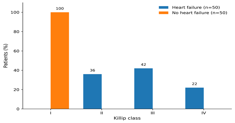
Figure 1. Distribution of Killip class among STEMI patients with and without heart failure.
Left Ventricular Ejection Fraction
The distribution of left ventricular ejection fraction (LVEF) is shown in Table 2 and Figure 2.
Among patients with heart failure, 39 (78.0%) had an LVEF of <40%, while 11 (22.0%) had an LVEF of 40–49%. None of the cases had an LVEF >50%.
Among controls, 4 (8.0%) had an LVEF of 40–49%, whereas 46 (92.0%) had an LVEF >50%. None of the controls had an LVEF <40%.
The mean LVEF was markedly lower among cases (34.78 ± 4.62%) than among controls (56.92 ± 5.39%), giving a mean difference of −22.14 percentage points. The difference between the two groups was highly statistically significant (p <0.001).
Table 2. Distribution and comparison of LVEF among STEMI patients with and without heart failure
|
LVEF (%) |
Cases (n=50), n (%) |
Controls (n=50), n (%) |
|
<40 |
39 (78.0) |
0 (0.0) |
|
40–49 |
11 (22.0) |
4 (8.0) |
|
>50 |
0 (0.0) |
46 (92.0) |
|
Total |
50 (100.0) |
50 (100.0) |
|
Mean ± SD |
34.78 ± 4.62 |
56.92 ± 5.39 |
Mean difference: −22.14 percentage points p <0.001.
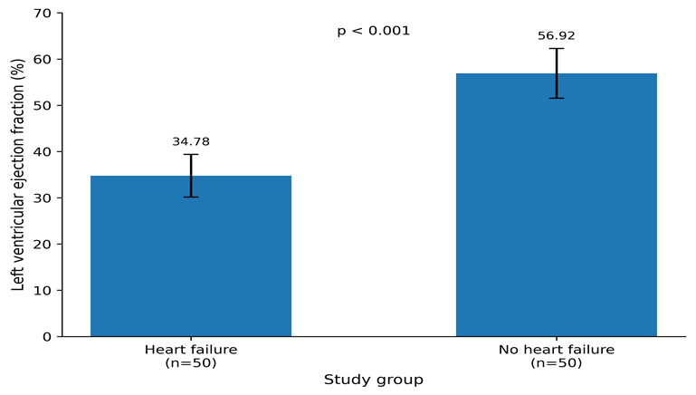
Figure 2. Distribution of LVEF among STEMI patients with and without heart failure.
Neutrophil-to-Lymphocyte Ratio
The distribution of neutrophil-to-lymphocyte ratio (NLR), the principal variable of the study, is presented in Table 3 and Figure 3.
A pronounced difference in NLR distribution was observed between patients with and without heart failure.
Among cases, 40 (80.0%) had an NLR >5.0, while 10 (20.0%) had an NLR between 3.5 and 5.0. None of the patients with heart failure had an NLR <3.5.
In contrast, all 50 controls (100.0%) had an NLR <3.5. None of the controls had an NLR between 3.5 and 5.0 or >5.0.
The mean NLR was 6.06 among cases compared with 2.61 among controls, demonstrating substantially higher NLR values among STEMI patients with heart failure.
Table 3. Distribution of NLR among STEMI patients with and without heart failure
|
NLR category |
Cases (n=50), n (%) |
Controls (n=50), n (%) |
|
<3.5 |
0 (0.0) |
50 (100.0) |
|
3.5–5.0 |
10 (20.0) |
0 (0.0) |
|
>5.0 |
40 (80.0) |
0 (0.0) |
|
Total |
50 (100.0) |
50 (100.0) |
|
Mean NLR |
6.06 |
2.61 |
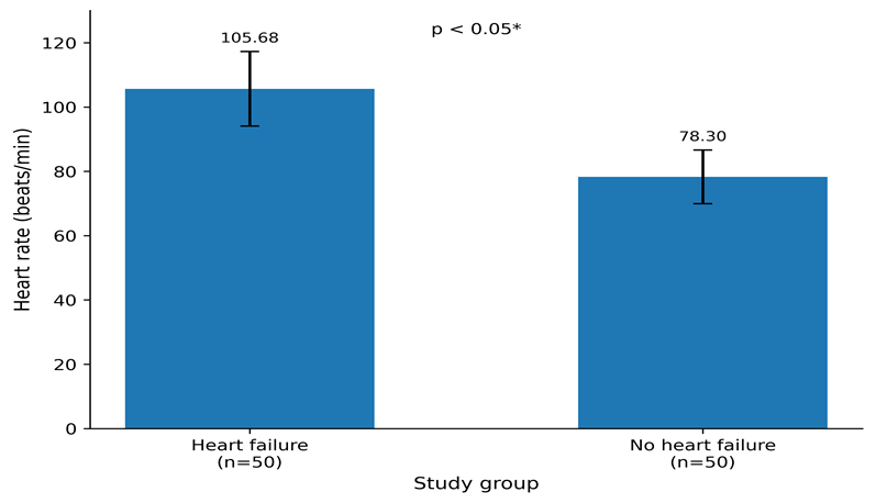
Figure 3. Distribution of neutrophil-to-lymphocyte ratio among STEMI patients with and without heart failure.
The distribution demonstrated a clear separation between the two groups, with higher NLR categories occurring exclusively among patients with heart failure.
Comparison of Heart Rate
The comparison of heart rate between the two study groups is presented in Table 4 and Figure 4.
The mean heart rate was significantly higher among patients with heart failure (105.68 ± 11.58 beats/min) compared with patients without heart failure (78.30 ± 8.33 beats/min).
The mean difference in heart rate between the groups was approximately 27.38 beats/min. The difference was statistically significant according to the study analysis (p <0.05).
Table 4. Comparison of heart rate among STEMI patients with and without heart failure
|
Parameter |
Cases (n=50) |
Controls (n=50) |
|
Mean heart rate (beats/min) |
105.68 |
78.30 |
|
SD |
11.58 |
8.33 |
|
Mean difference |
27.38 |
— |
|
p-value |
<0.05 |
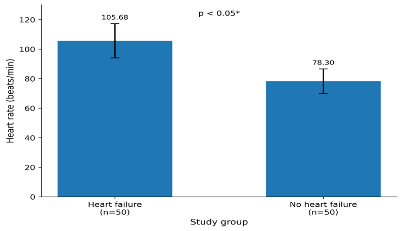
Figure 4. Comparison of mean heart rate between STEMI patients with and without heart failure.
Overall Comparison of Principal Study Variables
The principal clinical, inflammatory, and echocardiographic findings are summarized in Table 5.
Patients with heart failure had a substantially higher mean NLR than controls (6.06 vs 2.61). Similarly, 80.0% of cases had NLR >5.0, whereas all controls had NLR <3.5.
Clinical severity was greater among cases, with all patients classified as Killip Classes II–IV. Killip Class III was the most frequent category, accounting for 42.0% of cases.
Left ventricular systolic function was markedly impaired among patients with heart failure. The mean LVEF was 34.78 ± 4.62% among cases compared with 56.92 ± 5.39% among controls (p <0.001). Furthermore, 78.0% of cases had LVEF <40%, whereas none of the controls had LVEF <40%.
Heart rate was also higher among cases than controls (105.68 ± 11.58 vs 78.30 ± 8.33 beats/min; p <0.05).
Table 5. Summary comparison of principal variables between STEMI patients with and without heart failure
|
Variable |
Cases (n=50) |
Controls (n=50) |
|
Mean age (years) |
58.18 |
50.76 |
|
Male sex, n (%) |
36 (72.0) |
36 (72.0) |
|
Mean BMI (kg/m²) |
27.52 |
25.18 |
|
Mean systolic BP (mmHg) |
158.40 |
132.56 |
|
Mean diastolic BP (mmHg) |
94.08 |
83.62 |
|
Mean heart rate (beats/min) |
105.68 ± 11.58 |
78.30 ± 8.33 |
|
Mean NLR |
6.06 |
2.61 |
|
NLR >5.0, n (%) |
40 (80.0) |
0 (0.0) |
|
NLR 3.5–5.0, n (%) |
10 (20.0) |
0 (0.0) |
|
NLR <3.5, n (%) |
0 (0.0) |
50 (100.0) |
|
LVEF <40%, n (%) |
39 (78.0) |
0 (0.0) |
|
LVEF 40–49%, n (%) |
11 (22.0) |
4 (8.0) |
|
LVEF >50%, n (%) |
0 (0.0) |
46 (92.0) |
|
Mean LVEF (%) |
34.78 ± 4.62 |
56.92 ± 5.39 |
Summary of Findings
The study demonstrated a marked association between elevated NLR and heart failure among patients with acute STEMI. Patients with heart failure had substantially higher NLR values than controls, with a mean NLR of 6.06 compared with 2.61. Notably, 80.0% of cases had NLR >5.0, whereas 100.0% of controls had NLR <3.5.
The presence of heart failure was also associated with greater clinical severity. All cases had Killip Classes II–IV, with Killip Class III being the most common category (42.0%), while all controls had Killip Class I.
Patients with heart failure demonstrated substantial impairment of left ventricular systolic function. Seventy-eight percent had LVEF <40%, and the mean LVEF was significantly lower among cases than controls (34.78 ± 4.62% vs 56.92 ± 5.39%; p <0.001).
In addition, cases had a substantially higher mean heart rate than controls (105.68 ± 11.58 vs 78.30 ± 8.33 beats/min; p <0.05).
Taken together, the findings demonstrate that higher NLR was associated with the presence of heart failure, greater Killip class severity, and impaired left ventricular systolic function in patients with acute STEMI.
DISCUSSION:
The present hospital-based case-control study evaluated the association between neutrophil-to-lymphocyte ratio (NLR) and heart failure among patients with acute ST-elevation myocardial infarction (STEMI). Among 100 patients, comprising 50 with heart failure and 50 without heart failure, NLR was substantially higher in the heart-failure group. The mean NLR was 6.06 in cases compared with 2.61 in controls, and 80.0% of cases had NLR >5.0, whereas none of the controls had NLR >3.5. These findings demonstrate a strong association between elevated NLR and heart failure in patients with acute STEMI.
The biological basis for this association is plausible. Acute myocardial infarction triggers an inflammatory response characterized by neutrophil activation and recruitment, while physiological stress may produce relative lymphopenia. The resulting increase in NLR may therefore reflect the systemic inflammatory response and severity of myocardial injury. Because NLR is calculated from routine complete blood count parameters, it represents a simple and readily available inflammatory marker.³
The present findings are consistent with previous studies demonstrating an association between elevated NLR and adverse outcomes in STEMI. Karaca et al. reported that higher NLR was associated with worse in-hospital outcomes among STEMI patients undergoing primary PCI, while Tavares et al. found an association between elevated NLR and mortality and major adverse cardiovascular events in patients managed with a pharmaco-invasive strategy.⁷˒⁸ Similarly, a systematic review and meta-analysis by Ul Hussain et al. reported an association between increased NLR and adverse cardiovascular outcomes following STEMI.⁹ The present study extends these observations by demonstrating a marked difference in NLR between STEMI patients with and without clinically evident heart failure.
A substantial difference in left ventricular systolic function was also observed. Mean LVEF was 34.78 ± 4.62% in cases compared with 56.92 ± 5.39% in controls (p <0.001), and 78.0% of cases had LVEF <40%, compared with none of the controls. This finding is consistent with the expected reduction in ventricular function among patients with heart failure following myocardial infarction. Although inflammation may contribute to myocardial injury and adverse remodeling, the present case-control design cannot establish that elevated NLR directly causes reduced LVEF.¹²
Killip classification further demonstrated the greater clinical severity of the case group. Among cases, 36.0% were Killip Class II, 42.0% Class III, and 22.0% Class IV, whereas all controls were Class I. The predominance of higher Killip classes among cases confirms clinically significant cardiac dysfunction. The coexistence of elevated NLR and higher Killip class suggests that increased inflammatory activity may accompany greater clinical severity; however, an independent association cannot be established without multivariable analysis.
Cases also had higher mean heart rate (105.68 ± 11.58 vs 78.30 ± 8.33 beats/min) and were older than controls (58.18 vs 50.76 years). These differences may reflect greater physiological stress and disease severity but may also represent potential confounding factors. Although males constituted an identical proportion of both groups (72.0%), other unmeasured clinical factors may have influenced the observed association.
An important practical advantage of NLR is that it can be obtained from routine complete blood count testing without additional specialized investigations. Therefore, it may have potential as an adjunctive marker for early clinical risk assessment in STEMI patients, particularly in resource-limited settings.
However, the findings should be interpreted cautiously. The study does not establish NLR as an independent predictor or diagnostic test for heart failure. No multivariable logistic regression analysis was performed to adjust for potential confounders, and no ROC analysis was available to determine sensitivity, specificity, or an optimal cutoff value. Similarly, the NLR categories of <3.5, 3.5–5.0, and >5.0 should be regarded as study-specific categories rather than validated clinical thresholds.
The study has several limitations, including its single-center design, relatively small sample size, observational case-control methodology, and lack of multivariable analysis. In addition, NLR is a nonspecific inflammatory marker and may be influenced by other inflammatory, infectious, hematological, or physiological conditions. The close relationship between heart failure, Killip class, and LVEF also means that these variables should be interpreted primarily as measures of clinical severity rather than independent predictors.
Overall, the present study demonstrates a strong association between elevated NLR and heart failure among patients with acute STEMI. Patients with heart failure had higher NLR, higher Killip class, greater heart rate, and substantially reduced LVEF compared with controls. NLR may therefore be a useful adjunctive, inexpensive, and readily available marker of clinical severity in acute STEMI. Larger prospective multicenter studies with multivariable and ROC-based analyses are required to determine its independent prognostic value and establish a clinically meaningful cutoff.
CONCLUSION:
The present study demonstrated a strong association between elevated neutrophil-to-lymphocyte ratio (NLR) and heart failure in patients with acute STEMI. Patients with heart failure had a higher mean NLR (6.06 vs 2.61) and greater clinical severity, as reflected by higher Killip class and markedly reduced LVEF (34.78% vs 56.92%, p <0.001).
NLR is an inexpensive, readily available, and easily calculated inflammatory marker that may serve as a useful adjunct for early assessment of STEMI patients at increased risk of heart failure. However, the present findings do not establish NLR as an independent predictor. Larger prospective multicenter studies with multivariable and ROC analyses are required to validate its prognostic value and determine an appropriate clinical cutoff.
REFERENCES:
1. Akbar H, Mountfort S. Acute ST-Segment Elevation Myocardial Infarction (STEMI). In: StatPearls [Internet]. Treasure Island (FL): StatPearls Publishing; 2025.
2. Bueno H, Martín-Asenjo R. Acute heart failure after STEMI. Still a problem, still an opportunity for improving care quality. Int J Cardiol. 2017;248:274–275. doi:10.1016/j.ijcard.2017.08.048.
3. Buonacera A, Stancanelli B, Colaci M, Malatino L. Neutrophil to Lymphocyte Ratio: An Emerging Marker of the Relationships between the Immune System and Diseases. Int J Mol Sci. 2022;23(7):3636.
4. Karthikeyan T, Raja M, Radha D, Gaur TA, Geetha J, Sakthivadivel V. Risk factors and inflammatory markers in acute coronary syndrome-ST elevation myocardial infarction (STEMI). Horm Mol Biol Clin Investig. 2023;44(2):115–120. doi:10.1515/hmbci-2021-0106.
5. Gazi E, Bayram B, Gazi S, Temiz A, Kirilmaz B, Altun B, Barutcu A. Prognostic Value of the Neutrophil-Lymphocyte Ratio in Patients With ST-Elevated Acute Myocardial Infarction. Angiology. 2015;66(2):155–159. doi:10.1177/1076029613492011.
6. Rostami A, Tajlil A, Separham A, Sohrabi B, Pourafkari L, Roshanravan N, et al. Association between Neutrophil-to-Lymphocyte Ratio and the Systemic Inflammatory Immunologic Index and the Angiographic SYNTAX Score and the TIMI Flow Grade in Acute STEMI: A Cohort Study. J Tehran Heart Cent. 2021;16(4):147–155.
7. Karaca G, Ekmekci A, Kimiaei A, Safaei S, Özer N, Tayyareci G. The Impact of the Neutrophil-to-Lymphocyte Ratio on In-Hospital Outcomes in Patients With Acute ST-Segment Elevation Myocardial Infarction. Cureus. 2024;16(2):e54418. doi:10.7759/cureus.54418.
8. Tavares F, Moraes PIM, Souza JM, Barbosa AH, Santos EM, Marcondes JA, et al. Prognostic role of neutrophil-to-lymphocyte ratio in patients with ST-elevation myocardial infarction undergoing to pharmaco-invasive strategy. Cardiovasc Revasc Med. 2022;34:99–103.
9. Ul Hussain H, Kumar KA, Zahid M, Husban Burney M, Khan Z, Asif M, et al. Neutrophil to lymphocyte ratio as a prognostic marker for cardiovascular outcomes in patients with ST-segment elevation myocardial infarction after percutaneous coronary intervention: A systematic review and meta-analysis. Medicine (Baltimore). 2024;103(26):e38692.
10. Hou AC, Zhao JY, Wei YJ, Ou ZH, Liu CF. The neutrophil-to-lymphocyte ratio is associated with in-hospital heart failure in patients with ST-segment elevation myocardial infarction treated with primary percutaneous coronary intervention. Heliyon. 2024;10(21):e39761.
11. Yoon GS, Choi SH, Woo SI, Baek YS, Park SD, Shin SH, et al. Neutrophil-to-Lymphocyte Ratio at Emergency Room Predicts Mechanical Complications of ST-segment Elevation Myocardial Infarction. J Korean Med Sci. 2021;36(19):e131.
12. Gho JMIH, Postema PG, Conijn M, Bruinsma N, de Jong JSSG, Bezzina CR, et al. Heart failure following STEMI: a contemporary cohort study of incidence and prognostic factors. Open Heart. 2017;4(2):e000551.