Diagnostic Accuracy of High-Resolution Ultrasonography Compared with Magnetic Resonance Imaging for Supraspinatus Tendon Tears: A Cross-Sectional Study from a Tertiary-Care Hospital in Lahore, Pakistan.
- Dr. Zunair Maqsood , MBBS, FCPS Radiology, Assistant Professor of Radiology, Department of Diagnostic Radiology & Medical Imaging, Mayo Hospital / King Edward Medical University, Lahore
- Dr. Shehzad Karim Bhatti , MBBS, FCPS radiology, Associate Professor of Radiology, Department of Diagnostic Radiology & Medical Imaging, Mayo Hospital / King Edward Medical University, Lahore
- Dr. Irum Shahzad , FCPS Radiology, Senior Registrar Radiology, Department of Diagnostic Radiology & Medical Imaging, Mayo Hospital, Lahore
- Dr. Maryam Inayat , MBBS, FCPS, Head of Department Anesthesia and Pain Management, Doctors Hospital, Lahore
- Dr. Amir Rafiq , MBBS, MD Radiology, Department of Diagnostic Radiology & Medical Imaging, Mayo Hospital, Lahore
- Sana Zaheer Qureshi , B.Sc. Medical Imaging Technology, Lecturer MIT Radiology, Department of Diagnostic Radiology & Medical Imaging, Mayo Hospital / King Edward Medical University, Lahore
- Noor Shahzad , Noor Shahzad, Bhatti, BS Honors Computer Sciences, PGD Public Health, Health Services Academy, Islamabad
Article Information:
Abstract:
Background: High-resolution ultrasonography (HRUS) is accessible, radiation-free and allows real-time assessment of the rotator cuff, but its accuracy against magnetic resonance imaging (MRI) for full- and partial-thickness supraspinatus tears needs local evaluation. Objective: To determine the sensitivity, specificity, positive predictive value (PPV), negative predictive value (NPV) and accuracy of HRUS for full-thickness and partial-thickness supraspinatus tendon tears, using MRI as the reference standard. Methods: In this cross-sectional diagnostic-accuracy study at the Department of Diagnostic Radiology, Mayo Hospital, Lahore, Pakistan, 59 patients aged 18–85 years with suspected supraspinatus tears were recruited by convenience sampling and underwent both HRUS and MRI. Diagnostic performance was calculated separately for each tear type, and the association between HRUS and MRI findings was tested with the Pearson χ² test. Results: Mean age was 54.7 ± 12.6 years; 40 patients (68%) were male. HRUS identified a partial-thickness tear in 21 patients (35.6%), a full-thickness tear in 32 (54.2%) and no tear in 6 (10.2%). For full-thickness tears, sensitivity was 92.5%, specificity 98.3%, PPV 93.6%, NPV 97.8% and accuracy 95.4%. For partial-thickness tears, the corresponding values were 88.7%, 94.2%, 90.3%, 92.5% and 91.5%. The χ² statistic was 5.72 (1 degree of freedom; p = 0.017). Conclusions: HRUS showed high diagnostic performance against MRI, particularly for full-thickness tears. It is a reasonable initial examination for suspected supraspinatus tears, with MRI reserved for equivocal or complex cases, but equivalence to MRI is not shown. Larger blinded studies with confidence intervals and arthroscopic verification are needed.
Keywords:
Article :
INTRODUCTION:
Rotator cuff disorders are a common cause of shoulder pain and functional limitation, and the supraspinatus is the tendon most often affected. Tears are classified as partial-thickness or full-thickness according to the extent of tendon disruption [1]. Ultrasound screening of a general population has shown that full-thickness tears are frequent, increase with age and are often asymptomatic [2], so imaging must be interpreted together with the clinical picture. Accurate characterisation of a tear informs decisions about conservative management, further imaging and surgical referral.
MRI provides detailed assessment of the rotator cuff, including tendon retraction and muscle quality. HRUS is widely available, does not use ionising radiation and is performed in real time, but its performance depends on operator experience, patient positioning and image quality, and it has recognised pitfalls and artefacts [3]. Partial-thickness tears are particularly challenging because small or surface-specific defects can be subtle [4].
Systematic reviews and meta-analyses indicate that ultrasound performs well for full-thickness tears, whereas accuracy for partial-thickness tears is lower and more variable [5–10]. A Cochrane review found no statistically significant difference between ultrasound and MRI for full-thickness tears, although indirect comparisons suggested that ultrasound may be less sensitive for partial-thickness tears [8]. MRI is itself imperfect, with pooled sensitivity of 0.80 and specificity of 0.95 for partial-thickness tears and 0.91 and 0.97 for full-thickness tears [11]. Primary studies comparing ultrasound with MRI report similar full-thickness sensitivity (93.7–93.8%) but widely differing partial-thickness sensitivity (52.9–75.6%) [12,13], and a Pakistani study of 169 patients reported an overall sensitivity of 92.86% and specificity of 90.59% for rotator cuff pathology [14]. Ultrasound is regarded as reliable and accurate but operator dependent [15]. Data from tertiary-care departments in Pakistan remain limited.
The aim of this study was to determine the diagnostic accuracy of HRUS for full-thickness and partial-thickness supraspinatus tendon tears, with MRI as the reference standard, in patients referred to a tertiary-care radiology department in Lahore. This report follows the STARD 2015 recommendations [16,17].
MATERIALS AND METHODS:
Study design and setting
This cross-sectional diagnostic-accuracy study was conducted in the Department of Diagnostic Radiology, Mayo Hospital, Lahore, Pakistan, between January 2024 to August 2024.
Participants and sample size
Patients were recruited by non-probability convenience sampling from outpatient and inpatient referrals. Eligible participants were men and women aged 18–85 years referred for shoulder MRI with a suspected supraspinatus tear and restricted shoulder movement. Patients were excluded if they had undergone previous shoulder surgery, had a shoulder implant, cardiac pacemaker, prosthetic heart valve, aneurysm clip or other ferromagnetic material, had claustrophobia, or were unable to cooperate with the study. The sample size of 59 was calculated for the sensitivity of HRUS for full-thickness tears, using a 95% confidence level, 10% absolute precision, an expected sensitivity of 93.7% [12], an expected specificity of 100% and an expected prevalence of 39%. Participant flow is shown in Figure 1.
Index test: high-resolution ultrasonography
HRUS was performed with a multifrequency 6–13 MHz linear-array transducer, with focus and frequency adjusted to the depth of the tendon. The patient sat, and the supraspinatus was examined in the Crass position (shoulder extended, adducted and internally rotated, elbow flexed, palm directed away from the body and fingers against the opposite scapula), which brings the tendon from beneath the acromion. When the bicipital groove was not seen, the modified Crass position (hand on the hip or in the back pocket with the elbow drawn back) was used. The long head of the biceps tendon was used to localise the anterior edge of the supraspinatus. The tendon was scanned in longitudinal and transverse planes, along its whole length, to avoid false-negative findings. Tears were classified as partial-thickness, full-thickness or absent.
Reference standard: magnetic resonance imaging
MRI was the reference standard for classifying supraspinatus tears as full-thickness, partial-thickness or absent.
Outcomes and statistical analysis
The primary outcomes were sensitivity, specificity, PPV, NPV and accuracy of HRUS for full-thickness and partial-thickness tears. Continuous variables are presented as mean ± standard deviation and categorical variables as number (percentage). The association between HRUS and MRI findings was assessed with the Pearson χ² test, with p < 0.05 considered statistically significant. Analyses were performed with SPSS 2026.
Ethical considerations
The study was approved by King Edward Medical University, Lahore Institutional Review Board. Written informed consent was obtained from all participants, and the study followed the Declaration of Helsinki.
RESULTS:
Table 1. Characteristics of the study participants (n = 59)
|
Characteristic |
Value |
|
Participants, n |
59 |
|
Age, years, mean ± SD |
54.7 ± 12.6 |
|
Age, years, range |
18–85 |
|
Sex, n (%) |
|
|
Male |
40 (68) |
|
Female |
19 (32) |
|
Clinical presentation, % |
|
|
Shoulder pain |
87 |
|
Restricted shoulder movement |
75 |
|
Difficulty with abduction |
55 |
|
HRUS classification, n (%) |
|
|
Partial-thickness tear |
21 (35.6) |
|
Full-thickness tear |
32 (54.2) |
|
No tear |
6 (10.2) |
HRUS, high-resolution ultrasonography; SD, standard deviation.
Participants
Fifty-nine patients underwent both HRUS and MRI (Figure 1).
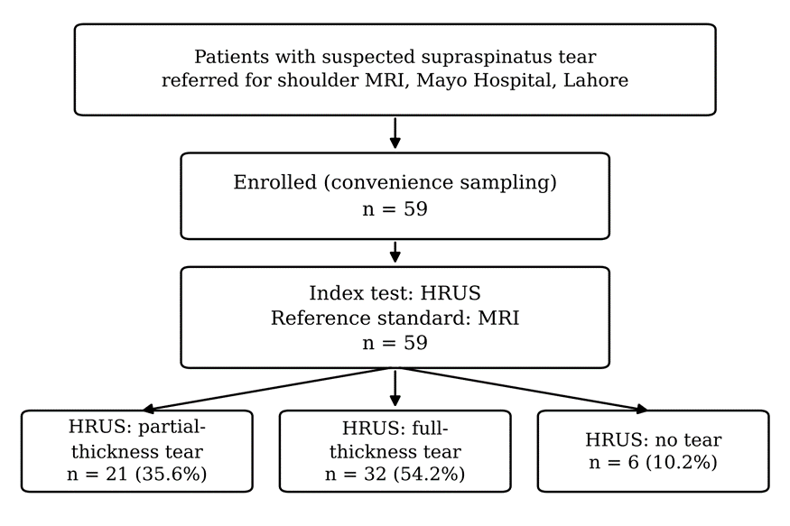
Figure 1. Flow of participants through the study. HRUS, high-resolution ultrasonography; MRI, magnetic resonance imaging.
The mean age was 54.7 ± 12.6 years (range, 18–85 years); 40 (68%) were male and 19 (32%) were female. Shoulder pain was reported by 87% of patients, restricted shoulder movement by 75% and difficulty with abduction by 55% (Table 1). HRUS classified 21 patients (35.6%) as having a partial-thickness tear, 32 (54.2%) as having a full-thickness tear and 6 (10.2%) as having no tear. These are HRUS classification counts and should not be read as MRI-defined disease prevalence.
Diagnostic performance of HRUS
Diagnostic performance against MRI is summarised in Table 2 and Figure 2.
Table 2. Diagnostic performance of HRUS for supraspinatus tendon tears, with MRI as the reference standard
|
Measure |
Full-thickness tear |
Partial-thickness tear |
|
Sensitivity, % |
92.5 |
88.7 |
|
Specificity, % |
98.3 |
94.2 |
|
Positive predictive value, % |
93.6 |
90.3 |
|
Negative predictive value, % |
97.8 |
92.5 |
|
Accuracy, % |
95.4 |
91.5 |
HRUS, high-resolution ultrasonography; MRI, magnetic resonance imaging. Pearson χ² = 5.72, 1 degree of freedom, p = 0.017.
For full-thickness tears, sensitivity was 92.5%, specificity 98.3%, PPV 93.6%, NPV 97.8% and accuracy 95.4%. For partial-thickness tears, the corresponding values were 88.7%, 94.2%, 90.3%, 92.5% and 91.5%.
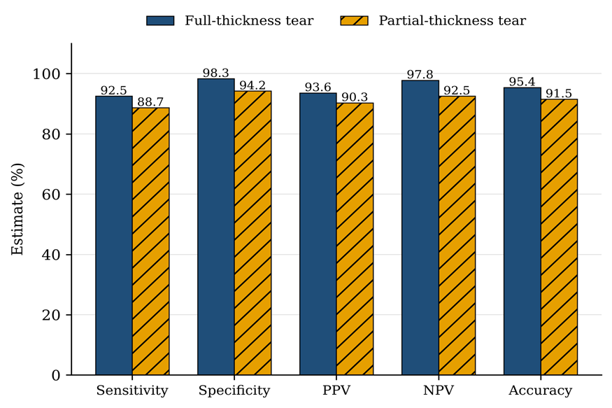
Figure 2. Diagnostic performance of HRUS for full-thickness and partial-thickness supraspinatus tears against MRI. PPV, positive predictive value; NPV, negative predictive value.
Every estimate was numerically higher for full-thickness than for partial-thickness tears.
Association between HRUS and MRI findings
The Pearson χ² test showed a statistically significant association between HRUS and MRI findings (χ² = 5.72, 1 degree of freedom, p = 0.017).
DISCUSSION:
In this single-centre study of 59 patients, HRUS showed high diagnostic performance against MRI for both full-thickness and partial-thickness supraspinatus tears, with higher estimates for full-thickness tears. This pattern is clinically plausible: full-thickness defects are usually more conspicuous on ultrasound, whereas small articular-sided, bursal-sided or intrasubstance defects can be subtle and depend more on technique and operator skill [3,4,15].
Our full-thickness estimates (sensitivity 92.5%, specificity 98.3%) are close to those of Aminzadeh et al. (93.7% and 100%; 48 patients) [12] and Agarwal et al. (93.8% and 100%; 50 patients) [13], and to meta-analytic estimates for ultrasound of 0.96 and 0.93 [6] and 92% and 93% [8]. A systematic review reported median full-thickness values of 0.88 and 0.93 [10], and individual studies against surgical findings report similar results [18,19]. For partial-thickness tears, our sensitivity (88.7%) and specificity (94.2%) are higher than the pooled 0.84 and 0.89 [6], the range of 0.67–0.83 for partial tears and tendinopathy [5] and the median of 0.65 and 0.86 [10], and far above the 52.9% and 75.6% reported by Aminzadeh et al. [12] and Agarwal et al. [13]. They do fall within the 72–94% range of sensitivities reported across 13 studies [9]. Meta-analytic work has found no significant difference between ultrasound and MRI for either tear type [7], and a smaller Pakistani study of 40 patients detected tears with ultrasound in 17 patients compared with 19 by MRI [20]. Our partial-thickness estimates should nevertheless be interpreted cautiously: only 21 patients were classified as having such tears, no confidence intervals are available, and differences in case mix, reader experience and MRI criteria could explain the higher values.
These results support the use of HRUS as an accessible first-line examination for suspected supraspinatus tears when performed by trained operators, particularly where MRI is costly or has long waiting times. MRI remains important when ultrasound findings are equivocal, when symptoms and ultrasound findings disagree, when a partial-thickness tear is suspected despite a negative ultrasound, and when information on tendon retraction, muscle atrophy or associated intra-articular pathology is needed for surgical planning.
This study has limitations. First, the sample was small and recruited by convenience sampling at a single tertiary-care centre, which limits precision and generalisability and carries a risk of spectrum and selection bias. Second, confidence intervals for the diagnostic estimates are not presented; with 59 participants they are expected to be wide. Third, ultrasound is operator dependent, and inter-reader reliability was not assessed. Fourth, MRI is not a perfect reference standard [11]; arthroscopic or surgical verification was not available, so the estimates describe agreement with MRI rather than with the true anatomical state. Fifth, the χ² test indicates association only. It does not quantify agreement or demonstrate equivalence between the tests; agreement is better described with a chance-corrected statistic such as kappa [21], and paired differences in sensitivity and specificity with paired confidence-interval methods [22]. Finally, only the supraspinatus was evaluated, so the findings do not extend to the other rotator cuff tendons.
Future studies should recruit consecutive patients, prespecify ultrasound and MRI criteria, use blinded independent readers and standardised protocols, verify findings arthroscopically where possible, report complete 2 × 2 tables with 95% confidence intervals, and plan sample size around the desired precision of sensitivity and specificity. Risk of bias and applicability can be appraised with QUADAS-2 [23], and reporting should follow STARD 2015 [16,17]. Larger multicentre studies and the evaluation of contrast-enhanced or artificial-intelligence-assisted ultrasound could further clarify performance for subtle partial-thickness tears.
CONCLUSION:
In this single-centre sample, HRUS showed high diagnostic performance against MRI for supraspinatus tendon tears, with better results for full-thickness than for partial-thickness tears. HRUS is a reasonable initial examination in suitable settings, particularly where MRI access is limited, but the present evidence is insufficient to conclude that it is equivalent to MRI or can replace it. Confirmation in larger, blinded studies with arthroscopic verification and confidence intervals is warranted.
REFERENCES:
1. Moosikasuwan JB, Miller TT, Burke BJ. Rotator cuff tears: clinical, radiographic, and US findings. Radiographics. 2005;25(6):1591-607. doi:10.1148/rg.256045203
2. Minagawa H, Yamamoto N, Abe H, Fukuda M, Seki N, Kikuchi K, et al. Prevalence of symptomatic and asymptomatic rotator cuff tears in the general population: from mass-screening in one village. J Orthop. 2013;10(1):8-12. doi:10.1016/j.jor.2013.01.008
3. Rutten MJCM, Jager GJ, Blickman JG. US of the rotator cuff: pitfalls, limitations, and artifacts. Radiographics. 2006;26(2):589-604. doi:10.1148/rg.262045719
4. Jacobson JA, Lancaster S, Prasad A, van Holsbeeck MT, Craig JG, Kolowich P. Full-thickness and partial-thickness supraspinatus tendon tears: value of US signs in diagnosis. Radiology. 2004;230(1):234-42. doi:10.1148/radiol.2301020418
5. Roy JS, Braën C, Leblond J, Desmeules F, Dionne CE, MacDermid JC, et al. Diagnostic accuracy of ultrasonography, MRI and MR arthrography in the characterisation of rotator cuff disorders: a systematic review and meta-analysis. Br J Sports Med. 2015;49(20):1316-28. doi:10.1136/bjsports-2014-094148
6. Smith TO, Back T, Toms AP, Hing CB. Diagnostic accuracy of ultrasound for rotator cuff tears in adults: a systematic review and meta-analysis. Clin Radiol. 2011;66(11):1036-48. doi:10.1016/j.crad.2011.05.007
7. de Jesus JO, Parker L, Frangos AJ, Nazarian LN. Accuracy of MRI, MR arthrography, and ultrasound in the diagnosis of rotator cuff tears: a meta-analysis. AJR Am J Roentgenol. 2009;192(6):1701-7. doi:10.2214/AJR.08.1241
8. Lenza M, Buchbinder R, Takwoingi Y, Johnston RV, Hanchard NCA, Faloppa F. Magnetic resonance imaging, magnetic resonance arthrography and ultrasonography for assessing rotator cuff tears in people with shoulder pain for whom surgery is being considered. Cochrane Database Syst Rev. 2013;(9):CD009020. doi:10.1002/14651858.CD009020.pub2
9. Hekmatnia F, Jamalipour Soufi G, Zarei AP, Shafieioun S, Hekmatnia A, Sadraei N, et al. Diagnostic accuracy of ultrasound compared to magnetic resonance imaging for rotator cuff tears: a systematic review. Int J Burns Trauma. 2026;16(2):33-44. doi:10.62347/VJLM3737
10. Farooqi AS, Lee A, Novikov D, Kelly AM, Li X, Kelly JD 4th, et al. Diagnostic accuracy of ultrasonography for rotator cuff tears: a systematic review and meta-analysis. Orthop J Sports Med. 2021;9(10):23259671211035106. doi:10.1177/23259671211035106
11. Smith TO, Daniell H, Geere JA, Toms AP, Hing CB. The diagnostic accuracy of MRI for the detection of partial- and full-thickness rotator cuff tears in adults. Magn Reson Imaging. 2012;30(3):336-46. doi:10.1016/j.mri.2011.12.008
12. Aminzadeh B, Najafi S, Moradi A, Abbasi B, Farrokh D, Emadzadeh M. Evaluation of diagnostic precision of ultrasound for rotator cuff disorders in patients with shoulder pain. Arch Bone Jt Surg. 2020;8(6):689-95. doi:10.22038/abjs.2020.42894.2166
13. Agarwal A, Vani K, Batta A, Verma K, Chumber S. Can ultrasound suffice for triaging patients requiring surgical correction of rotator cuff tears: a comparative evaluation of ultrasound and magnetic resonance imaging. Egypt J Radiol Nucl Med. 2021;52(1):119. doi:10.1186/s43055-021-00477-2
14. Aziz S, Nigar F, Baloch SK, Latif A, Shafiq M, Mallhi EA. The diagnostic accuracy of shoulder ultrasound for rotator cuff pathology compared to magnetic resonance imaging as a gold standard. Pak Armed Forces Med J. 2025;75(Suppl 7):S1062-6. doi:10.51253/pafmj.v75iSUPPL-7.11608
15. Okoroha KR, Fidai MS, Tramer JS, Davis KD, Kolowich PA. Diagnostic accuracy of ultrasound for rotator cuff tears. Ultrasonography. 2019;38(3):215-20.
16. Bossuyt PM, Reitsma JB, Bruns DE, Gatsonis CA, Glasziou PP, Irwig L, et al. STARD 2015: an updated list of essential items for reporting diagnostic accuracy studies. BMJ. 2015;351:h5527. doi:10.1136/bmj.h5527
17. Cohen JF, Korevaar DA, Altman DG, Bruns DE, Gatsonis CA, Hooft L, et al. STARD 2015 guidelines for reporting diagnostic accuracy studies: explanation and elaboration. BMJ Open. 2016;6(11):e012799.
18. Teefey SA, Rubin DA, Middleton WD, Hildebolt CF, Leibold RA, Yamaguchi K. Detection and quantification of rotator cuff tears: comparison of ultrasonographic, magnetic resonance imaging, and arthroscopic findings in seventy-one consecutive cases. J Bone Joint Surg Am. 2004;86(4):708-16. doi:10.2106/00004623-200404000-00007
19. Naqvi GA, Jadaan M, Harrington P. Accuracy of ultrasonography and magnetic resonance imaging for detection of full thickness rotator cuff tears. Int J Shoulder Surg. 2009;3(4):94-7. doi:10.4103/0973-6042.63218
20. Qayyum A, Ilyas M, Adil R. Role of ultrasound in rotator cuff tears. Pak Armed Forces Med J. 2009;59(3):304-9.
21. Landis JR, Koch GG. The measurement of observer agreement for categorical data. Biometrics. 1977;33(1):159-74.
22. Newcombe RG. Improved confidence intervals for the difference between binomial proportions based on paired data. Stat Med. 1998;17(22):2635-50.
23. Whiting PF, Rutjes AWS, Westwood ME, Mallett S, Deeks JJ, Reitsma JB, et al. QUADAS-2: a revised tool for the quality assessment of diagnostic accuracy studies. Ann Intern Med. 2011;155(8):529-36.
24. International Committee of Medical Journal Editors. Recommendations for the conduct, reporting, editing, and publication of scholarly work in medical journals [Internet]. Updated January 2026 [cited 2026 Oct 26]. Available from: https://www.icmje.org/recommendations/