Acute renal failure in haemotoxic snake envenomation: a prospective observational study from a tertiary care hospital in South India.
- Dr. Sanket Patil G , Assistant Professor, Department of General Medicine, Raichur institute of medical sciences (Rims), Raichur, Karnataka, India
- Dr. Sharanabasavaraj Devareddy , Senior Resident, Department of General Medicine, Sri Siddhartha Institute of Medical Sciences & Research Centre, Bangalore, Karnataka, India.
Article Information:
Abstract:
Background: Acute renal failure (ARF) is a leading cause of death and long-term morbidity after viperine envenomation in South Asia, yet the clinical factors that identify patients at risk on admission remain incompletely defined. We aimed to determine the incidence, timing, probable aetiology, risk factors and outcome of ARF in adults with haemotoxic snake envenomation. Methods: In this prospective observational study at , Raichur institute of medical sciences (Rims), Raichur, Karnataka, India (July 2006 to June 2007), 107 consecutive adolescents and adults with haemotoxic envenomation were followed from admission to discharge or death. Associations with ARF were expressed as risk ratios (RR) with 95% confidence intervals (CI) and tested with Fisher's exact test. Results: ARF developed in 25 of 107 patients (23.4%; 95% CI 16.4–32.2%), 20 of whom were oliguric. Onset occurred at a mean of 25 hours after the bite and within 24 hours in 16 patients (64%). ARF was more frequent with systemic bleeding (51.6% vs 11.8%; RR 4.36, 95% CI 2.16–8.79; p<0.001), antivenom started more than 24 hours after the bite (83.3% vs 19.8%; RR 4.21, 2.47–7.16; p=0.003) and pre-existing anaemia (90.9% vs 15.6%; RR 5.82, 3.53–9.60; p<0.001). Sex, age above 50 years and tourniquet use were not associated with ARF. Disseminated intravascular coagulation contributed in 19 patients (76%), and direct nephrotoxicity was presumed in six (24%). Fourteen patients (56%) recovered with supportive therapy and five of seven treated with haemodialysis recovered. Three patients died (2.8% overall; 12.0% of those with ARF), all with ARF and coagulopathy. Conclusion: Nearly one in four patients with haemotoxic envenomation developed ARF, usually within the first day. Systemic bleeding, delayed antivenom and anaemia identify a high-risk group that warrants serial renal monitoring and early access to dialysis; tourniquets offered no renal protection.
Keywords:
Article :
INTRODUCTION:
Snakebite envenoming is a neglected tropical disease that falls most heavily on agricultural communities in South Asia, sub-Saharan Africa and Latin America. Literature-based modelling has estimated at least 421,000 envenomings and 20,000 deaths each year worldwide, and possibly as many as 1.8 million envenomings and 94,000 deaths, with South Asia carrying the largest share1. More recent estimates put annual deaths above 100,000 and permanent disability above 400,0002. India is the epicentre of this burden. A nationally representative verbal-autopsy study estimated about 1.2 million snakebite deaths in India between 2000 and 2019, an average of 58,000 deaths a year, most of them among rural people of working age bitten at home or in the fields3. These deaths are only part of the burden, because survivors may be left with amputation, chronic kidney disease or endocrine failure.
Most medically important bites in peninsular India are inflicted by the 'big four': the spectacled cobra, the common krait, Russell's viper (Daboia russelii) and the saw-scaled viper. The two vipers cause haemotoxic envenoming. Russell's viper venom contains potent procoagulants, including factor X and factor V activators, together with phospholipase A2 isoenzymes, metalloproteinases and other enzymes that produce defibrination, spontaneous haemorrhage, shock, intravascular haemolysis and acute renal failure (ARF)4. In Russell's viper envenoming, ARF is common, often severe, and one of the main causes of death among patients who survive the first hours after the bite4,5.
The kidney is vulnerable to snake venom for several reasons. It receives a fifth of the cardiac output, concentrates filtered toxins in the tubular lumen, and has an outer medulla that operates close to hypoxia even in health. The pathogenesis of snakebite-associated ARF is multifactorial. Haemodynamic injury follows bleeding, vasodilatation and capillary leak. Consumptive coagulopathy and thrombotic microangiopathy cause microvascular occlusion, while haemolysis and rhabdomyolysis release pigments toxic to the tubules. Direct cytotoxicity of venom components on tubular and glomerular cells adds to the injury6,7,8. On histology, acute tubular necrosis predominates, followed by acute interstitial nephritis and acute cortical necrosis. Cortical necrosis carries the worst prognosis9. A recent South Asian review emphasised that the relative weight of these mechanisms differs between species and settings and that progression to chronic kidney disease is a major concern among survivors10.
Estimates of how often ARF complicates envenoming vary widely. A 2026 systematic review and meta-analysis of 30 studies (8,612 participants) reported a pooled incidence of 23% (95% CI 17–33%), rising to 37% after Russell's viper bites, with substantial heterogeneity between studies11. Hospital series from southern India report ARF in about one-fifth of envenomed adults, of whom more than a third require dialysis12. In the Malabar region of Kerala, haemotoxic bites predominated in a series of 1,500 envenomings, and acute kidney injury was among the factors associated with poor outcome13. This variation reflects differences in snake species, referral pathways, delays before antivenom, and above all the definition of renal failure, which ranged from ad hoc creatinine cut-offs in older series to the RIFLE, AKIN and, since 2012, KDIGO criteria14.
The consequences of ARF extend well beyond the acute admission. Survivors of snakebite-associated ARF face a substantial risk of incomplete recovery: in a Sri Lankan nephrology cohort, 37% had developed chronic kidney disease one year after the bite, and the duration of renal replacement therapy predicted non-recovery15. A systematic review of the long-term effects of envenoming confirmed that progression from acute to chronic renal failure after Russell's viper bites has been reported repeatedly from India and Sri Lanka16. In India, the burden falls on farm labourers for whom snakebite is an occupational hazard and who often reach hospital late, after first aid or traditional treatment17. Dialysis capacity in public hospitals is limited, and the World Health Organization's goal of halving snakebite deaths by 2030 will not be met unless India makes substantial progress3. Identifying patients at highest risk of ARF early, using information available at the bedside, is therefore a practical priority for district and tertiary hospitals alike.
Several gaps remain in the evidence. First, most Indian series are retrospective, are restricted to patients already referred for dialysis, or report only patients who developed ARF, which makes it impossible to compare patients with and without renal failure within the same envenomed population. Second, clinically accessible predictors that can be recognised at the bedside on admission — the bite-to-antivenom interval, systemic bleeding, the clotting time, pre-existing anaemia and first-aid practices such as tourniquet application — have seldom been examined together in one prospective cohort. Third, the time course of renal failure after the bite, which determines how long patients must be monitored, is poorly described. Fourth, tourniquets remain a widely used first-aid measure in rural India despite little evidence of benefit.
We therefore studied a prospective cohort of adolescents and adults with haemotoxic envenomation admitted to a large public teaching hospital in Raichur,Karnataka. We hypothesised that ARF would be more frequent among patients with systemic bleeding, delayed antivenom therapy and pre-existing anaemia, and that tourniquet application would not protect against renal failure.
OBJECTIVES
The primary objective was to determine the incidence of ARF among patients admitted with haemotoxic snake envenomation.
The secondary objectives were: (i) to examine the association of ARF with age, sex, tourniquet application, the interval from bite to antivenom therapy, systemic bleeding, the clotting time and pre-existing anaemia; (ii) to describe the time of onset of ARF after the bite and its probable aetiology; (iii) to evaluate the outcome of supportive therapy, haemodialysis and peritoneal dialysis; (iv) to describe bleeding manifestations and other complications, including unusual presentations; and (v) to determine the case fatality rate and the probable causes of death.
METHODOLOGY:
Study design and setting
This was a prospective observational cohort study conducted in the medical wards of Raichur institute of medical sciences (Rims), Raichur, Karnataka, India. The hospital is a tertiary referral centre for a largely agrarian catchment in Karnataka. Patients were enrolled over 12 months, from July 2020, and followed until discharge or death. The study is reported in accordance with the STROBE statement18.
Participants
All patients aged 13 years or above admitted to the medical wards with a history of snakebite during the study period were screened for eligibility; the number of patients screened and excluded was not recorded. Patients were included if they had (i) a history of snakebite with local cellulitis together with bleeding manifestations, a prolonged clotting time, or both; or (ii) a history of snakebite with oliguria, irrespective of the interval since the bite. Patients with neurotoxic features (ptosis, external ophthalmoplegia or respiratory failure) and those without a visible bite mark were excluded. Children were managed in the paediatric department and were not enrolled. Snake species was not identified routinely, and inclusion was based on the clinical haemotoxic syndrome.
Data collection
A structured proforma was completed for every patient by the principal investigator. The history recorded the time, number and anatomical site of the bite; any first aid, including tourniquet application; the onset and type of bleeding (bite site, gums, haematemesis, melaena, haemoptysis, haematuria, epistaxis); pre-existing illness, with particular attention to anaemia; and urine volume and colour, including the time of onset of oliguria or anuria. Examination recorded the bite mark, local blistering, necrosis, regional lymphadenitis and cellulitis; pulse, blood pressure, temperature, respiratory rate and level of consciousness; and pallor, jaundice, oedema and signs of uraemia. Fluid intake and urine output were charted daily.
Investigations on admission, repeated as clinically indicated, included urinalysis (albumin, sugar, red cells, bile salts and pigments); total and differential leucocyte count, haemoglobin and erythrocyte sedimentation rate; bleeding time and whole blood clotting time; platelet count, peripheral smear for fragmented red cells, reticulocyte count and plasma fibrinogen; blood urea, serum creatinine and electrolytes; electrocardiography; and abdominal ultrasonography. Cranial computed tomography was performed when neurological signs developed.
Treatment
All patients received intravenous polyvalent antisnake venom (ASV; 10 mL per reconstituted vial), repeated until the clotting time normalised. The interval from bite to the first ASV dose and the total number of vials were recorded. Patients with ARF received supportive therapy consisting of careful fluid replacement, intravenous furosemide, dopamine, blood transfusion for anaemia or haemorrhage, treatment of hypotension, and avoidance of nephrotoxic drugs. Patients who did not respond to supportive therapy underwent haemodialysis or peritoneal dialysis. The number of dialysis sessions and the outcome were recorded.
Definitions
The primary outcome was ARF, defined as a rise in blood urea and serum creatinine above the upper limit of the laboratory reference range during admission, with or without oliguria. Oliguria was defined as urine output below 400 mL in 24 hours, and ARF without oliguria was classified as non-oliguric. Onset of ARF was the interval in hours from the bite to the first documented oliguria or biochemical rise. The whole blood clotting time was measured at the bedside, and a clotting time exceeding 20 minutes was taken to indicate venom-induced coagulopathy. Systemic bleeding was any bleeding away from the bite site. Pre-existing anaemia was severe anaemia (haemoglobin <8 g/dL) attributed by history and examination to a cause predating the bite. Hypotension was a blood pressure below the normal range on examination.
The probable aetiology of ARF was assigned clinically at the end of each admission. Disseminated intravascular coagulation (DIC) was diagnosed when systemic bleeding or incoagulable blood was accompanied by thrombocytopenia, low fibrinogen or fragmented red cells. DIC was further classified as occurring alone or with hypotension, shock and capillary leak syndrome, or shock and sepsis. Direct nephrotoxicity was presumed when ARF developed without evidence of DIC, hypotension or sepsis. Acute-on-chronic renal failure was diagnosed when ultrasonographic or biochemical evidence indicated pre-existing renal disease.
Study size and bias
No formal sample size calculation was performed. The study enrolled all eligible patients admitted during a fixed 12-month period, a pragmatic approach that captures a full seasonal cycle of snakebite. With 107 patients, the incidence of ARF could be estimated with a 95% confidence interval of about 16 percentage points (Wilson method), which was judged adequate for a descriptive primary objective. To limit selection bias, consecutive patients were enrolled throughout the year rather than only during working hours. To limit information bias, a single investigator completed every proforma using predefined fields. The primary outcome depended on laboratory measurements that did not require subjective judgement, but the assignment of probable aetiology was clinical and could have been influenced by knowledge of the clinical course.
Statistical analysis
Data were entered in a master chart and summarised as frequencies and percentages. Continuous data were available only as group-level summaries and are reported as means and ranges. The original study plan was descriptive. For this report, associations between candidate risk factors and ARF were quantified post hoc from the prospectively recorded counts as unadjusted risk ratios (RR) with 95% confidence intervals calculated by the Katz log method, applying a 0.5 continuity correction to tables containing a zero cell. Differences in proportions were tested with the two-sided Fisher's exact test because several expected cell counts were below five. Linear trends across ordered categories of age and bite-to-ASV interval were tested with the Cochran–Armitage test, and 95% confidence intervals for single proportions were calculated by the Wilson score method. Multivariable adjustment was not attempted because 25 outcome events could not support a stable model containing the candidate predictors. No data were missing for the variables analysed. A two-sided p value below 0.05 was considered statistically significant. Analyses were performed in Python version 3.13 using SciPy version 1.18.
Ethical considerations
The study protocol was approved by the Institutional Ethical Committee of , Raichur institute of medical sciences (Rims), Raichur, Karnataka, India. before patient enrolment. Informed consent was obtained from each patient or, where the patient could not consent or was under 18 years of age, from the next of kin or guardian. Patients were identified only by serial number in the analysis dataset.
RESULTS:
Participants and baseline characteristics
A total of 107 patients with haemotoxic envenomation were enrolled and followed to discharge or death . Of these, 75 (70.1%) were men and 32 (29.9%) were women. Ages ranged from 13 to 75 years, and 53 patients (49.5%) were aged 30 years or younger. The bite was on the lower limb in 88 patients (82.2%), the upper limb in 18 (16.8%) and the back in one. A tourniquet had been applied as first aid in 67 patients (62.6%). The clotting time was prolonged on admission in 101 patients (94.4%). ASV was started within 12 hours of the bite in 89 patients (83.2%), between 13 and 24 hours in 12 (11.2%), between 25 and 48 hours in four (3.7%) and after 48 hours in two (1.9%). A mean of 13 vials (range 4–25) was required to normalise the clotting time. Baseline characteristics are shown in Table 1.
Bleeding manifestations
Systemic bleeding occurred in 31 patients (29.0%). Its onset ranged from 30 minutes to 40 hours after the bite. It began within 6 hours in 21 of these patients (67.7%) and within 24 hours in 30 (96.8%). Bleeding from the gums was the most frequent manifestation (14 patients, 45.2%), followed by bleeding from multiple sites (8, 25.8%), haematemesis (4, 12.9%), haematuria (2, 6.5%), melaena (2, 6.5%) and haemoptysis (1, 3.2%). Of the 31 patients with systemic bleeding, 27 (87.1%) also had a prolonged clotting time.
Incidence of acute renal failure
ARF developed in 25 of 107 patients (23.4%; 95% CI 16.4–32.2%). Twenty of these patients (80.0%) were oliguric and five (20.0%) non-oliguric. ARF occurred in 17 of 75 men (22.7%) and 8 of 32 women (25.0%).
Factors associated with acute renal failure
Unadjusted associations are presented in Table 2 and Figure 1. ARF developed in 16 of 31 patients with systemic bleeding (51.6%) compared with 9 of 76 without it (11.8%) (RR 4.36, 95% CI 2.16–8.79; p<0.001). Among the 11 patients with pre-existing severe anaemia, 10 developed ARF (90.9%), compared with 15 of 96 patients without anaemia (15.6%) (RR 5.82, 95% CI 3.53–9.60; p<0.001). The risk of ARF rose with the interval from bite to ASV (Figure 2). ARF developed in 18 of 89 patients (20.2%) treated within 12 hours, 2 of 12 (16.7%) treated between 13 and 24 hours, 3 of 4 (75.0%) treated between 25 and 48 hours, and both patients treated after 48 hours (Cochran–Armitage z=2.97, p=0.003). Overall, ARF occurred in 5 of 6 patients (83.3%) whose ASV was started more than 24 hours after the bite, compared with 20 of 101 (19.8%) treated within 24 hours (RR 4.21, 95% CI 2.47–7.16; p=0.003). Contrary to expectation, ARF was more frequent among the six patients whose clotting time was normal on admission (4 of 6, 66.7%) than among the 101 with a prolonged clotting time (21 of 101, 20.8%) (RR 3.21, 95% CI 1.62–6.34; p=0.026). On case-level review of the master chart, two of these four patients had received ASV more than 24 hours after the bite. The proportion with ARF was 33.3% (7 of 21) among patients older than 50 years and 20.9% (18 of 86) among those aged 50 years or younger (RR 1.59, 95% CI 0.77–3.31; p=0.255). ARF occurred in both patients older than 60 years, but there was no significant linear trend across the five age bands (p=0.354). Sex (RR 1.10, 95% CI 0.53–2.29; p=0.807) and tourniquet application (23.9% vs 22.5%; RR 1.06, 95% CI 0.52–2.17; p=1.000) were not associated with ARF.
Onset, aetiology and management of acute renal failure
The interval from bite to onset of ARF ranged from 1.5 to 56 hours (mean 25 hours). Onset occurred within 12 hours in five patients (20%), between 13 and 24 hours in 11 (44%), between 25 and 36 hours in four (16%), between 37 and 48 hours in three (12%) and after 48 hours in two (8%). Thus, ARF developed within 24 hours of the bite in 16 of 25 patients (64%) (Table 3). The probable aetiology was DIC alone in 12 patients (48%), DIC with hypotension in four (16%), DIC with shock and capillary leak syndrome in two (8%), and DIC with shock and sepsis in one (4%). DIC therefore contributed to ARF in 19 of 25 patients (76%). In the remaining six patients (24%), ARF developed without evidence of DIC, hypotension or sepsis and was attributed to direct nephrotoxicity. All 25 patients received supportive therapy, with which renal function recovered in 14 (56.0%; 95% CI 37.1–73.3%). Of the 11 patients who did not respond, seven underwent haemodialysis (mean 6 sessions, range 2–12), and five of them recovered (71.4%). The two patients who did not recover were found to have acute-on-chronic renal failure. Four patients underwent peritoneal dialysis, and none recovered. One of them was found to have acute-on-chronic renal failure, and three died with ARF and other complications of envenomation.
Other complications and mortality
Complications other than ARF and bleeding comprised pre-existing anaemia (11 patients, 10.3%), hypotension or shock (8, 7.5%), compartment syndrome (5, 4.7%) and gangrene (2, 1.9%). Intracranial haemorrhage, sepsis, jaundice, ischaemic stroke and vitreous haemorrhage each occurred in one patient (0.9%) (Table 4). Two presentations were unusual. An 18-year-old man had multisite bleeding with left vitreous haemorrhage and ARF, recovered renal function after haemodialysis and partially regained vision in that eye. A 40-year-old man developed right hemiparesis, upper motor neuron facial palsy and Wernicke's aphasia within 4 hours of the bite; cranial computed tomography showed a left temporoparietal infarct, and his renal function remained normal. Three patients died (2.8%; 95% CI 1.0–7.9%). All were men, all had ARF and all had DIC, so the case fatality rate among patients with ARF was 12.0% (95% CI 4.2–30.0%). The probable causes of death were DIC with intracranial haemorrhage and ARF; DIC, circulatory failure, ARF and sepsis; and DIC, circulatory failure, ARF and capillary leak syndrome. Death occurred 36 to 84 hours after the bite (mean 64 hours), although all three patients had received ASV within 3 to 5 hours of the bite (mean 4 hours). Tourniquet use was not associated with mortality (1 of 67 vs 2 of 40; RR 0.30, 95% CI 0.03–3.19; p=0.554).
Table 1. Baseline characteristics of patients with haemotoxic snake envenomation (n = 107)
|
Characteristic |
n (%) |
|
Sex |
|
|
Male |
75 (70.1) |
|
Female |
32 (29.9) |
|
Age group (years) |
|
|
13–30 |
53 (49.5) |
|
31–40 |
18 (16.8) |
|
41–50 |
15 (14.0) |
|
51–60 |
17 (15.9) |
|
>60 |
4 (3.7) |
|
Site of bite |
|
|
Lower limb |
88 (82.2) |
|
Upper limb |
18 (16.8) |
|
Back |
1 (0.9) |
|
Tourniquet applied as first aid |
67 (62.6) |
|
Prolonged clotting time on admission |
101 (94.4) |
|
Systemic bleeding |
31 (29.0) |
|
Pre-existing severe anaemia (Hb <8 g/dL) |
11 (10.3) |
|
Interval from bite to first ASV dose (hours) |
|
|
1–12 |
89 (83.2) |
|
13–24 |
12 (11.2) |
|
25–48 |
4 (3.7) |
|
>48 |
2 (1.9) |
|
ASV vials to normalise clotting time, mean (range) |
13 (4–25) |
ASV, antisnake venom; Hb, haemoglobin. Percentages are of 107 patients. Age range 13–75 years.
Table 2. Unadjusted association of clinical factors with acute renal failure (n = 107)
|
Factor |
Exposed: ARF/n (%) |
Unexposed: ARF/n (%) |
RR (95% CI) |
p value |
|
Female vs male sex |
8/32 (25.0) |
17/75 (22.7) |
1.10 (0.53–2.29) |
0.807 |
|
Age >50 vs ≤50 years |
7/21 (33.3) |
18/86 (20.9) |
1.59 (0.77–3.31) |
0.255 |
|
Tourniquet applied vs not |
16/67 (23.9) |
9/40 (22.5) |
1.06 (0.52–2.17) |
1.000 |
|
Systemic bleeding vs none |
16/31 (51.6) |
9/76 (11.8) |
4.36 (2.16–8.79) |
<0.001 |
|
Normal vs prolonged clotting time |
4/6 (66.7) |
21/101 (20.8) |
3.21 (1.62–6.34) |
0.026 |
|
ASV >24 h vs ≤24 h after bite |
5/6 (83.3) |
20/101 (19.8) |
4.21 (2.47–7.16) |
0.003 |
|
Pre-existing anaemia vs none |
10/11 (90.9) |
15/96 (15.6) |
5.82 (3.53–9.60) |
<0.001 |
ARF, acute renal failure; ASV, antisnake venom; CI, confidence interval; RR, risk ratio. RR with 95% CI by the Katz log method; p values by two-sided Fisher's exact test. Trend tests (Cochran–Armitage): five age bands, p = 0.354; four bite-to-ASV bands, p = 0.003. Estimates are unadjusted.
Table 3. Onset, probable aetiology, management and outcome of acute renal failure (n = 25)
|
Variable |
n (%) |
|
Type |
|
|
Oliguric |
20 (80.0) |
|
Non-oliguric |
5 (20.0) |
|
Interval from bite to onset (hours); mean 25, range 1.5–56 |
|
|
≤12 |
5 (20.0) |
|
13–24 |
11 (44.0) |
|
25–36 |
4 (16.0) |
|
37–48 |
3 (12.0) |
|
>48 |
2 (8.0) |
|
Probable aetiology |
|
|
DIC alone |
12 (48.0) |
|
DIC with hypotension |
4 (16.0) |
|
DIC with shock and capillary leak syndrome |
2 (8.0) |
|
DIC with shock and sepsis |
1 (4.0) |
|
Presumed direct nephrotoxicity |
6 (24.0) |
|
Management and outcome |
|
|
Supportive therapy (all patients) |
25 (100) |
|
Recovered with supportive therapy alone |
14 (56.0) |
|
Haemodialysis (mean 6 sessions, range 2–12) |
7 (28.0) |
|
Recovered |
5 of 7 |
|
Acute-on-chronic renal failure |
2 of 7 |
|
Peritoneal dialysis |
4 (16.0) |
|
Died |
3 of 4 |
|
Acute-on-chronic renal failure |
1 of 4 |
|
Deaths among patients with ARF |
3 (12.0) |
ARF, acute renal failure; DIC, disseminated intravascular coagulation. Supportive therapy comprised fluid replacement, furosemide, dopamine, blood transfusion and avoidance of nephrotoxic drugs. Recovery refers to renal function at discharge.
Table 4. Complications and deaths in haemotoxic snake envenomation (n = 107)
|
Complication |
n (%) |
|
Systemic bleeding |
31 (29.0) |
|
Acute renal failure |
25 (23.4) |
|
Pre-existing severe anaemia |
11 (10.3) |
|
Hypotension or shock |
8 (7.5) |
|
Compartment syndrome |
5 (4.7) |
|
Gangrene |
2 (1.9) |
|
Intracranial haemorrhage |
1 (0.9) |
|
Sepsis |
1 (0.9) |
|
Jaundice |
1 (0.9) |
|
Ischaemic stroke |
1 (0.9) |
|
Vitreous haemorrhage |
1 (0.9) |
|
Deaths |
3 (2.8) |
|
DIC, intracranial haemorrhage, ARF (death 72 h; ASV at 4 h) |
1 |
|
DIC, circulatory failure, ARF, sepsis (death 36 h; ASV at 5 h) |
1 |
|
DIC, circulatory failure, ARF, capillary leak syndrome (death 84 h; ASV at 3 h) |
1 |
ARF, acute renal failure; ASV, antisnake venom; DIC, disseminated intravascular coagulation. Times are hours after the bite. Patients may have had more than one complication.
FIGURE LEGENDS
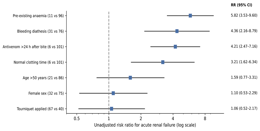
Figure 1. Unadjusted risk ratios for acute renal failure. Squares show risk ratios and horizontal lines their 95% confidence intervals (Katz log method) on a logarithmic scale; the dashed line marks no association. Group sizes (exposed vs unexposed) are shown in parentheses.
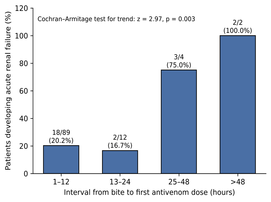
Figure 2. Incidence of acute renal failure by interval from bite to first antivenom dose. Bars show the percentage of patients in each interval who developed acute renal failure; numbers above the bars are events/patients. Linear trend tested by the Cochran–Armitage test.
DISCUSSION:
In this prospective cohort of 107 patients with haemotoxic snake envenomation, nearly one in four developed ARF, and in almost two-thirds of them onset occurred within 24 hours of the bite. Systemic bleeding, initiation of ASV more than 24 hours after the bite and pre-existing severe anaemia were each associated with a four- to six-fold higher risk of ARF. Tourniquet application, sex and age were not significantly associated with it. DIC contributed to most cases. Renal function recovered with supportive therapy alone in over half of the patients with ARF. Every death occurred in a patient with ARF and coagulopathy, even though all three had received ASV within five hours of the bite.
Incidence of acute renal failure
The incidence of 23.4% lies close to the pooled estimate of 23% in the most recent meta-analysis11 and within the range reported by Indian centres using comparable inclusion criteria. In Vellore, 21.2% of 866 envenomed adults developed AKI12, and a Chennai series of 769 envenomed patients reported 20.7%19. Lower figures, such as 13.4% in Sichuan20 and 8% in south-east Anatolia21, come from regions where Russell's viper is absent. Paediatric cohorts from southern and eastern India have reported higher rates of 37% to 46%, partly reflecting more sensitive staging criteria such as AKIN22,23. Our cohort was restricted to clinically haemotoxic bites, which in Karnataka are predominantly due to Russell's viper and the saw-scaled viper. This probably explains why the incidence was higher than in mixed series but lower than the 37% pooled estimate for confirmed Russell's viper envenoming11. The predominance of oliguric ARF (80%) matches the 60% to 82% reported from Hyderabad and Shimla17,24. It probably reflects the delayed, oliguric presentation typical of public referral hospitals, where non-oliguric ARF is detected only if creatinine is measured serially.
Coagulopathy, bleeding and the mechanism of renal injury
The strongest clinical correlate of ARF was systemic bleeding, and DIC was implicated in three-quarters of the cases. This is consistent with the pathophysiology of Russell's viper venom, whose procoagulant enzymes consume fibrinogen and clotting factors, deposit fibrin in the glomerular microvasculature and, together with haemorrhage, reduce renal perfusion4,8. In a prospective paediatric series, bleeding manifestations were associated with AKI after Russell's viper bites22, and in Turkey DIC was more than three times as frequent in patients who developed AKI (25% vs 7.1%)21. Among Indian patients with snakebite AKI who required dialysis, DIC and hypotension on presentation independently predicted death25. Recent work has also shown that part of what was historically labelled DIC is venom-induced thrombotic microangiopathy, with thrombocytopenia, microangiopathic haemolysis and severe AKI26,27. We recorded fragmented red cells on the peripheral smear but did not apply formal criteria for thrombotic microangiopathy, so some of our 'DIC' cases may belong to that entity. The distinction matters because thrombotic microangiopathy responds poorly to additional antivenom and plasma exchange has not shown consistent benefit27.
The higher incidence of ARF among patients with a normal clotting time on admission (4 of 6) was unexpected. In the original analysis it was interpreted as evidence of direct venom nephrotoxicity, a mechanism well supported experimentally through metalloproteinase and phospholipase A2 injury to tubular epithelium6,7. Two other explanations deserve equal weight. First, two of these four patients presented more than 24 hours after the bite, by which time the coagulopathy may have resolved spontaneously while renal injury persisted. Second, the bedside whole blood clotting test has imperfect sensitivity: a meta-analysis estimated its sensitivity at 0.72 for detecting fibrinogen below 100 mg/dL, and it is particularly insensitive to mild coagulopathy28. A normal clotting time should therefore not reassure the clinician about the kidney. Patients bitten by vipers need serial creatinine and urine-output monitoring regardless of the clotting result.
Timing of antivenom therapy
The step-wise increase in ARF with longer bite-to-ASV intervals supports the principle that antivenom neutralises circulating venom but cannot reverse injury already established. In a prospective Brazilian cohort of Crotalus bites, patients who developed ARF had received antivenom at a median of 12 hours compared with 2 hours in those who did not29. In Taiwan, specific antivenom given 3 to 6 hours after systemic envenoming reduced renal damage compared with delayed treatment30. A Chinese multivariable analysis identified delayed antivenom as an independent risk factor for AKI20, and the meta-analysis listed delayed presentation among the common predictors11. The observation that all three deaths occurred despite ASV within 5 hours is a counterweight. Early antivenom is necessary but is not always sufficient after massive envenoming, when capillary leak, shock and intracranial bleeding are already under way. In a series of 1,500 venomous bites from the Malabar region of Kerala, capillary leak syndrome accounted for 80% of deaths and was often refractory to treatment13. Our mean requirement of 13 vials to normalise clotting is comparable to Indian practice. A randomised trial from Kerala found no advantage of a fixed 12-vial regimen over 6 vials in dialysis requirement or mortality31, which suggests that the timing of ASV matters more than escalating the dose.
Anaemia, age and sex
Pre-existing severe anaemia showed the strongest association with ARF (RR 5.82), with 10 of 11 anaemic patients affected. A biological rationale exists: anaemia lowers oxygen delivery to an outer medulla already at the margin of hypoxia, and venom-induced hypotension and haemolysis compound the deficit. In a Chinese cohort, haemoglobin also differed significantly between patients who did and did not develop AKI20. This finding must nevertheless be interpreted cautiously. Distinguishing anaemia that predates the bite from early blood loss and haemolysis is difficult on admission, so some 'pre-existing' anaemia may itself have been a marker of severe envenoming. With only 11 exposed patients, the confidence interval is also wide. The association merits confirmation in a study that measures haemoglobin before significant bleeding and adjusts for severity of envenoming.
Older age is a consistent predictor of snakebite AKI in larger cohorts11,20 and of adverse long-term renal outcome among survivors19. ARF was more frequent in our patients older than 50 years and occurred in both patients older than 60 years, but neither the dichotomised comparison nor the trend test was significant. With only 21 patients older than 50, the study lacked power to exclude a clinically important effect, so the result should be read as inconclusive rather than negative. The male predominance (70%) mirrors every Indian series and reflects the occupational exposure of men in agriculture3,17. The similar ARF incidence in women indicates that sex influences exposure to bites rather than susceptibility to renal failure.
Tourniquet application
Nearly two-thirds of our patients arrived with a tourniquet, yet ARF was equally frequent with or without one. This finding agrees with a controlled comparison from Belo Horizonte, where tourniquets applied in the field did not reduce plasma venom concentrations, ARF, respiratory failure or deaths after Crotalus bites32. The lower mortality in the tourniquet group (1.5% vs 5%) was based on three deaths and was not statistically significant. It should not be interpreted as evidence of protection, and the conclusion of the original thesis to that effect is not supported. Arterial tourniquets cause ischaemia, can worsen local tissue damage, delay transfer to hospital, and on release may deliver a bolus of venom into the circulation. The persistence of tourniquet use in rural Tamil Nadu shows the need for community education on evidence-based first aid: immobilisation and rapid transport to a facility that stocks ASV.
Course and outcome of acute renal failure
ARF began within 24 hours in 64% of patients and within 56 hours in all, which is consistent with a Sri Lankan biomarker study in which most renal injury markers were already elevated within 8 hours of Russell's viper bites33. This supports a minimum observation period of 48 to 72 hours, with twice-daily urine output and daily creatinine, for every patient with haemotoxic envenoming, including those who seem well on admission. The case fatality rate among patients with ARF (12.0%) lies within the 6% to 15% reported by Indian tertiary centres17,19,24 and close to the pooled 10%11. It is considerably lower than the 29.7% reported among patients who needed dialysis in West Bengal, a group selected for severity25. Haemodialysis achieved recovery in five of seven patients. Peritoneal dialysis failed in all four patients in whom it was used, but these were among the sickest patients, with circulatory failure, sepsis or intracranial bleeding, so the comparison reflects severity and the choice of modality when haemodialysis was unavailable rather than any inferiority of peritoneal dialysis. Earlier haemodialysis in patients who fail to respond to fluids and diuretics within 24 to 48 hours is nevertheless a reasonable inference.
Three patients were found to have acute-on-chronic renal failure. This highlights that chronic kidney disease, which is common and often undiagnosed in rural India, can both predispose to and be unmasked by envenoming. Equally important is chronic kidney disease as a consequence of envenoming. In Sri Lanka, 37% of patients with snakebite AKI had not recovered renal function at one year15, and in Chennai 48.8% of survivors who attended follow-up had reduced eGFR, hypertension or proteinuria19. Systematic reviews of long-term effects confirm progression to chronic kidney disease after Russell's viper bites in India and Sri Lanka16. Our study did not follow patients after discharge, and the 'recovery' we report refers to recovery of renal function at discharge only.
Unusual complications
Vitreous haemorrhage and ischaemic stroke are rare manifestations of viperine envenoming. They illustrate the dual haemorrhagic and thrombotic potential of venom acting on haemostasis8. Ischaemic stroke after a viper bite probably results from procoagulant-induced thrombosis or vasculitic endothelial injury, which can occur even when the clotting time is prolonged. Endocrine sequelae, notably hypopituitarism, are more likely in patients who develop AKI, capillary leak or DIC34. Survivors of severe haemotoxic envenoming therefore need follow-up that extends beyond the kidney.
Strengths and limitations
The study has several strengths. It used a prospective design with a standardised proforma, enrolled consecutive patients at a high-volume public referral hospital where most bites in the region are treated, and followed every patient to discharge or death without loss. It examined several bedside predictors together in a single envenomed population that included patients with and without ARF, which most published Indian series of AKI-only patients cannot do.
Its limitations should be considered when interpreting the results. First, the data were collected in 2020, before the KDIGO classification14 and the current national snakebite management protocol, so practice in ASV dosing and access to dialysis may have changed since. Second, ARF was defined by a rise above the laboratory reference range rather than by staged creatinine criteria, which probably under-ascertains milder AKI and limits direct comparison with recent cohorts. Third, snake species was not identified by the patient, by specimen or by venom immunoassay, so haemotoxic envenoming from different vipers was analysed together. Fourth, with 25 outcome events, the associations reported are unadjusted; bleeding, delay to ASV and anaemia are likely to be correlated markers of severity, and their independent contributions could not be estimated. Several exposure groups (normal clotting time, ASV after 24 hours, anaemia) contained six to eleven patients, so the confidence intervals are wide. Fifth, statistical testing was applied after data collection to the recorded counts and was not pre-specified. Sixth, aetiology was assigned clinically without renal biopsy or formal criteria for thrombotic microangiopathy. Seventh, there was no follow-up after discharge. Finally, this was a single-centre study, and its findings may not generalise to regions with different snake fauna or health-care access.
Clinical implications and future research
For clinicians, the findings support three practical measures: treating antivenom as a time-critical therapy and decentralising it to primary health centres; monitoring renal function and urine output serially for at least 48 to 72 hours in every haemotoxic bite, regardless of the initial clotting time; and identifying patients with systemic bleeding, delayed presentation or severe anaemia for early referral to centres with haemodialysis. Public health messages should discourage tourniquets. Future studies should use KDIGO staging and venom-specific species confirmation, recruit enough patients for multivariable risk modelling, evaluate early biomarkers such as cystatin C and urinary NGAL33, and follow survivors for chronic kidney disease.
CONCLUSION:
Acute renal failure complicated nearly one-quarter of haemotoxic snake envenomations in this South Indian cohort, typically within the first 24 hours of the bite and predominantly through mechanisms involving DIC. Systemic bleeding, delay in antivenom beyond 24 hours and pre-existing severe anaemia identified patients at markedly higher risk, whereas tourniquet application conferred no renal benefit. Early antivenom, vigilant renal monitoring during the first 72 hours and timely haemodialysis are the practical priorities for reducing renal morbidity and mortality after viperine bites.
These findings come from a single centre and from a period before current AKI staging criteria, and the associations are unadjusted. They should therefore be regarded as hypothesis-generating rather than definitive. Even so, the three markers of risk identified here (systemic bleeding, delayed presentation and anaemia) are simple to recognise on admission and require no laboratory beyond a bedside clotting test and a haemoglobin estimate. They offer a practical basis for triage in district hospitals that must decide which patients to refer to centres with dialysis. Community education that replaces tourniquets with immobilisation and prompt transport, together with wider availability of antivenom at primary care level, is likely to prevent more renal failure than any intervention delivered after it has developed.
REFERENCES:
1. Kasturiratne A, Wickremasinghe AR, de Silva N, Gunawardena NK, Pathmeswaran A, Premaratna R, et al. The global burden of snakebite: a literature analysis and modelling based on regional estimates of envenoming and deaths. PLoS Med. 2008;5(11):e218. doi:10.1371/journal.pmed.0050218 PMID: 18986210.
2. Gutiérrez JM, Calvete JJ, Habib AG, Harrison RA, Williams DJ, Warrell DA. Snakebite envenoming. Nat Rev Dis Primers. 2017;3:17063. doi:10.1038/nrdp.2017.63 PMID: 28905944.
3. Suraweera W, Warrell D, Whitaker R, Menon G, Rodrigues R, Fu SH, et al. Trends in snakebite deaths in India from 2000 to 2019 in a nationally representative mortality study. Elife. 2020;9. doi:10.7554/eLife.54076 PMID: 32633232.
4. Warrell DA. Snake venoms in science and clinical medicine. 1. Russell's viper: biology, venom and treatment of bites. Trans R Soc Trop Med Hyg. 1989;83(6):732-40. doi:10.1016/0035-9203(89)90311-8 PMID: 2533418.
5. Pinho FMO, Yu L, Burdmann EA. Snakebite-induced acute kidney injury in Latin America. Semin Nephrol. 2008;28(4):354-62. doi:10.1016/j.semnephrol.2008.04.004 PMID: 18620958.
6. Sitprija V. Animal toxins and the kidney. Nat Clin Pract Nephrol. 2008;4(11):616-27. doi:10.1038/ncpneph0941 PMID: 18813235.
7. Sarkar S, Sinha R, Chaudhury AR, Maduwage K, Abeyagunawardena A, Bose N, et al. Snake bite associated with acute kidney injury. Pediatr Nephrol. 2021;36(12):3829-3840. doi:10.1007/s00467-020-04911-x PMID: 33559706.
8. Larréché S, Chippaux JP, Chevillard L, Mathé S, Résière D, Siguret V, et al. Bleeding and Thrombosis: Insights into Pathophysiology of Venom-Related Hemostasis Disorders. Int J Mol Sci. 2021;22(17). doi:10.3390/ijms22179643 PMID: 34502548.
9. Meena P, Bhargava V, Gupta P, Panda S, Bhaumik S. The kidney histopathological spectrum of patients with kidney injury following snakebite envenomation in India: scoping review of five decades. BMC Nephrol. 2024;25(1):112. doi:10.1186/s12882-024-03508-y PMID: 38515042.
10. Rao PSK, Priyamvada PS, Bammigatti C. Snakebite envenomation-associated acute kidney injury: a South-Asian perspective. Trans R Soc Trop Med Hyg. 2025;119(7):780-787. doi:10.1093/trstmh/trae114 PMID: 39749490.
11. Goumboundi IA, Taylor S, Ayerkain NL, Asum K, Oti MG, Afrifa J, et al. Snake envenomation and acute kidney injury: a systematic review and meta-analysis. BMC Nephrol. 2026;27(1). doi:10.1186/s12882-026-04823-2 PMID: 41776424.
12. George TK, Toms AG, Fenn BN, Kumar V, Kavitha R, Georgy JT, et al. Renal outcomes among snake-envenomed patients with acute kidney injury in southern India. Natl Med J India. 2019;32(1):5-8. doi:10.4103/0970-258X.272106 PMID: 31823930.
13. Kumar KS, Narayanan S, Udayabhaskaran V, Thulaseedharan NK. Clinical and epidemiologic profile and predictors of outcome of poisonous snake bites - an analysis of 1,500 cases from a tertiary care center in Malabar, North Kerala, India. Int J Gen Med. 2018;11:209-216. doi:10.2147/IJGM.S136153 PMID: 29892202.
14. Khwaja A. KDIGO clinical practice guidelines for acute kidney injury. Nephron Clin Pract. 2012;120(4):c179-84. doi:10.1159/000339789 PMID: 22890468.
15. Herath HM, Wazil AW, Abeysekara DT, Jeewani ND, Weerakoon KG, Ratnatunga NV, et al. Chronic kidney disease in snake envenomed patients with acute kidney injury in Sri Lanka: a descriptive study. Postgrad Med J. 2012;88(1037):138-42. doi:10.1136/postgradmedj-2011-130225 PMID: 22282736.
16. Waiddyanatha S, Silva A, Siribaddana S, Isbister GK. Long-term Effects of Snake Envenoming. Toxins (Basel). 2019;11(4). doi:10.3390/toxins11040193 PMID: 30935096.
17. Pulimaddi R, Parveda AR, Brahmanpally B, Kalakanda PM, Ramakrishna K, Chinnapaka VRD. Incidence & prognosis of acute kidney injury in individuals of snakebite in a tertiary care hospital in India. Indian J Med Res. 2017;146(6):754-758. doi:10.4103/ijmr.IJMR_1581_16 PMID: 29664034.
18. von Elm E, Altman DG, Egger M, Pocock SJ, Gøtzsche PC, Vandenbroucke JP. The Strengthening the Reporting of Observational Studies in Epidemiology (STROBE) statement: guidelines for reporting observational studies. Lancet. 2007;370(9596):1453-7. doi:10.1016/S0140-6736(07)61602-X PMID: 18064739.
19. Kumar M, Arcot Thanjan M, Gopalakrishnan N, Jeyachandran D, Thanigachalam D, Ramanathan S. Snake envenomation-induced acute kidney injury: prognosis and long-term renal outcomes. Postgrad Med J. 2021;98(1158):264-268. doi:10.1136/postgradmedj-2020-139021 PMID: 33692155.
20. Li W, Chen F, Wu S. The Related Risk Factors Analysis of Snake-Bite Induced Acute Kidney Injury. Med Sci Monit. 2016;22:2335-9. doi:10.12659/msm.899072 PMID: 27377078.
21. Danis R, Ozmen S, Celen MK, Akin D, Ayaz C, Yazanel O. Snakebite-induced acute kidney injury: data from Southeast Anatolia. Ren Fail. 2008;30(1):51-5. doi:10.1080/08860220701742021 PMID: 18197543.
22. Krishnamurthy S, Gunasekaran K, Mahadevan S, Bobby Z, Kumar AP. Russells Viper Envenomation-associated Acute Kidney Injury in Children in Southern India. Indian Pediatr. 2015;52(7):583-6. doi:10.1007/s13312-015-0679-x PMID: 26244951.
23. Das K, Das S, Mohakud NK, Pradhan SK, Sahu SK. Risk factors and outcome of acute kidney injury in children with snake envenomation. Trop Doct. 2023;53(4):441-443. doi:10.1177/00494755231192684 PMID: 37545383.
24. Vikrant S, Jaryal A, Gupta D, Parashar A. Epidemiology and outcome of acute kidney injury due to venomous animals from a subtropical region of India. Clin Toxicol (Phila). 2018;57(4):240-245. doi:10.1080/15563650.2018.1513526 PMID: 30306815.
25. Mukhopadhyay P, Mishra R, Mukherjee D, Mishra R, Kar M. Snakebite mediated acute kidney injury, prognostic predictors, oxidative and carbonyl stress: A prospective study. Indian J Nephrol. 2016;26(6):427-433. doi:10.4103/0971-4065.175987 PMID: 27942175.
26. Wijewickrama ES, Gooneratne LV, Gnanathasan A, Gawarammana I, Gunatilake M, Isbister GK. Severe acute kidney injury following Sri Lankan Hypnale spp. envenoming is associated with thrombotic microangiopathy. Clin Toxicol (Phila). 2020;59(4):296-302. doi:10.1080/15563650.2020.1810695 PMID: 32870056.
27. Noutsos T, Isbister GK. Snakebite-associated thrombotic microangiopathy: a spotlight on pharmaceutical interventions. Expert Rev Clin Pharmacol. 2023;16(6):559-574. doi:10.1080/17512433.2023.2220963 PMID: 37259708.
28. Lamb T, Abouyannis M, de Oliveira SS, Shenoy K R, Geevar T, Zachariah A, et al. The 20-minute whole blood clotting test (20WBCT) for snakebite coagulopathy-A systematic review and meta-analysis of diagnostic test accuracy. PLoS Negl Trop Dis. 2021;15(8):e0009657. doi:10.1371/journal.pntd.0009657 PMID: 34375338.
29. Pinho FM, Zanetta DM, Burdmann EA. Acute renal failure after Crotalus durissus snakebite: a prospective survey on 100 patients. Kidney Int. 2005;67(2):659-67. doi:10.1111/j.1523-1755.2005.67122.x PMID: 15673314.
30. Hung DZ, Yu YJ, Hsu CL, Lin TJ. Antivenom treatment and renal dysfunction in Russell's viper snakebite in Taiwan: a case series. Trans R Soc Trop Med Hyg. 2005;100(5):489-94. doi:10.1016/j.trstmh.2005.07.020 PMID: 16325876.
31. Paul V, Pratibha S, Prahlad KA, Earali J, Francis S, Lewis F. High-dose anti-snake venom versus low-dose anti-snake venom in the treatment of poisonous snake bites--a critical study. J Assoc Physicians India. 2004;52:14-7. PMID: 15633711.
32. Amaral CF, Campolina D, Dias MB, Bueno CM, Rezende NA. Tourniquet ineffectiveness to reduce the severity of envenoming after Crotalus durissus snake bite in Belo Horizonte, Minas Gerais, Brazil. Toxicon. 1998;36(5):805-8. doi:10.1016/s0041-0101(97)00132-3 PMID: 9655642.
33. Ratnayake I, Mohamed F, Buckley NA, Gawarammana IB, Dissanayake DM, Chathuranga U, et al. Early identification of acute kidney injury in Russell's viper (Daboia russelii) envenoming using renal biomarkers. PLoS Negl Trop Dis. 2019;13(7):e0007486. doi:10.1371/journal.pntd.0007486 PMID: 31260445.
34. Bhattacharya S, Krishnamurthy A, Gopalakrishnan M, Kalra S, Kantroo V, Aggarwal S, et al. Endocrine and Metabolic Manifestations of Snakebite Envenoming. Am J Trop Med Hyg. 2020;103(4):1388-1396. doi:10.4269/ajtmh.20-0161 PMID: 32602439.