To Study Clinical Profile of Allergic Bronchopulmonary Aspergillosis in Uncontrolled Asthma Patients in Tertiary Care Centre.
- Anupam Prakash , Resident, Department of Respiratory Medicine, MGM Medical College and Hospital, Chh. Sambhajinager, Maharashtra, India, India
- Aamir Saleem , Junior Resident, Department of Respiratory Medicine, MGM Medical College and Hospital, Chh. Sambhajinager, Maharashtra, India
- Sunil Jadhav , Professor, Department of Respiratory Medicine, MGM Medical College and Hospital, Chh. Sambhajinager, Maharashtra, India
- Ashish S. Deshmukh , Professor and Head, Department of Respiratory Medicine, MGM Medical College and Hospital, Chh. Sambhajinager, Maharashtra, India
- Hafiz Deshmukh , Associate Professor, Department of Respiratory Medicine, MGM Medical College and Hospital, Chh. Sambhajinager, Maharashtra, India
- Shivprasad Kasat , Assistant Professor, Department of Respiratory Medicine, MGM Medical College and Hospital, Chh.Sambhajinager, Maharashtra, India
Article Information:
Abstract:
Background: Bronchial asthma represents a major global public health problem. Despite advances in pharmacotherapy, including inhaled corticosteroids, long-acting bronchodilators and biologic therapies, a substantial proportion of patients experience poor disease control and exacerbations. Methods: A prospective observational study was conducted over a period of two years at MGM Medical College and Hospital, Aurangabad, Maharashtra. A total of 49 patients with clinical suspicion of uncontrolled asthma, fulfilling the inclusion and exclusion criteria and giving consent, were included in the study. Patients were assessed for co-existing allergic bronchopulmonary aspergillosis (ABPA) and treated accordingly. Assessment was done using the ABPA profile (total IgE, serum IgG and serum IgE specific to Aspergillus fumigatus) and sputum examination for fungal hyphae. Results: The majority of patients belonged to the 31–40 years age group (34.69%), followed by 41–50 years (20.41%) and 21–30 years (18.37%). Females constituted the majority, with dust allergy being the most common allergy history. The majority had no family history. 34.69% of patients had 1–5 years of inhaled steroid use, followed by 6–10 years (28.57%) and 11–20 years (26.53%). Most patients showed elevated IgE levels, supporting an allergic etiology of the disease. Immunological sensitization to Aspergillus fumigatus supports its role in the pathogenesis of allergic airway disease pathogenesis. Direct fungal detection in sputum was relatively uncommon. Conclusion: The study emphasizes the importance of early diagnosis and appropriate management of early diagnosis and appropriate management ABPA in patients with allergic airway disease to improve long-term outcomes.
Keywords:
Article :
INTRODUCTION:
Bronchial asthma is a chronic inflammatory disorder of the airways characterized by variable airflow obstruction, bronchial hyperresponsiveness and persistent airway inflammation. In India it affects 2–4% of the adult population [1], and the burden of uncontrolled asthma is disproportionately high, particularly in tertiary care and referral centres. An under-recognized contributor to uncontrolled asthma is fungal sensitization, particularly allergic bronchopulmonary aspergillosis (ABPA).
ABPA is an immunologically mediated pulmonary disorder that occurs in bronchial asthma and, less commonly, in cystic fibrosis [2]. Globally, the prevalence of ABPA among patients with asthma ranges from 1% to 11%. If not recognized and treated early, ABPA can lead to irreversible and progressive lung damage. The revised ISHAM-ABPA working group guidelines (2024) have refined diagnostic thresholds and incorporated updated classification and management recommendations [3–5]. Radiological evaluation, especially HRCT chest, plays a pivotal role in the diagnosis and prognosis of ABPA [6].
A comprehensive understanding of its epidemiology, clinical profile, radiological features and diagnostic criteria is essential for improving patient outcomes.
Pathophysiology
Interleukin (IL)-4, IL-5 and IL-13 are secreted as a result of CD4+ T-cell development into Th2 cells and antigen-driven dendritic cell activation in classical allergic (or “Type 2 high”) asthma. These cytokines cause episodic wheezing, dyspnoea, coughing and tightness in the chest by promoting IgE class switching, eosinophilic recruitment and survival, mucus hypersecretion, airway smooth muscle contraction and vascular permeability [7].
Persistent inflammation causes airway remodelling, which includes goblet cell hyperplasia, smooth muscle hypertrophy, subepithelial fibrosis, basement membrane thickening, angiogenesis and epithelial damage [7,8].
Allergic bronchopulmonary aspergillosis is a complicated lung condition that primarily affects those with cystic fibrosis or asthma. Exaggerated Type I and Type III hypersensitivity reactions, including increased total IgE, Aspergillus-specific IgE and IgG antibodies, eosinophilia and Th2-dominant inflammation, are caused by Aspergillus fumigatus colonization of the airways.
Aim
To assess the co-existing occurrence of allergic bronchopulmonary aspergillosis in uncontrolled asthma patients in a tertiary care centre.
Objectives
1. To find out the co-existence of ABPA as a cause of uncontrolled asthma.
2. To treat the patients who are diagnosed with ABPA accordingly.
MATERIALS AND METHODS:
STUDY AREA: Department of Pulmonary Medicine, MGM Medical College & Hospital Chh. Sambhajinagar.
STUDY DESIGN: Prospective Observational study.
STUDY POPULATION: It includes the Patients with clinical suspicion of uncontrolled asthma attending OPD
and/or getting admitted at MGM Medical College & Hospital Chh. Sambgajinagar and fulfilling the inclusion
criteria and giving consent will be included in the study.
STUDY DURATION: Two years [April 2024 to March 2026]
APPROVAL FOR STUDY: Approval for the study will be obtained from the Institutional Ethical Committee
of MGM Medical College and Hospital, Chh. Sambgajinagar. [MS] India.
SELECTION CRITERIA:
INCLUSION CRITERIA:
• Patients with uncontrolled bronchial asthma as per GINA guidelines
1. Poor asthma control (frequent symptoms or reliever use, activity limited by asthma, night
waking due to asthma)
2. Frequent exacerbations (≥ 2/year) requiring OCS or serious exacerbations (≥ 1/year)
requiring hospitalization.
EXCLUSION CRITERIA:
· Patients with other coexisting respiratory diseases like bronchiectasis due to another etiology
· Patients with COPD and other obstructive airway diseases.
SAMPLE SIZE: 49
STATISTICAL ANALYSIS
Data was entered in Microsoft Excel and analysed using SPSS version 24.0th Mean and SD was calculated for quantitative variables and proportions was calculated for categorical variables. Also, data was represented in form of visual impression like bar-diagram etc. Chi-square test was applied to check significant association between different attributes. P-value was check at 5% level of significant.
METHODS:
· Those patients who satisfying inclusion and exclusion criteria was enrolled in study.
· All the patients were screened before enrolment in the study.
· After eliciting detailed history, they have complete medical and laboratory examinations.
· A voluntary written assent/parental informed consent was taken from all the eligible. Parents were
· explained the benefit and harm of joining the study.
STUDY TOOLS:
All patients selected for study require following investigations:
1. Chest X-ray PA View
2. Pulmonary Function Test
3. HRCT CHEST
4. CBC and Peripheral Eosinophil count
5. ABPA Profile (Total serum IgE level, Serum IgG and Serum IgE specific to A. fumigatus).
6. Sputum for fungal hyphae.
RESULTS:
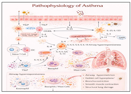
Fig1: This diagram illustrates the immunopathogenesis of asthma, depicting epithelial–immune interactions, Type-2 cytokine–mediated inflammation, and downstream airway hyperresponsiveness and structural remodelling [8].
A total of 49 patients were included in the present study. Demographic, clinical, immunological, and pulmonary function parameters were analysed and presented in tables and graphs. The study evaluated age, sex distribution, allergy history, family history, duration of inhaled steroid use, difficulty in tapering steroids, Total IgE, Specific IgE, Specific IgG, peripheral eosinophil count, sputum fungal elements, PFT results, diagnosis, and ABPA subtype.
1. PATIENT DISTRIBUTION ACCORDING TO AGE
|
Age Group (Years) |
Number of Patients |
Percentage (%) |
|
11-20 |
2 |
4.08% |
|
21-30 |
9 |
18.37% |
|
31-40 |
17 |
34.69% |
|
41-50 |
10 |
20.41% |
|
51-60 |
7 |
14.29% |
|
61-70 |
3 |
6.12% |
|
71-80 |
3 |
6.12% |
Table 1: Patient Distribution According to Age
The majority of patients belonged to the 31–40 years age group (34.69%), followed by 41–50 years (20.41%) and 21–30 years (18.37%). Fewer patients were observed in older age groups, with 71–80 years accounting for 6.12% of cases. This indicates that most patients in the present study were young to middle-aged adults, with peak occurrence in the third and fourth decades of life.
2. SEX-WISE DISTRIBUTION OF STUDY POPULATION
|
Sex |
Number of Patients |
Percentage (%) |
|
Male |
20 |
40.82% |
|
Female |
29 |
59.18% |
Table 2: Sex-Wise Distribution of Study Population
In the present study, female patients constituted the majority (59.18%), while male patients accounted for 40.82% of the study population, showing a female predominance. The higher proportion of female patients suggests that the condition under study may be more commonly observed among females or that females seek medical attention more frequently compared to males in the study population.
3. DISTRIBUTION OF ALLERGY HISTORY AMONG STUDY POPULATION
|
Allergy History |
Number of Patients |
Percentage (%) |
|
Dust Allergy |
25 |
51.02% |
|
Dust Allergy and Recurrent Rhinitis |
15 |
30.61% |
|
Dust and Cold Allergy |
8 |
16.33% |
|
Cold Allergy |
1 |
2.04% |
Table 3: Distribution of Allergy History Among Study Population
In the present study, dust allergy was the most common allergy history observed (51.02%), followed by dust allergy with recurrent rhinitis (30.61%). Dust and cold allergy were reported in 16.33% of patients, while isolated cold allergy was observed in 2.04% of cases. These findings indicate that dust exposure is the predominant allergen among the study population. The substantial proportion of patients with recurrent rhinitis associated with dust allergy suggests chronic allergic airway inflammation, which may contribute to disease persistence and recurrence in affected individuals.
4. DISTRIBUTION OF FAMILY HISTORY AMONG STUDY POPULATION
|
Family History |
Number of Patients |
Percentage (%) |
|
Yes |
4 |
8.16% |
|
No |
45 |
91.84% |
Table 4: Distribution of Family History Among Study Population
In the present study, the majority of patients (91.84%) had no family history, while 8.16% of patients reported a positive family history. These findings suggest that most cases in the present study were sporadic rather than familial. The low proportion of positive family history indicates that environmental factors, allergen exposure, and acquired risk factors may play a more significant role than genetic predisposition in the development of the condition.
5. DISTRIBUTION OF DURATION OF INHALED STEROID USE
|
Duration of Inhaled Steroid Use |
Number of Patients |
Percentage (%) |
|
< 1 year |
2 |
4.08% |
|
1–5 years |
17 |
34.69% |
|
6–10 years |
14 |
28.57% |
|
11–20 years |
13 |
26.53% |
|
> 20 years |
3 |
6.12% |
Table 5: Distribution of Duration of Inhaled Steroid Use
In the present study, the majority of patients had 1–5 years of inhaled steroid use (34.69%), followed by 6–10 years (28.57%) and 11–20 years (26.53%). A smaller proportion of patients had more than 20 years of steroid use (6.12%), while only 4.08% had less than 1 year of steroid exposure. These findings indicate that most patients in the study were on long-term inhaled steroid therapy, suggesting chronic disease severity and prolonged treatment requirement.
6. DISTRIBUTION OF TOTAL IgE LEVELS AMONG STUDY POPULATION
|
Total IgE Level (IU/mL) |
Number of Patients |
Percentage (%) |
|
< 100 |
6 |
12.24% |
|
100–500 |
12 |
24.49% |
|
501–1000 |
10 |
20.41% |
|
1001–5000 |
15 |
30.61% |
|
> 5000 |
6 |
12.24% |
Table 6: Distribution of Total IgE Levels Among Study Population
In the present study, the majority of patients had total IgE levels between 1001–5000 IU/mL (30.61%), followed by 100–500 IU/mL (24.49%) and 501–1000 IU/mL (20.41%). A considerable proportion of patients also demonstrated very high IgE levels (>5000 IU/mL) in 12.24% of cases, indicating a severe allergic response. Only 12.24% of patients had IgE levels below 100 IU/mL, suggesting that most patients exhibited elevated IgE levels, supporting the allergic etiology of the disease. These findings indicate that elevated total IgE levels were common among the study population, reflecting significant allergic sensitization and immune activation.
7. DISTRIBUTION OF SPECIFIC IgE (ASPERGILLUS FUMIGATUS) AMONG STUDY POPULATION
|
Specific IgE (A. fumigatus) (kUA/L) |
Number of Patients |
Percentage (%) |
|
< 0.35 |
33 |
67.35% |
|
0.35 – 1.0 |
3 |
6.12% |
|
1.01 – 5.0 |
6 |
12.24% |
|
5.01 – 10.0 |
2 |
4.08% |
|
> 10.0 |
5 |
10.20% |
Table 7: Distribution of Specific IgE (Aspergillus fumigatus) Among Study Population
In the present study, the majority of patients (67.35%) had specific IgE levels <0.35 kUA/L, indicating absence of significant fungal sensitization in most cases. However, 32.65% of patients showed elevated specific IgE levels, suggesting sensitization to Aspergillus fumigatus. Among sensitized patients, 12.24% had levels between 1.01–5.0 kUA/L, while 10.20% demonstrated markedly elevated levels (>10.0 kUA/L), indicating strong fungal sensitization. These findings suggest that a considerable proportion of patients exhibited immunological sensitization to Aspergillus fumigatus, supporting its role in allergic airway disease pathogenesis.
8. DISTRIBUTION OF SPECIFIC IgG (ASPERGILLUS FUMIGATUS) AMONG STUDY POPULATION
|
Specific IgG (A. fumigatus) |
Number of Patients |
Percentage (%) |
|
< 10 |
18 |
36.73% |
|
10 – 25 |
15 |
30.61% |
|
26 – 50 |
11 |
22.45% |
|
51 – 100 |
2 |
4.08% |
|
> 100 |
3 |
6.12% |
Table 8: Distribution of Specific IgG (Aspergillus fumigatus) Among Study Population
In the present study, the majority of patients had specific IgG levels below 25, with 36.73% having <10 and 30.61% having 10–25, indicating a mild to moderate immune response in most cases. However, 22.45% of patients demonstrated IgG levels between 26–50, while 10.20% showed markedly elevated levels (>50), suggesting chronic exposure and immune sensitization to Aspergillus fumigatus. The presence of elevated specific IgG levels in a subset of patients supports ongoing antigen exposure and a chronic inflammatory response, which is consistent with allergic fungal airway disease.
9. DISTRIBUTION OF PERIPHERAL EOSINOPHIL COUNT AMONG STUDY POPULATION
|
Peripheral Eosinophil Count (cells/µL) |
Number of Patients |
Percentage (%) |
|
< 100 |
12 |
24.49% |
|
100 – 300 |
11 |
22.45% |
|
301 – 500 |
13 |
26.53% |
|
501 – 1000 |
12 |
24.49% |
|
> 1000 |
1 |
2.04% |
Table 9: Distribution of Peripheral Eosinophil Count Among Study Population
These findings indicate that most patients demonstrated mild to moderate eosinophilia, suggesting underlying allergic and inflammatory processes. Elevated eosinophil counts in a substantial proportion of patients support allergic airway inflammation and immunological activation.
10. DISTRIBUTION OF PATIENTS BASED ON SPUTUM FUNGAL ELEMENTS
|
Sputum Fungal Elements |
Number of Patients |
Percentage (%) |
|
Yes |
4 |
8.16% |
|
No |
45 |
91.84% |
Table 10: Distribution of Patients Based on Sputum Fungal Elements
In the present study, fungal elements in sputum were detected in 8.16% of patients, while 91.84% of patients showed no fungal elements on sputum examination. The low proportion of positive sputum fungal elements suggests that direct fungal detection in sputum was relatively uncommon, despite clinical and immunological evidence of fungal sensitization. This may be due to intermittent fungal shedding, prior treatment, or limitations of sputum microscopy.
11. DISTRIBUTION OF PULMONARY FUNCTION TEST (PFT) RESULTS AMONG STUDY POPULATION
|
PFT Result |
Number of Patients |
Percentage (%) |
|
Normal |
7 |
14.29% |
|
Mild |
19 |
38.78% |
|
Moderate |
17 |
34.69% |
|
Severe |
6 |
12.24% |
Table 11: Distribution of Pulmonary Function Test (PFT) Results Among Study Population
In the present study, mild airway obstruction was the most common PFT finding (38.78%), followed by moderate obstruction (34.69%). Severe obstruction was observed in 12.24% of patients, while normal PFT findings were noted in 14.29% of cases. These findings indicate that most patients had mild to moderate airflow limitation, suggesting chronic airway inflammation and bronchial obstruction. The presence of severe obstruction in a subset of patients reflects advanced disease severity and prolonged airway involvement.
12. DISTRIBUTION OF DIAGNOSIS (ABPA VS NON-ABPA) AMONG STUDY POPULATION
|
Diagnosis |
Number of Patients |
Percentage (%) |
|
ABPA |
10 |
20.41% |
|
Non-ABPA |
39 |
79.59% |
Table 12: Distribution of Diagnosis (ABPA vs Non-ABPA) Among Study Population
In the present study, 20.41% of patients were diagnosed with ABPA, while 79.59% were classified as non-ABPA. This indicates that ABPA constituted nearly one-fifth of the study population. The higher proportion of non-ABPA patients suggests that although allergic fungal sensitization is common, only a subset fulfils the diagnostic criteria for ABPA. This highlights the importance of comprehensive immunological, radiological and clinical evaluation in differentiating ABPA from other allergic airway diseases.
13. DISTRIBUTION OF ABPA TYPE AMONG STUDY POPULATION
|
Categories of ABPA |
Number of Patients |
Percentage (%) |
|
ABPA-S (Serological) |
7 |
14.29% |
|
ABPA-CT (Bronchiectasis) |
3 |
6.12% |
|
Non-ABPA |
39 |
79.59% |
Table 13: Distribution of ABPA Type Among Study Population
In the present study, serological ABPA was observed in 14.29% of patients, while ABPA with bronchiectasis was present in 6.12% of cases. The majority of patients (79.59%) did not have ABPA. Among ABPA patients, serological ABPA was more common than ABPA with bronchiectasis, indicating that most cases were detected in earlier disease stages before structural lung damage developed. The smaller proportion of bronchiectasis-associated ABPA suggests that early diagnosis and intervention may help prevent progression to irreversible airway damage.
14. ASSOCIATION BETWEEN SPUTUM FUNGAL ELEMENTS AND ABPA DIAGNOSIS
|
Category |
Number of Patients |
|
ABPA with Sputum Positive |
3 |
|
Non-ABPA with Sputum Positive |
1 |
|
Total Sputum Positive |
4 |
Table 14: Association Between Sputum Fungal Elements and ABPA Diagnosis
Among the 4 patients with sputum fungal element positivity, 3 patients (75%) were diagnosed with ABPA, while 1 patient (25%) belonged to the non-ABPA group. This suggests that sputum fungal element positivity was more frequently associated with ABPA patients, supporting fungal colonization as an important feature in ABPA diagnosis. In the present study evaluating patients with allergic fungal airway disease, the majority were young to middle-aged adults with a female predominance and a high prevalence of dust-related allergy and long-term inhaled steroid use. Elevated total IgE levels, increased specific IgE and IgG to Aspergillus fumigatus, and mild to moderate eosinophilia were observed in a substantial proportion of patients, supporting underlying allergic and immunological mechanisms. Pulmonary function testing predominantly demonstrated mild to moderate airflow limitation, consistent with chronic airway inflammation. Based on comprehensive clinical, immunological and functional assessment, a subset of patients was diagnosed with ABPA, with serological ABPA being more common than ABPA with bronchiectasis, suggesting detection at relatively earlier stages of disease. Sputum fungal element positivity was more frequently observed among patients diagnosed with ABPA, indicating an association between fungal colonization and disease manifestation. Additionally, difficulty in tapering steroids was noted in some patients, including those with ABPA, suggesting persistent inflammation and steroid dependence in selected cases. These findings emphasize the importance of early screening using immunological markers, sputum examination and clinical evaluation in patients with allergic airway disease. Early identification and appropriate management may help prevent disease progression and reduce the risk of irreversible airway damage such as bronchiectasis.
DISCUSSION:
In the present study on 49 patients, the majority of patients belonged to the 31–40 years age group (34.69%), followed by 41–50 years (20.41%) and 21–30 years (18.37%). These findings suggest that uncontrolled asthma and ABPA predominantly affect young and middle-aged adults. The female predominance (59.18%) could be attributed to an increased prevalence of allergic airway disease among females. Dust allergy was the most common allergic trigger observed (51.02%), followed by dust allergy with recurrent rhinitis (30.61%), supporting the role of environmental allergens in uncontrolled asthma and the development of ABPA. Chronic allergen exposure, including dust and environmental pollutants, leads to persistent airway inflammation and increased fungal sensitization [9,10]. The present study also suggests that environmental factors play a more significant role than genetic predisposition in the development of ABPA. Elevated total IgE levels were observed in the majority of patients, with 30.61% showing IgE levels between 1001–5000 IU/mL and 12.24% showing levels greater than 5000 IU/mL. Elevated IgE is a hallmark of ABPA and reflects an exaggerated type 2 immune response to Aspergillus antigens. ABPA diagnosis primarily relies on immunological and radiological parameters rather than direct fungal detection on microscopy. Early detection is important because untreated ABPA may progress to bronchiectasis, pulmonary fibrosis and chronic respiratory failure; early diagnosis therefore improves clinical outcomes, particularly in regions with high fungal exposure such as India [3,4].
Limitations: It was conducted at a single tertiary care centre with a relatively small sample size, which may limit the generalizability of the findings; patients presenting to tertiary care often have more severe disease, which may introduce referral bias. Patients were not followed up long term, so disease progression, relapse rates and treatment outcomes could not be assessed. Most patients had received long-term inhaled corticosteroids before evaluation, which may have altered immunological parameters such as IgE levels and eosinophil counts. The absence of a control group of well-controlled asthma patients limited comparison of clinical and laboratory parameters.
CONCLUSION:
The present study emphasizes the importance of early diagnosis and appropriate management of ABPA in patients with allergic airway disease to prevent irreversible airway damage, reduce steroid dependency and improve long-term outcomes. The study highlights the need for routine screening for ABPA in patients with uncontrolled asthma in tertiary care settings.
REFERENCES:
1. Aggarwal AN, Chaudhry K, Chhabra SK, D’Souza GA, Gupta D, Jindal SK, et al. Prevalence and risk factors for bronchial asthma in Indian adults: a multicentre study. Indian J Chest Dis Allied Sci. 2006;48(1):13–22.
2. Patterson K, Strek ME. Allergic bronchopulmonary aspergillosis. Proc Am Thorac Soc. 2010;7(3):237–44.
3. Agarwal R, Chakrabarti A, Shah A, Gupta D, Meis JF, Guleria R, et al. Allergic bronchopulmonary aspergillosis: review of literature and proposal of new diagnostic and classification criteria. Clin Exp Allergy. 2013;43(8):850–73.
4. Agarwal R, Sehgal IS, Muthu V, Denning DW, Chakrabarti A, Soundappan K, et al. Revised ISHAM-ABPA working group clinical practice guidelines for diagnosing, classifying and treating allergic bronchopulmonary aspergillosis/mycoses. Eur Respir J. 2024;63(4):2400061.
5. Agarwal R, Sehgal IS, Muthu V. The 2024 International Society for Human and Animal Mycology (ISHAM)-ABPA working group guidelines: transforming diagnosis and management of ABPA. Lung India. 2025;42(2):79–83.
6. Agarwal R, Khan A, Garg M, Aggarwal AN, Gupta D. Pictorial essay: allergic bronchopulmonary aspergillosis. Indian J Radiol Imaging. 2011;21(4):242–52.
7. Wenzel SE. Asthma phenotypes: the evolution from clinical to molecular approaches. Nat Med. 2012;18(5):716–25.
8. Barnes PJ. Immunology of asthma and chronic obstructive pulmonary disease. Nat Rev Immunol. 2008;8(3):183–92.
9. Denning DW, Pashley C, Hartl D, Wardlaw A, Godet C, Del Giacco S, et al. Fungal allergy in asthma – state of the art and research needs. Clin Transl Allergy. 2014;4:14.
10. Agarwal R. Severe asthma with fungal sensitization. Curr Allergy Asthma Rep. 2011;11(5):403–13.