Development and Preliminary Validation of a Multiplex PCR Based Diagnosis Method for Rapid Identification of Major Bacterial Pathogens Associated with Chronic Suppurative Otitis Media.
- Dr. Monalisa Patsani , Associate Professor, Department of ENT and Head and Neck Surgery, Srirama Chandra Bhanja Medical College and Hospital, Cuttack, Odisha, India
- Dr. Sasmita Khatua , Associate Professor, Department of Microbiology, Pandit Raghunath Murmu Medical College and Hospital, Baripada, Odisha, India
- Dr. Subhalaxmi Routray , Associate Professor, Department of ENT and Head and Neck Surgery, Srirama Chandra Bhanja Medical College and Hospital, Cuttack, Odisha, India.
- Dr. Subrat Kumar Behera , Professor, Department of ENT and Head and Neck Surgery, Srirama Chandra Bhanja Medical College and Hospital, Cuttack, Odisha, India.
Article Information:
Abstract:
Background: Chronic Suppurative Otitis Media (CSOM) is a major health problem especially throughout the world mainly in underdeveloped and developing countries including India. It is amongst the most common childhood diseases and is associated with multiple microbial pathogens including Escherichia coli, Pseudomonas aeruginosa, Staphylococcus aureus and Klebsiella pneumoniae within the middle ear. In order to continue effective treatment, the authentic identification of the causative organisms is critical. While culture-based techniques remain as the gold standard for detecting the major pathogens, molecular methods are changing the ways for exploring the complex polymicrobial dynamics of CSOM. Methods: To develop the method for multiplex PCR, species specific genes were targeted for PCR amplification from four major pathogens. Primer sets were designed either using bioinformatics tools or directly picked from already published research articles. The standardization of PCR condition for amplification of the species-specific genes and 16S rRNA gene was done by colony PCR using specific bacterial culture. Results: The species specific amplification of the PCR products were observed for all the primer sets for each bacterial culture. The 16S rRNA gene was chosen as the control, which showed amplified bands from all the four above mentioned bacteria. Conclusion: Our study provides a foundation to design and develop methods for a rapid and cost-effective detection of causative organisms in CSOM using Multiplex PCR, so that directed patient treatment can be initiated and possible complications can thus be avoided.
Keywords:
Article :
INTRODUCTION:
Chronic Suppurative Otitis Media (CSOM) is one of the leading causes of preventable hearing loss worldwide. In most cases it is a sequela of improperly attended acute otitis media (AOM). It is associated with chronic inflammation of the middle ear and is characterized by persistent perforation of the tympanic membrane with recurrent otorrhoea. According to the World Health Organization (WHO), it affects anywhere between 65 and 330 million people worldwide, but mainly in the developing countries. Along with its high prevalence, extracranial complications like mastoid abscess and facial nerve palsy, as well as life-threatening intracranial complications like brain abscess and meningitis have also been associated with CSOM (Orji 2013, Yorgancılar, Yıldırım et al. 2013).1,2 Despite appropriate antibiotic therapy, AOM may progress to CSOM (Котлукова 2004)3. Although the pathogenesis of AOM is well studied, very limited research is available in relation to CSOM.
Bacteria and fungi both have the potential to cause CSOM (Juyal, Negi et al. 2014).4 However, bacterial pathogens are most commonly isolated and are responsible for initiating the infection (Alsaimary, Alabbasi et al. 2010).5 CSOM is a polymicrobial disease and varies with respect to time and geographical region. Staphylococcus aureus, Pseudomonas aeruginosa, Escherichia coli, Proteus vulgaris, Proteus mirabilis, Klebsiella pneumoniae and some anaerobic bacteria are responsible for causing CSOM (Poorey 2002).6 However, Escherichia coli, Pseudomonas aeruginosa, Staphylococcus aureus and Klebsiella pneumoniae are the major bacterial pathogens of the disease. With the irrational use of antibiotics, there has been increased incidence of multi drug resistance which is another hindrance as they are related to increased complications associated with CSOM (Park, Jung et al. 2008, Choi, Park et al. 2010).7,8 Pseudomonas aeruginosa and Staphylococcus aureus are the most common isolates found in the pus swab culture of CSOM patients, and poses a significant challenge for disease management due to their resistance against commonly used antibiotics (Choi, Park et al. 2010).7,8 Rapid and authentic detection of causative group seems to be the only solution to prevent the further complications of the disease and emergence of multi drug resistant bacteria.
Conventional methods for detection of bacterial pathogens includes culture based and immunological methods. While culture based method cannot detect various fastidious organisms and needs days to produce initial results (Váradi, Luo et al. 2017)9, immunological methods, even if they produce fast results but may give false positive result due to interaction with non-specific matter or cross reactivity with a wide range of organisms (Kfir and Genthe 1993, Peruski and Peruski 2003).10,11 However, molecular based techniques are considered to be more rapid and specific, as they makes use of specific nucleic acid sequences to detect the presence of any pathogen (Houpikian and Raoult 2002).12 Also, multiplex PCR poses a significant capability in detecting multiple pathogens simultaneously and has also been used in disease management of AOM by isolating DNA from the pus sample of infected ear (Billal, Hotomi et al. 2006).13
In order to prevent the severe complications of CSOM, we need to control the non-specific use of antibiotics for treatment of the disease. So, for precision medicine, it requires a clear knowledge of the microbial groups that are responsible for the disease. The antibiotics are prescribed on the basis of the type of Otitis Media the patient is suffering from. As CSOM is a result of multiple bacterial infection, it becomes crucial for a clinician to know the true causative group, while prescribing antibiotics or developing treatment strategies. To meet this challenge, we proposed to design a method, using multiplex PCR that could detect the presence of bacterial pathogens of CSOM that are predominant in any infected patient, directly from their pus sample. As we would be detecting the four pathogens simultaneously using four specific primers and have eliminated the DNA isolation step, we believe that this would provide a rapid, cost-effective and more specific results and would contribute in reducing the disease burden and developing effective treatment strategies.
MATERIALS AND METHODS:
I) Bacterial strains and growth conditions
Bacterial strains used in this study includes: Escherichia coli, Pseudomonas aeruginosa, Staphylococcus aureus and Klebsiella pneumoniae. All the bacterial strains were grown on Luria Bertani (LB) agar at 370C with required incubation time
II) Primer design: In order to specifically amplify one gene, each from four targeted bacterial strains, we have used four primer sets against the specific-specific genes of each organism. As we aimed to detect the bacterial pathogens of CSOM, we have considered 16s rRNA gene to be our control gene that is to be amplified along with the four species-specific genes. The primer set, Endo16sF 5'CGTGGAGCATGTGTTTA3' and Endo16sR 5'CCATTGTAGCACTGTGT3', against the 16srRNA gene was designed which gives an amplicon of 300bp. For specific detection of Escherichia coli, primer set, DNATopoAF 5'CATAATAGTTGACTCCCCAGC3' and DNATopAR 5'AACCGACCTTGACCGCGAA3' was using primer 3 Input (version 0.4.0), against the topoisomerase A gene that would produce an amplicon of length 330bp. To specifically detect Pseudomonas aeruginosa, an already reported primer set for the O-antigen acetylase gene of Pseudomonas aeruginosa PA01 was selected. Primers for PCR, PA431CF 5'CTGGGTCGAAAGGTGGTTGTTATC3' and PA431CR 5'GCGGCTGGTGCGGCTGAGTC3' were used for amplification and detection of a 232bp fragment (Choi, Kim et al. 2013).14 For specific detection of Staphylococcus aureus, we chose an already reported primer set, EAPCON1F 5'CTGTGTGTGGCAAGCAAGTT3'and EAPCON2R 5'TTGCCAGCGACAATATTTGA3' (Hussain, von Eiff et al. 2008)15, for the amplification of eap gene of Staphylococcus aureus. Further analysis of the primer sets were done using OligoAnalyzer and their species-specificity were assessed using NCBI-BLAST searches (http://www.ncbi.nlm.nih.gov/). For our target organisms, we searched for genes that are specific to only one organism and did not show any significant match with the other three organisms.
III) DNA preparation
As our aim was to detect the bacterial pathogens without isolating their DNA, therefore, we used the bacterial cultures directly and performed colony PCR for standardizing the PCR conditions required for amplification of the four species-specific genes along with internal control. For all the four bacterial strains, we picked a single colony from the culture plate and suspended it in a 1.5µl microcentrifuge tube containing 9µl of Nuclease free water (NFW) followed by 10 mins of denaturation in water bath. And then we directly used it as our template for the PCR amplification.
IV) PCR analysis
For conventional PCR assay, the reaction mixture in each PCR tube contained 10µl of Taq pre mix, from Takara (dNTP, Taq polymerase, 10x buffer, NFW), 0.5µl of forward primer, 0.5µl of reverse primer and 9µl of template. Optimum annealing temperature for each primer was standardized using different temperature gradients. Different PCR conditions used for the amplifications of all the species-specific genes and control gene is given below (Table 1 and 2).
Table 1: Species-specific genes and different annealing temperatures used for their standardization
|
Genes |
Annealing temperatures |
|
16s rRNA |
51℃, 53℃, 55℃ |
|
Topoisomerase A |
56℃, 58℃, 60℃, 62℃, 64℃, 66℃ |
|
O-antigen acetylase |
57℃, 59℃, 61℃, 63, ℃, 65℃ |
|
EAP |
52℃, 54℃, 56℃ |
Table 2: PCR steps and different conditions used to amplify all the specific genes
|
PCR steps |
Conditions |
No of Cycles |
|
Initial denaturation |
95℃, 5mins |
1 |
|
Denaturation |
95℃, 30 secs |
35 cycles |
|
Annealing |
Optimized temperature, 30secs |
|
|
Extension |
72℃, 30secs/45secs |
|
|
Final extension |
72℃, 10mins |
1 |
|
Hold |
4℃ |
|
V) Multiplex PCR from mixed cultures
In order to analyse the efficiency of the primers against the specific genes of Escherichia coli, Staphylococcus aureus and Pseudomonas aeruginosa, we used all the three primer sets for amplification of the target genes at an annealing temperature 56℃ which was chosen on the basis of previous standardizations. For template, single colonies were picked from the three bacterial culture and suspended in 9µl of NFW.
RESULTS:
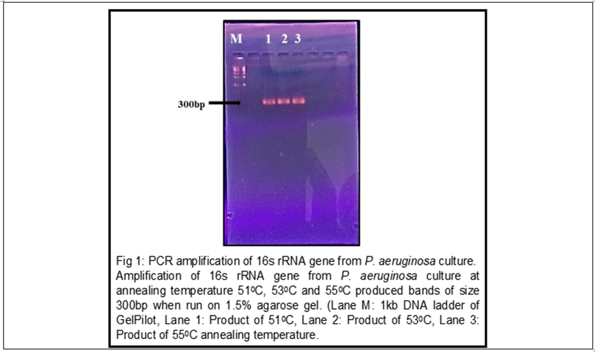
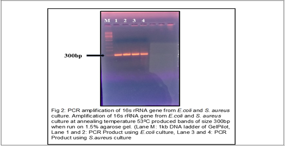
I) Standardization of the annealing temperature of primer for the amplification of 16s rRNA gene: Amplified bands of size around 300bp were observed at all the three temperatures using P. aeruginosa crude culture as template (explained in the method section) and 53℃ was considered as the optimum temperature for amplification of the gene from the remaining three target organisms (Fig 1). Bacterial cultures of Escherichia coli and Staphylococcus aureus together when used to amplify the 16s rRNA gene at the optimized annealing temperature, 53℃ it showed successful amplification of 300bp fragment (Fig 2).
II) Standardization of the annealing temperature of primers for the amplification of species-specific genes
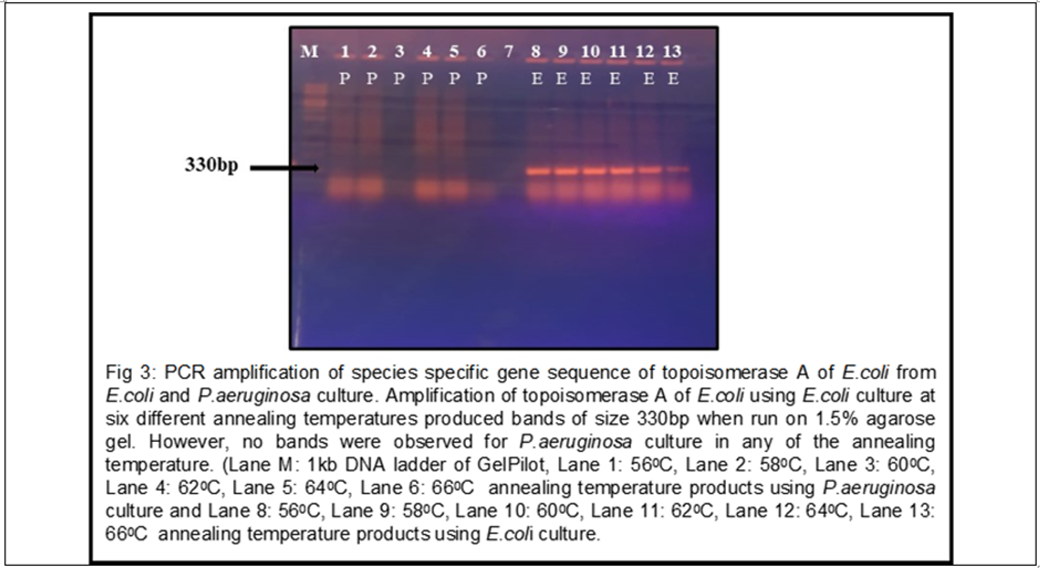
The amplification of the species- specific gene of Escherichia coli: topoisomerase A was done using the Escherichia coli and Pseudomonas aeruginosa culture with six different annealing temperatures, 56℃, 58℃, 60℃, 62℃, 64℃ and 66℃. Amplified bands were observed at all the six temperatures only for Escherichia coli culture and not for Pseudomonas aeruginosa, which indicates the specificity of the primer set. For future experiments, 58℃ was taken as the optimum temperature for amplification of topoisomerase A (Fig 3).
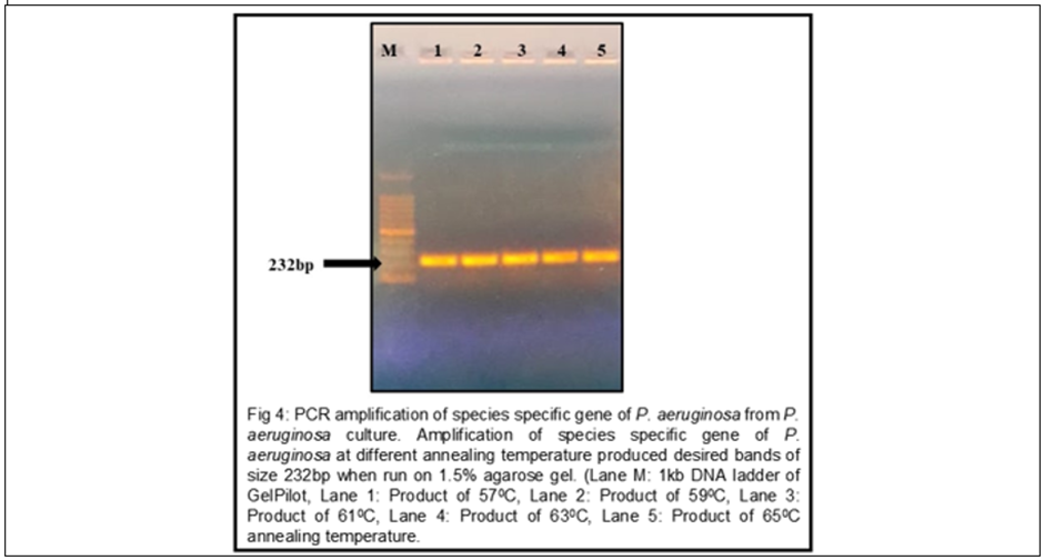
Amplification of the species-specific gene of Pseudomonas aeruginosa: O-antigen acetylase, was done using the specific bacterial culture with five different annealing temperatures 57℃, 59℃, 61℃, 63℃ and 65℃. All the five temperatures produced amplified bands (Fig 4) and 57℃ was taken as the optimum annealing temperature. Amplification of the species-specific gene from Staphylococcus aureus was done using specific bacterial culture at three different annealing temperatures 52℃, 54℃ and 56℃. Faint amplified bands were observed at all three temperatures (Fig 5) and 56℃ was taken as the optimum annealing temperature.
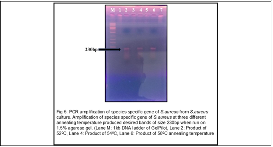
III) Detection of sensitivity of the species specific primer sets
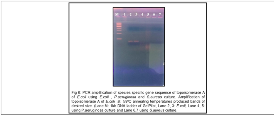
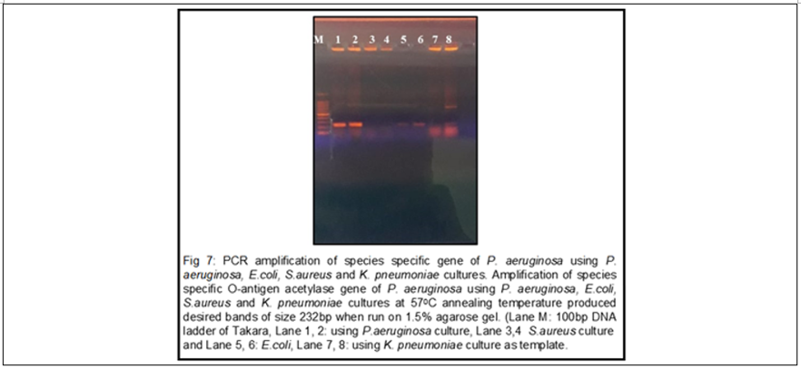
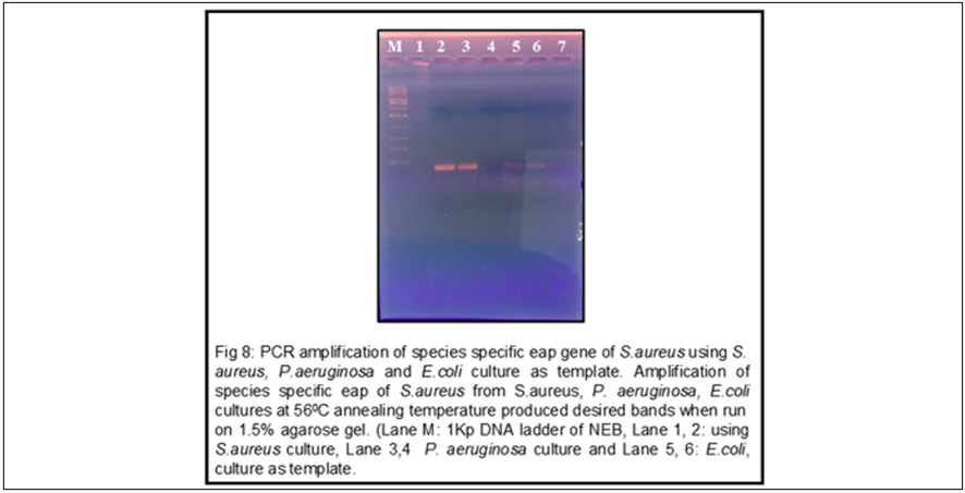
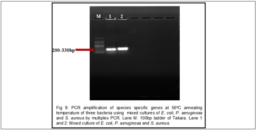
When the species-specific gene of Escherichia coli, topoisomerase A was amplified from Escherichia coli, Pseudomonas aeruginosa and Staphylococcus aureus culture at 58℃, only the Escherichia coli culture showed the desired size of band (Fig 6). When the species-specific gene of Pseudomonas aeruginosa was amplified for Pseudomonas aeruginosa, Escherichia coli, Staphylococcus aureus and Klebsiella pneumoniae at 57℃, the desired size of bands were observed in Pseudomonas aeruginosa and faint bands of same size was also observed in Escherichia coli. Also, a band of size around 700bp was observed in one sample of Klebsiella pneumoniae (Fig 7). When the species-specific gene of Staphylococcus aureus eap gene was amplified from Staphylococcus aureus, Escherichia coli and Pseudomonas aeruginosa culture at 56℃, the desired size of band was observed in Staphylococcus aureus and in one sample each from Escherichia coli and Pseudomonas aeruginosa (Fig 8). The eap gene of Staphylococcus aureus when again amplified from Escherichia coli and Pseudomonas aeruginosa culture at 56℃. A band desired size of band was observed in both Escherichia coli and Pseudomonas aeruginosa as noticed earlier (Fig 8). Multiplex PCR from mixed culture of three bacteria produced two thick bands between 200-330bp when amplified at annealing temperature, 56℃. (Fig 9)
DISCUSSION:
We aimed to design a set of primers against the species-specific genes from each of our four target organisms (Escherichia coli, Pseudomonas aeruginosa, Staphylococcus aureus and Klebsiella pneumoniae) and use those primers for multiplex PCR, which would provide the authentic knowledge of the causative group of CSOM present in the pus sample of infected ears. Due to time constraints, we could only able to standardize the PCR condition for amplification of the 16sr RNA gene (control) from all the four target organisms and species-specific genes of three target organisms (Escherichia coli, Pseudomonas aeruginosa and Staphylococcus aureus), which also needs further alterations. We have also detected the sensitivity of the primer sets, specific for these three organisms and have performed multiplex PCR for the same three organisms.
The primer for 16srRNA gene worked successfully and produced a 300bp fragment from all the four organisms. PCR using the primers for specific genes of Escherichia coli, Pseudomonas aeruginosa and Staphylococcus aureus also produced the desired size of bands 330bp, 232bp and 230bp respectively. However, the issue with these primers was their sensitivity. Out of the three primer sets, two sets of primer (primers for eap and O-antigen acetylase gene) showed the desired size of bands in their specific organisms and also in other two organisms. Also, the primer for 0-antigen acetylase showed a band of different size in Klebsiella pneumoniae. Thus, this could be a hindrance while detecting the pathogens simultaneously and may produce false positive result. From the multiplex PCR result, we observed that since the size of our amplified fragments is very close to each other it could not produce three separate bands for three organisms. Hence, the two primer sets showed reduced sensitivity and the amplicon size produced by them could not meet our objective. Therefore, few more primers needs to be standardized before using those in multiplex PCR for the identification of the causative organism of CSOM.
As CSOM is a complex infectious disease and its causative group of organism varies with respect to time and geographical region, therefore, it is essential to know about its major pathogens. The standard method for detection of bacterial pathogens use culture-based technique. However, we are utilising the molecular-based technique for the detection of our target organisms (Escherichia coli, Pseudomonas aeruginosa, Staphylococcus aureus, Klebsiella pneumoniae), which provides more rapid and truthful results. As this disease is more prevalent in low socioeconomic groups, we intended to design our method in a cost-effective manner by eliminating the DNA extraction steps using any DNA extraction kit. While molecular method such as multiplex PCR by isolating DNA from the ear discharge of infected patient, have been used for detection of pathogens from AOM (Kaur, Adlowitz et al. 2010)16 no such method has been reported for CSOM. Despite appropriate antibiotic therapy, AOM may progress to chronic suppurative OM (CSOM) (Schousboe, Rasmussen et al. 2001).17 As multiple factors are responsible for the development of CSOM, it becomes difficult to identify the predominant pathogen responsible for the development of the disease. Insufficient knowledge of the causative groups leads to the use of nonspecific antibiotics. It has also been reported that, chances of acquiring fungal pathogen increases with prolong use of ototopical antibiotics (Schousboe, Rasmussen et al. 2001).17 Thus, there is an urgent need for this type of multiplex based PCR diagnosis methods to detect the causative groups of CSOM from various regions, prior to any antibiotic treatment in order to prevent the irrational use of antibiotics and further complications of the disease.
CONCLUSION:
Our study provides the foundation for the development of multiplex PCR based disgnosis tools for rapid and pecise identification of the causatice organisms of chronic suppurative otitis media (CSOM). Acurate and timely identification of the pathogen can facilitate targeted antimicrobial therapy, thereby reducing the indiscriminate and irrational use of antibiotics and helping to prevent the generation of new Multi-drug resistance strains of bacteria.
REFERENCES:
1. Orji F. A survey of the burden of management of chronic suppurative otitis media in a developing country. Annals of Medical and Health Sciences Research 2013;3(3):598-612.
2. Yorgancılar E, Yildirim M, Gun R, et al. Complications of chronic suppurative otitis media: a retrospective review. European Archives of Oto-Rhino-Laryngology 2013;270(1):69-76.
3. Котлукова Т. Лечение синегнойной инфекции у взрослых и детей. Фарматека, 2004;94(17):23–30.
4. Juyal, D. Significance of fungal flora in chronic suppurative otitis media. Annals of Tropical Medicine and Public Health 2014;7(2):120.
5. Alsaimary IE, Alabbasi AM, Najim JM. Antibiotics susceptibility of bacterial pathogens associated with otitis media. African Journal of Bacteriology Research 2010;2(4):41-50.
6. Poorey V. Study of bacterial flora in CSOM and its clinical significance. Indian Journal of Otolaryngology and Head and Neck Surgery 2002;54(2):91-5.
7. Choi HG, Park KH, Park SN, et al. The appropriate medical management of methicillin-resistant Staphylococcus aureus in chronic suppurative otitis media. Acta Oto-Laryngologica 2010;130(1):42-6.
8. Park, MK, Jung MH, Kang HJ, et al. The changes of MRSA infections in chronic suppurative otitis media. Otolaryngology–Head and Neck Surgery 2008;139(3):395-8.
9. Váradi L, Luo JL, Hibbs DE, et al. Methods for the detection and identification of pathogenic bacteria: Past, present, and future. Chemical Society Reviews, 2017;46(16):4818032.
10. Kfir R, Genthe B. Advantages and disadvantages of the use of immunodetection techniques for the enumeration of microorganisms and toxins in water. Water Science and Technology 1993;27(3-4):243-52.
11. Peruski AH, Peruski LF. Immunological methods for detection and identification of infectious disease and biological warfare agents. Clinical and Diagnostic Laboratory Immunology, 2003;10(4):506-13.
12. Houpikian P, Raoult D. Traditional and molecular techniques for the study of emerging bacterial diseases: One laboratory’s perspective. Emerging Infectious Diseases 2002;8(2):122.
13. Billal, DS, Hotomi M, Tasnim S, et al. Evaluation of serotypes of Streptococcus pneumoniae isolated from otitis media patients by multiplex polymerase chain reaction. ORL 2006;68(3):135-8.
14. Choi HJ, Kim MH, Cho MS, et al. Improved PCR for identification of Pseudomonas aeruginosa. Applied Microbiology and Biotechnology 2013;97(8):3643-51.
15. Hussain M, von Eiff C, Sinha B, Joost I, Herrmann M, Peters G, Becker K. eap Gene as novel target for specific identification of Staphylococcus aureus. J Clin Microbiol 2008;46(2):470-6.
16. Kaur R, Adlowitz DG, Casey JR, et al. Simultaneous assay for four bacterial species including Alloiococcus otitidis using multiplex-PCR in children with culture-negative acute otitis media. The Pediatric Infectious Disease Journal, 2010;29(8):741-5.
17. Schousboe LP, Ovesen T, Eckhardt L, et al. How does endotoxin trigger inflammation in otitis media with effusion? The Laryngoscope 2001;111(2):297-300.