Clinical Profile, Hydrostatic Reduction Outcomes, and Factors Associated with Surgical Intervention in Pediatric Intussusception.
- Dr. Chintha Salam A. , Associate Professor, Department of Pediatric Surgery, Institute of Child Health, Government Medical College, Kottayam, Kerala, India.
- Dr. Beena S.V. , Professor, Department of Pediatric Surgery, Institute of Child Health, Government medical College, Kottayam, Kerala, India..
Article Information:
Abstract:
Background: Intussusception is the most common cause of intestinal obstruction in infants and toddlers. Non-operative hydrostatic reduction is the standard first-line therapy for pediatric intussusception, but early identification of children at risk of reduction failure is vital to prevent ischemic bowel complications and timely operative intervention. Objective: To characterize the clinical presentation, outcomes of ultrasound-guided hydrostatic reduction, and clinical factors associated with reduction failure and surgical intervention in children under two years of age. Methods: A prospective study of 110 consecutive children aged <24 months presenting with intussusception confirmed on ultrasonography. All patients underwent initial saline hydrostatic reduction under ultrasound guidance. Clinical, demographic, dietary, and anatomical parameters were compared between children successfully managed non-operatively and those requiring surgical intervention using the Wilcoxon rank-sum test and Fisher’s exact test. Results: The median age was 9.0 months (IQR: 6.0–17.0), with 70 boys (63.6%) and 40 girls (36.4%). Ileocolic intussusception occurred in 105 patients (95.5%) and colocolic in 5 (4.5%). Frequent presenting symptoms included abdominal pain in 108 patients (98.2%), vomiting in 76 (69.1%), and bloody stools in 38 (34.5%). Hospitalization delay (≥1 day after onset) was present in 74 children (67.3%). Hydrostatic reduction was successful in 106 patients (96.4%). Four patients (3.6%) failed hydrostatic reduction and required surgical intervention. Among these 4 surgical cases, 1 required bowel resection (0.9% overall; 25.0% of surgical cases) and 2 had an identifiable pathological lead point (50.0% of surgical cases). Reduction failure requiring surgery was significantly associated with younger age (mean 3.3±1.5vs. 11.3±5.9months; p=0.002), age ≤6months (4/4, 100.0% vs. 27/106, 25.5%; p=0.005). Delay in hospitalization, sex, vomiting, and bloody stools were not significantly associated with reduction failure. Post-reduction recurrence was documented in 4 patients (3.6%). Conclusions: Hydrostatic reduction demonstrated an excellent primary success rate exceeding 96%. Young infant age (≤6 months, and particularly <3months) is the primary factor associated with reduction failure, pathological lead points, and intestinal resection. In hemodynamically stable children without signs of peritonitis, presentation delay alone should not preclude an initial attempt at hydrostatic reduction under surgical standby.
Keywords:
Article :
INTRODUCTION:
Intussusception is the most common cause of intestinal obstruction in infants and toddlers. It is an acquired invagination of proximal bowel into the distal bowel. The reported incidence is approximately 1 to 4 in 2000 infants and children.[1] Highest incidence is seen in between ages 4 to 9 months2. Most affected are healthy well thriving infants and two -third are males. Intussusception can be idiopathic or with a pathological lead point. The incidence of lead point is 1.5% to 20%.[2] The classical triad is with intermittent, cramping abdominal pain, red current jelly stools and palpable abdominal mass. But this triad is seen in less than a fourth of children.[2] Abdominal ultrasonography is the diagnostic modality of choice which demonstrates a "target" or "doughnut" sign on transverse imaging and a "pseudokidney" appearance on longitudinal views. Non operative method of reduction is the first line of management, provided there is no features of intestinal perforation, peritonitis and refractory hypotension. Successful reduction with USS guided hydrostatic reduction in uncomplicated patients is seen in 85% of cases, the range being 42% to 95%.[2] Operative management is indicated when the nonoperative management is contraindicated or failed. Identification of early predictors of failed non operative reduction may help in timely surgical decision making and reduce complications. Identifying early clinical and disease-related factors associated with reduction failure is essential for surgical decision-making. This will prevent progressive intestinal ischemia and reduce the necessity for bowel resection. This study aimed to evaluate the clinical profile, therapeutic outcomes of ultrasound-guided hydrostatic reduction, and clinical factors associated with surgical intervention in pediatric intussusception.
MATERIALS AND METHODS:
Study Design and Setting
This prospective observational study was conducted in the Department of Paediatric Surgery GMCH Kottayam, a tertiary care referral and teaching hospital in Kerala, India, between December 2020 and December 2022.The study protocol was approved by the Institutional Ethics Committee (IRB108/2020), and written informed consent was obtained from the parents or legal guardians of all participants prior to enrollment.
All consecutive children younger than 2 years of age presenting with a clinical diagnosis of acute intussusception that was confirmed on abdominal ultrasonography. A total of 110 children met these criteria and formed the study cohort. Following adequate intravenous fluid resuscitation, all 110 children underwent a standardized trial of hydrostatic reduction under real-time ultrasonographic visualization. Saline was instilled through a wide-bore rectal catheter under controlled hydrostatic column pressure. Successful reduction was defined sonographically by the complete disappearance of the intussusception and free reflux of fluid across the ileocecal valve into multiple terminal ileal loop. Operative intervention was done on failure to achieve reduction after three controlled hydrostatic attempts, or if signs of intestinal compromise during the procedure. Data were prospectively recorded using standardized case report forms.
Variables studied included-Demographic and Socioeconomic Characteristics like age in months, sex, Clinical Presentation, Duration of symptoms before presentation, delay in hospitalization (≥1 day after symptom onset), history of prior intussusception, specific symptoms (abdominal pain, vomiting, abdominal distension, fever, bleeding per rectum/bloody stools, diarrhea, palpable abdominal mass, and clinical peritonitis, and pre-existing chronic conditions. Regarding outcomes and operative Findings: Method of initial intervention-ultrasound guided hydrostatic reduction, procedural success or failure, post-reduction recurrence, anatomical location, intraoperative bowel viability (presence of gangrene or perforation), identification of pathological lead points, and the need for intestinal resection.
Statistical Analysis
Continuous variables were tested for distribution normality and are expressed as mean ± standard deviation (SD) or median with interquartile range (IQR). Categorical variables are summarized as absolute counts and percentages (n,%). Univariate comparisons between children managed successfully with hydrostatic reduction and those requiring surgical intervention were conducted using the Wilcoxon rank-sum test for continuous data and Fisher's exact test for categorical variables. A two-tailed p-value of <0.05 was considered statistically significant. All statistical analyses were conducted using standard statistical software.
RESULTS:
A total of 110 children younger than 2 years with confirmed intussusception were evaluated. The median patient age was 9.0 months (IQR: 6.0–17.0). 31 children (28.2%) were aged 0–6 months, 37 (33.6%) were aged 6–12 months, and 42 (38.2%) were aged 1–2 years. Boys represented 70 patients (63.6%), corresponding to a male-to-female ratio of 1.75:1 (Table 1). The median maternal age was 26.0 years (IQR: 25.0–28.0), and 55 mothers (50.0%) had completed university education or higher.
Table 1: Age and sex distribution of the study cohort (N = 110)
|
Characteristic |
Overall |
|
N = 1101 |
|
|
Age in months |
9.0 (6.0, 17.0) |
|
Age in months (Category) |
|
|
0 - 6 months |
31 (28%) |
|
6 - 12 months |
37 (34%) |
|
1 - 2 years |
42 (38%) |
|
Sex |
|
|
Male |
70 (64%) |
|
Female |
40 (36%) |
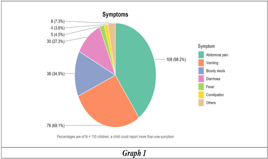
The anatomical type was ileocolic, observed in 105 patients (95.5%), colocolic intussusception in 5 (4.5%). No isolated ileoileal cases were noted. The most frequent presenting symptom was abdominal pain in 108 patients (98.2%), followed by vomiting in 76 (69.1%), bloody stools in 38 (34.5%), and diarrhea in 30 (27.3%). Fever was documented in only 5 children (4.5%) (Graph 1).
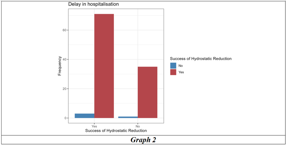
A delay in hospitalization of day after symptom onset was present in 74 patients (67.3%). Specifically, 36 patients (32.7%) presented on day 0, 43 (39.1%) on day 1, 23 (20.9%) on day 2, 4 (3.6%) on day 3, 2 (1.8%) on day 4, and 2 (1.8%) on day 7.
Hydrostatic reduction was attempted in all 110 children and succeeded in 106, yielding a success rate of 96.4%. Hydrostatic reduction failed in 4 patients (3.6%), all of whom required surgical intervention. Among these 4 surgical cases three patients had ileocolic intussusception (75.0%) and 1 had colocolic intussusception (25.0%). A pathological lead point was confirmed in 2 patients (50.0% of surgical interventions; 1.8% of the overall cohort). Intestinal resection was necessary in 1 patient (0.9% of the total cohort; 25.0% of surgical interventions) (Table 2) (Graph 2). This patient was a 2-month-old male infant. This child reported again at 1 year of age with adhesive intestinal obstruction that failed conservative management. Exploratory laparotomy revealed a closed-loop obstruction, necessitating adhesiolysis, repeat segmental bowel resection, and end-to-end anastomosis.
Table 2: Therapeutic management and procedural outcomes of hydrostatic reduction (N = 110)
|
Characteristic |
N = 1101 |
|
Hydrostatic reduction Attempted |
110 (100%) |
|
Success of Hydrostatic reduction |
106 (96%) |
|
Intussusception resolved by surgery |
4 (3.6%) |
|
Intestinal resection performed |
1 (0.9%) |
|
Lead point identified |
|
|
Missing |
106 (96%) |
|
Yes |
2 (1.8%) |
|
No |
2 (1.8%) |
Factors Associated with Surgical Intervention
Comparing patients with successful hydrostatic reduction ( ) to those requiring surgical intervention ( ) Children requiring surgery were significantly younger than those managed successfully with hydrostatic reduction (mean vs. months; ). All 4 surgical patients (100.0%) were aged 0–6 months, compared with 27 of 106 (25.5%) in the successfully reduced cohort ( ).
Complementary Solid foods had not been introduced in any of the 4 surgical cases (100.0%), compared with 44 of 106 patients (41.5%) in the successful reduction group (p=0.034). Hospitalisation delay of ≥1day was present in 3 surgical patients (75.0%) and in 71 successfully reduced patients (67.0%), demonstrating no significant difference ( ).
Table 3: Factor affecting Success of Hydrostatic Reduction
|
Characteristic |
Overall N = 1101 |
No N = 41 |
Yes N = 1061 |
p-value2 |
|
Age in months |
11.0 (6.0) |
3.3 (1.5) |
11.3 (5.9) |
0.002 |
|
Age in months (Category) |
|
|
|
0.005 |
|
0 - 6 months |
31 (28%) |
4 (100%) |
27 (25%) |
|
|
6 - 12 months |
37 (34%) |
0 (0%) |
37 (35%) |
|
|
1 - 2 years |
42 (38%) |
0 (0%) |
42 (40%) |
|
|
Sex |
|
|
|
0.3 |
|
Male |
70 (64%) |
4 (100%) |
66 (62%) |
|
|
Female |
40 (36%) |
0 (0%) |
40 (38%) |
|
|
Others |
0 (0%) |
0 (0%) |
0 (0%) |
|
|
Delay in hospitalisation |
|
|
|
>0.9 |
|
No |
36 (33%) |
1 (25%) |
35 (33%) |
|
|
Yes |
74 (67%) |
3 (75%) |
71 (67%) |
|
|
Fever |
|
|
|
>0.9 |
|
No |
105 (95%) |
4 (100%) |
101 (95%) |
|
|
Yes |
5 (4.5%) |
0 (0%) |
5 (4.7%) |
|
|
Vomiting |
|
|
|
0.3 |
|
No |
34 (31%) |
0 (0%) |
34 (32%) |
|
|
Yes |
76 (69%) |
4 (100%) |
72 (68%) |
|
|
Bloody stools |
|
|
|
0.6 |
|
No |
72 (65%) |
2 (50%) |
70 (66%) |
|
|
Yes |
38 (35%) |
2 (50%) |
36 (34%) |
|
|
Abdominal pain |
|
|
|
0.072 |
|
No |
2 (1.8%) |
1 (25%) |
1 (0.9%) |
|
|
Yes |
108 (98%) |
3 (75%) |
105 (99%) |
|
|
Chronic medical conditions |
|
|
|
0.11 |
|
No |
107 (97%) |
3 (75%) |
104 (98%) |
|
|
Yes |
3 (2.7%) |
1 (25%) |
2 (1.9%) |
|
|
Breastfed |
|
|
|
>0.9 |
|
No |
2 (1.8%) |
0 (0%) |
2 (1.9%) |
|
|
Yes |
108 (98%) |
4 (100%) |
104 (98%) |
|
|
Solid food introduced |
|
|
|
0.034 |
|
No |
48 (44%) |
4 (100%) |
44 (42%) |
|
|
Yes |
62 (56%) |
0 (0%) |
62 (58%) |
|
|
Mother's age |
26.38 (1.70) |
27.25 (2.06) |
26.35 (1.69) |
0.4 |
|
Mother's educational level |
|
|
|
0.7 |
|
Secondary |
7 (6.4%) |
0 (0%) |
7 (6.6%) |
|
|
Higher secondary |
48 (44%) |
1 (25%) |
47 (44%) |
|
|
University or above |
55 (50%) |
3 (75%) |
52 (49%) |
|
|
Age at Solid food started |
|
|
|
>0.9 |
|
0 - 6 months |
11 (18%) |
0 (NA%) |
11 (18%) |
|
|
6 - 24 months |
51 (82%) |
0 (NA%) |
51 (82%) |
|
|
Unknown |
48 |
4 |
44 |
|
|
1Mean (SD); n (%) |
||||
|
2Wilcoxon rank sum test; Fisher's exact test |
||||
Vomiting was present in all 4 surgical patients (100.0%) versus 72 of 106 (67.9%) in the success group ( ). Bloody stools occurred in 2 surgical patients (50.0%) compared with 36 (34.0%) in the success group ( ). Abdominal pain was documented in 3 surgical patients (75.0%) versus 105 (99.1%) in the success group ( ). Fever was not present in any surgical case (0.0%) and in 5 successfully reduced patients (4.7%; ). All 4 surgical patients were male (100.0%) compared with 66 of 106 (62.3%) in the successful cohort ( ).
Among the 62 children in whom complementary feeding had been initiated, 11 infants (17.7%) were started on solid foods before 6 months of age, while 51 (82.3%) initiated solids between 6 and 24 months. Despite high maternal educational attainment across the cohort (94.0% completing higher secondary education or above), this premature introduction of solid foods occurred in nearly one-fifth of weaned infants.
DISCUSSION:
Non-operative hydrostatic reduction under image guidance is the standard of care for uncomplicated pediatric intussusception. In our cohort, ultrasound-guided hydrostatic reduction achieved an overall success rate of 96.4% and the recurrence rate was 3.6%, aligning with the lower boundaries of international reports (where recurrence typically ranges from 5% to 12%). Our success rate exceeds the 82% to 92% success reported by Naomi A et al.[3] Soundharya S. et al[4] reported success rate of 90.2% and recurrence rate was 11.8%.[4] All recurrences were reduced by a repeated attempt of UHR
In our study 4 patients (3.6%) required surgical intervention. Intestinal resection was necessary in 1 patient (0.9% of the total cohort; 25.0% of surgical interventions. Selvakumar et al[5] reported an 80.6% success rate for non-operative reduction, with 19.4% of patients requiring surgical exploration; among their operative cases, 85.7% required bowel resection and anastomosis, while 14.3% were managed with manual reduction alone. The most important finding from our cohort was the strong link between younger infant age and failed hydrostatic reduction. Patients who required surgical intervention were younger than successfully reduced counterparts (3.3±1.5 vs. 11.3±5.9months; p=0.002). Every patient who failed reduction was 6 months of age or younger (p=0.005). Fallon et al[6] and Tota-Maharaj et al[7] found that an age <1 year was significantly associated with failed reduction. Multiple benchmark reports have identified age <6months-and particularly <3months-as an independent clinical risk factor for non-operative failure, irreducible intussusception, and intestinal resection. While intussusception in older infants and toddlers (6 to 24 months) is mostly idiopathic and caused by viral-induced lymphoid hyperplasia in pliable bowel, intussusception in early infancy presents distinct anatomic and physiological challenges. Young infants have narrower bowel lumens and fragile mesenteric vessels. Because of this, swelling and venous congestion set in quickly within a tight intussusception, making it much harder to reduce with hydrostatic pressure alone. In our study, the only baby who required bowel resection was a 2-month-old infant, which highlights just how vulnerable early infant bowel is to rapid ischemia and gangrene.
Hospitalisation delay of more than 1 day was present in 3 surgical patients (75.0%) and in 71 successfully reduced patients (67.0%), demonstrating no significant difference (p>0.9). Van den Ende et al,[8] Talabi et al,[9] Binkovitz et al,[10] found that the duration of symptoms had no association with the nonoperative reduction of intussusception. While Soundharya S et al[4] and Gadgade BD[11] reported higher chance of reduction in cases which are symptomatic for <48 h. Similarly, Reijnen et al[12] and Xiaolong et al[13] found that the children who presented after 24 h had an increased risk of failed nonoperative reduction. Historically, symptoms persisting beyond 24 to 48 hours were considered a relative contraindication due to concerns regarding bowel necrosis and perforation risk. However, contemporary publications in Pediatrics and the Journal of Pediatric Surgery challenge this dogma, showing that symptom duration alone does not reliably predict reduction failure or perforation when peritonitis, perforation, and hemodynamic instability are absent. Our findings support this as although 74 children (67.3%) presented after a delay of ≥1day, hospitalization delay was not significantly different between the surgical failure and successful reduction groups (75.0% vs. 67.0%; p>0.9). Likewise, bloody stools (present in 38 children, 34.5%), vomiting, and fever showed no predictive value for reduction failure. Hence clinically stable children presenting after a delay should still receive a careful trial of hydrostatic reduction under surgical standby.
In literature, pathological lead points occur in only 1.5% to 12% of intussusceptions.[1] However, among cohorts failing non-operative reduction, the reported frequency of PLPs rises to 20%–50%. This was mirrored in our series, where 2 of the 4 surgically explored patients (50.0%) had confirmed pathological lead points. Anthony D. Kaiser et al[14] reported pathologic lead points in 14%. In infants under 6 months, congenital lesions such as Meckel's diverticulum, intestinal duplication cysts, and ectopic tissue are disproportionately represented, and these fixed mechanical lesions are inherently irreducible by hydrostatic column pressure alone.
Limitations
This study is limited by the small number of failure events (4 surgical cases), which shows the high efficacy of the hydrostatic reduction but precluded multivariable regression analysis to adjust for potential confounders. Additionally, pathological lead points could only be definitively evaluated in patients who underwent surgical exploration.
CONCLUSION:
Ultrasound-guided hydrostatic reduction is a safe and highly effective first-line therapy for pediatric intussusception, achieving a success rate of 96.4% and a low recurrence rate of 3.6%. Age ≤6 months-and particularly <3 months-is the foremost factor associated with reduction failure, pathological lead points, and the need for surgical bowel resection. In clinically stable children without signs of peritonitis, presentation delay and bloody stools should not preclude a controlled ultrasound guided hydrostatic attempt, provided immediate surgical standby is maintained for young infants.
REFERENCES:
1. Coran AG, Adzick NS. Pediatric surgery. 7th edn. Philadelphia: Elsevier Mosby 2012:1093-110.
2. Ashcraft KW, Holcomb GW, Murphy JP, et al. Holcomb and Ashcraft’s Pediatric surgery, 36th edn. Philadelphia: Elsevier 2020:568-75.
3. Strubel NA, Barton K, Baran TM, et al. Multicenter analysis of time interval to intussusception reduction: Success and complication rates. J Pediatr Surg 2026;61(6):163023.
4. Soundharya S, Parelkar SV, Sanghvi BV, et al. Ultrasound-guided saline hydrostatic reduction in pediatric intussusception - experience from a tertiary care center in India. J Indian Assoc Pediatr Surg 2025;30(2):190-4.
5. Soundharya S, Parelkar SV, Sanghvi BV, et al. Ultrasound-guided saline hydrostatic reduction in pediatric intussusception - experience from a tertiary care center in India. J Indian Assoc Pediatr Surg 2025;30(2):190-4.
6. Fallon SC, Lopez ME, Zhang W, et al. Risk factors for surgery in pediatric intussusception in the era of pneumatic reduction. J Pediatr Surg 2013;48(5):1032-6.
7. Tota-Maharaj R, Rampersad B, Indalsingh R. Barium enema reduction of intussusception in a developing country. West Indian Med J 2010;59(5):535-9.
8. Van den Ende ED, Allema JH, Hazebroek FW, et al. Success with hydrostatic reduction of intussusception in relation to duration of symptoms. Arch Dis Child 2005;90:1071-2.
9. Talabi AO, Famurewa OC, Bamigbola KT, et al. Sonographic guided hydrostatic saline enema reduction of childhood intussusception: a prospective study BMC Emerg Med 2018;18:46.
10. Binkovitz LA, Kolbe AB, Orth RC, et al Pediatric ileocolic intussusception: New observations and unexpected implications Pediatr Radiol 2019;49:76-81.
11. Kumar N, Gadgade B, Radhakrishna V. Factors associated with a failed nonoperative reduction of intussusception in children. Journal of Indian Association of Pediatric Surgeons, 2021;26(6):421-6.
12. Reijnen JA, Festen C, van Roosmalen RP. Intussusception: factors related to treatment Arch Dis Child 1990;65:871-3.
13. Xiaolong X, Yang W, Qi W, et al. Risk factors for failure of hydrostatic reduction of intussusception in pediatric patients: a retrospective study Medicine (Baltimore) 2019;98:e13826.
14. Kaiser AD, Applegate KE, Ladd AP. Current success in the treatment of intussusception in children. Surgery 2007;142(4):469-77.