Study of histopathological spectrum of endometrial biopsies in a multispeciality hospital in Amravati, Maharashtra, India.
- Dr. Suvidhi Dhore , Consultant pathologist, Veritas pathology lab, Veritas heart and multispeciality hospital, Kalyan Nagar Amravati, Maharashtra, India.
- Dr. Shivangi Maru , Consultant pathologist, Veritas pathology lab, Veritas heart and multispeciality hospital, Kalyan Nagar Amravati, Maharashtra, India.
- Dr. Jeevika Ande , Consultant pathologist, Veritas pathology lab, Veritas heart and multispeciality hospital, Kalyan Nagar Amravati, Maharashtra, India.
Article Information:
Abstract:
Background: Endometrial biopsy and curettage are important diagnostic procedures for evaluating abnormal uterine bleeding and identifying functional, benign, premalignant, and malignant endometrial lesions. The histopathological spectrum of endometrial specimens varies according to age, reproductive status, hormonal influences, and associated clinical conditions. Histopathological examination remains essential for accurate diagnosis and appropriate patient management. Aim: To study the histopathological spectrum and age-wise distribution of endometrial lesions in endometrial biopsy and curettage specimens received at a multispeciality hospital in Amravati, Maharashtra, India. Materials and Methods: This retrospective, cross-sectional observational study included 122 endometrial biopsy and curettage specimens received at a multispeciality hospital in Amravati, Maharashtra, over a period of one year from January 2025 to December 2025. Relevant demographic, clinical, and radiological information was retrieved from available records. Specimens were fixed in 10% formalin, processed routinely, embedded in paraffin, sectioned, and stained with Hematoxylin and Eosin. Histopathological findings were categorized and analyzed according to age groups and available clinical/radiological diagnoses. Data were analyzed using descriptive and inferential statistical methods. Chi-square test and Fisher’s exact test were applied as appropriate, with p<0.05 considered statistically significant. Results: The mean age of participants was 35.9 ± 9.8 years. The 20-30-year age group constituted the largest proportion of cases (34.4%). Menorrhagia was the most common presenting complaint (46.7%), and dysfunctional uterine bleeding was the most frequent clinical/radiological diagnosis (54.1%). Proliferative endometrium was the most common histopathological finding (39.3%), followed by simple hyperplasia with cystic dilatation without atypia (26.2%), secretory endometrium (9.8%), benign endometrial polyps (8.2%), disordered proliferative endometrium (5.7%), and progestin effect (4.9%). Benign lesions accounted for 72.1% of cases, premalignant lesions for 27.0%, and malignant lesions for 0.8%. A statistically significant association was observed between age group and histopathological category (χ²=12.77, p=0.047). Clinical/radiological diagnoses showed a statistically significant association with final histopathological findings (χ²=294.66, p<0.001). Conclusion: Proliferative endometrium was the predominant histopathological pattern, while endometrial hyperplasia constituted the major premalignant lesion. Premalignant and malignant lesions were more clinically important with advancing age. The significant association of histopathological findings with age and clinical/radiological diagnoses emphasizes the diagnostic value of endometrial sampling. Histopathological examination of endometrial biopsy and curettage specimens remains an essential tool for accurate diagnosis and early detection of premalignant and malignant endometrial lesions.
Keywords:
Article :
INTRODUCTION:
The endometrium is a hormonally responsive tissue that undergoes cyclical morphological changes throughout the reproductive life of a woman. Alterations in hormonal stimulation, ovulatory function, inflammatory processes, drug exposure, and neoplastic transformation may produce a wide spectrum of histopathological patterns in endometrial biopsy specimens. Abnormal uterine bleeding (AUB) is one of the most common gynecological complaints and may occur in women of all age groups, ranging from adolescence to the postmenopausal period. It includes abnormalities in the frequency, regularity, duration, and volume of uterine bleeding and can significantly affect the physical, psychological, and social well-being of women [1].
The International Federation of Gynecology and Obstetrics (FIGO) introduced the PALM-COEIN classification system to provide a standardized approach for determining the causes of AUB. The PALM group includes structural causes such as polyp, adenomyosis, leiomyoma, malignancy, and hyperplasia, whereas the COEIN group comprises non-structural causes including coagulopathy, ovulatory dysfunction, endometrial disorders, iatrogenic causes, and conditions not otherwise classified [2]. The relative frequency of these etiologies varies according to the age and reproductive status of the patient. Hormonal disturbances and ovulatory dysfunction are commonly encountered during reproductive years, while endometrial hyperplasia and malignancy assume greater clinical importance in perimenopausal and postmenopausal women.
Endometrial biopsy and curettage are valuable diagnostic procedures for evaluating patients with AUB, infertility, postmenopausal bleeding, suspected endometrial hyperplasia, and malignancy. Histopathological examination of endometrial tissue enables the identification of physiological patterns, including proliferative and secretory endometrium, as well as pathological conditions such as disordered proliferative endometrium, endometritis, endometrial polyps, atrophic endometrium, endometrial hyperplasia, atypical hyperplasia/endometrial intraepithelial neoplasia, and carcinoma [3]. Accurate histopathological diagnosis is essential because clinical and radiological findings alone may not reliably differentiate functional endometrial changes from premalignant and malignant lesions.
The histopathological spectrum of endometrial lesions is influenced by the age of the patient, menstrual and reproductive status, exposure to endogenous and exogenous hormones, and associated clinical conditions. Evaluation of the age-wise distribution of histopathological patterns can provide valuable information regarding the occurrence of benign, premalignant, and malignant endometrial lesions in different groups of women [4]. Furthermore, timely detection of endometrial hyperplasia and atypical lesions is important because these abnormalities may precede the development of endometrial carcinoma.
Histopathological examination of endometrial biopsy specimens therefore remains an important, reliable, and relatively accessible method for evaluating endometrial pathology. Documentation of the local spectrum of endometrial lesions can help clinicians and pathologists understand the distribution of endometrial abnormalities and facilitate appropriate patient management [5].
AIM
To study the histopathological spectrum and age-wise distribution of endometrial lesions in endometrial biopsy and curettage specimens received at a multispeciality hospital in Amravati, Maharashtra, India.
OBJECTIVES
1. To determine the frequency and distribution of various histopathological patterns and lesions identified in endometrial biopsy and curettage specimens.
2. To assess the age-wise distribution of benign, premalignant, and malignant histopathological lesions of the endometrium.
3. To correlate the histopathological findings of endometrial specimens with the available clinical and radiological diagnoses of the patients.
MATERIALS AND METHODS:
Source of Data
The study data were obtained from the records of patients whose endometrial biopsy and/or endometrial curettage specimens were received for histopathological examination at the Department of Pathology/Veritas Pathology Laboratory, Veritas Multispeciality Hospital, Amravati, Maharashtra, India. Relevant clinical information, age, presenting complaints, clinical diagnosis, radiological findings, and histopathological diagnoses were retrieved from laboratory requisition forms, histopathology registers, patient records, and archived histopathology reports.
Study Design
The study was a retrospective, record-based, cross-sectional observational study in which endometrial biopsy and curettage specimens received during the defined study period were reviewed and analyzed.
Study Location
The study was conducted at the Veritas Pathology Laboratory, Veritas Multispeciality Hospital, Amravati, Maharashtra, India. Histopathological processing, staining, microscopic examination, reporting, and retrieval of archived records and slides were performed at the pathology laboratory of the study centre.
Study Duration
The study included endometrial biopsy and curettage specimens received over a period of one year, from January 2025 to December 2025.
Sample Size
A total of 122 endometrial biopsy and curettage specimens fulfilling the predefined eligibility criteria and received during the study period were included in the study.
Inclusion Criteria
1. Endometrial biopsy and endometrial curettage specimens received for histopathological examination during the study period.
2. Specimens obtained from women presenting with abnormal uterine bleeding due to suspected endometrial causes.
3. Specimens from patients undergoing endometrial evaluation for clinical or radiological suspicion of dysfunctional uterine bleeding, endometrial polyp, endometrial hyperplasia, or malignancy.
4. Cases for which relevant clinical information and histopathological records were available.
5. Specimens that were adequately preserved and suitable for routine histopathological processing and microscopic examination.
Exclusion Criteria
1. Women with abnormal uterine bleeding attributable to hematological disorders.
2. Women with abnormal uterine bleeding due to pelvic disorders originating from organs other than the uterus.
3. Products of conception and specimens obtained exclusively for the evaluation of pregnancy-related conditions.
4. Hysterectomy specimens without a corresponding endometrial biopsy or curettage specimen included in the study dataset.
5. Cases with incomplete clinical or histopathological records that prevented appropriate evaluation and classification.
Procedure and Methodology
After obtaining approval from the Institutional Ethics Committee and permission from the concerned authorities, records of all endometrial biopsy and curettage specimens received during the study period were retrieved from the pathology laboratory database, histopathology registers, requisition forms, and archived patient records.
The specimens had been obtained by conventional dilatation and curettage or endometrial biopsy procedures performed according to the clinical indications of individual patients. Relevant demographic and clinical information, including age, presenting complaints, menstrual status, clinical diagnosis, radiological diagnosis, and indication for endometrial sampling, was recorded whenever available.
Each eligible case was assigned a study identification number to maintain patient confidentiality. Histopathology reports and available archived slides were reviewed. The specimens were evaluated for adequacy and histomorphological features of the endometrial glands, stroma, inflammatory changes, hormonal effects, hyperplastic changes, atypia, and malignant transformation.
The histopathological findings were classified into different diagnostic categories, including proliferative endometrium, secretory endometrium, disordered proliferative endometrium, progestin-associated changes, benign endometrial polyps, atrophic endometrium, endometritis, endometrial hyperplasia without atypia, atypical hyperplasia/endometrial intraepithelial neoplasia, and endometrial carcinoma, as applicable.
The distribution of histopathological diagnoses was evaluated according to predefined age groups. Histopathological findings were also compared with the available clinical and radiological diagnoses to assess the relationship between pre-biopsy assessment and the final microscopic diagnosis.
Sample Processing
All endometrial biopsy and curettage specimens had been received in containers containing 10% neutral buffered formalin and were allowed adequate fixation. The specimens were examined grossly, and the amount, appearance, and nature of the tissue fragments were documented.
The entire specimen was submitted for histopathological processing whenever feasible. The tissue was processed using routine paraffin-embedding techniques. The specimens underwent dehydration through ascending grades of alcohol, clearing in xylene, impregnation with molten paraffin wax, and embedding in paraffin blocks.
Sections of approximately 3-5 µm thickness were cut from the paraffin blocks using a rotary microtome and mounted on clean glass slides. The sections were stained routinely with Hematoxylin and Eosin (H&E).
The stained slides were examined under a light microscope. Histopathological evaluation included assessment of endometrial gland architecture, gland-to-stroma relationship, cytological atypia, stromal morphology, inflammatory infiltrates, hormonal changes, metaplastic changes, evidence of hyperplasia, and features of malignancy.
The final histopathological diagnosis was recorded for each specimen. In cases where archived slides were available, the slides and corresponding reports were reviewed to confirm the histopathological classification. The uploaded study methodology similarly documents conventional dilatation and curettage/biopsy, fixation in 10% formalin, routine histological processing, and H&E staining.
Data Collection
Data were collected retrospectively using a predesigned and structured data collection sheet. The following information was recorded for each eligible case: age of the patient, age group, presenting complaint, menstrual and menopausal status whenever available, relevant clinical history, clinical diagnosis, radiological diagnosis, indication for endometrial sampling, type of specimen, adequacy of specimen, and final histopathological diagnosis.
Histopathological diagnoses were categorized into physiological endometrial patterns, benign endometrial lesions, inflammatory lesions, endometrial hyperplasia without atypia, atypical hyperplasia/endometrial intraepithelial neoplasia, and malignant lesions. The collected data were checked for completeness and consistency before entry into the statistical database.
Statistical Methods
The collected data were entered into Microsoft Excel and analyzed using an appropriate statistical software package. Descriptive statistics were used to summarize the demographic, clinical, radiological, and histopathological findings.
Continuous variables, such as age, were expressed as mean ± standard deviation (SD) or median and interquartile range, depending on the distribution of data. Categorical variables, including age groups, presenting complaints, clinical diagnoses, radiological diagnoses, and histopathological patterns, were presented as frequencies and percentages.
The Chi-square test was used to assess the association between categorical variables, such as age groups and histopathological diagnoses. Fisher’s exact test was applied when the expected frequency in any cell was less than five.
The agreement or association between clinical/radiological diagnoses and final histopathological diagnoses was evaluated using appropriate cross-tabulation and inferential statistical tests. Where suitable, Cohen’s kappa coefficient was calculated to assess agreement beyond chance between pre-biopsy diagnosis and histopathological diagnosis.
RESULTS:
Table 1: Overall clinico-demographic profile of study participants/specimens (N=122)
|
Variable |
Category / Mean ± SD |
n (%) / Mean ± SD |
Test of significance |
95% CI |
p-value |
|
Age |
Mean ± SD |
35.9 ± 9.8 years |
t = 40.50 |
34.15-37.66 |
<0.001* |
|
Age group |
20-30 years |
42 (34.4%) |
χ² = 18.66 |
26.6-43.2% |
<0.001* |
|
31-40 years |
38 (31.1%) |
23.6-39.8% |
|||
|
41-50 years |
31 (25.4%) |
18.5-33.8% |
|||
|
>50 years |
11 (9.0%) |
5.1-15.4% |
|||
|
Chief complaint |
Menorrhagia |
57 (46.7%) |
χ² = 96.03 |
38.1-55.5% |
<0.001* |
|
Dysmenorrhea |
42 (34.4%) |
26.6-43.2% |
|||
|
Postmenopausal bleeding |
17 (13.9%) |
8.9-21.2% |
|||
|
Vaginal discharge |
3 (2.5%) |
0.8-7.0% |
|||
|
Polymenorrhoea |
3 (2.5%) |
0.8-7.0% |
|||
|
Clinical/radiological diagnosis |
Dysfunctional uterine bleeding |
66 (54.1%) |
χ² = 103.82 |
45.3-62.7% |
<0.001* |
|
Infertility |
15 (12.3%) |
7.6-19.3% |
|||
|
Endometrial polyp |
12 (9.8%) |
5.7-16.4% |
|||
|
Endometrial hyperplasia |
28 (23.0%) |
16.4-31.2% |
|||
|
Malignancy |
1 (0.8%) |
0.1-4.5% |
Table 1 presents the overall clinico-demographic profile of the 122 study participants/specimens. The mean age of the participants was 35.9 ± 9.8 years (95% CI: 34.15-37.66), which was statistically significant (t=40.50, p<0.001). The largest proportion of participants belonged to the 20-30-year age group (34.4%), followed by 31-40 years (31.1%), 41-50 years (25.4%), and >50 years (9.0%). The difference in age-group distribution was statistically significant (χ²=18.66, p<0.001). Menorrhagia was the most common presenting complaint, observed in 46.7% of participants, followed by dysmenorrhea in 34.4%, postmenopausal bleeding in 13.9%, and vaginal discharge and polymenorrhoea in 2.5% each. There was a statistically significant difference in the distribution of presenting complaints (χ²=96.03, p<0.001). Regarding clinical and radiological diagnoses, dysfunctional uterine bleeding was the most frequent diagnosis, accounting for 54.1% of cases, followed by endometrial hyperplasia (23.0%), infertility (12.3%), endometrial polyp (9.8%), and malignancy (0.8%). The distribution of clinical/radiological diagnoses was statistically significant (χ²=103.82, p<0.001).
Table 2: Frequency and distribution of histopathological patterns/lesions (N=122)
|
Histopathological diagnosis |
n (%) |
Test of significance |
95% CI |
p-value |
|
Proliferative endometrium |
48 (39.3%) |
χ² = 178.98 |
31.1-48.2% |
<0.001* |
|
Simple hyperplasia with cystic dilatation without atypia |
32 (26.2%) |
19.2-34.7% |
||
|
Secretory endometrium |
12 (9.8%) |
5.7-16.4% |
||
|
Benign endometrial polyp |
10 (8.2%) |
4.5-14.4% |
||
|
Disordered proliferative endometrium |
7 (5.7%) |
2.8-11.4% |
||
|
Progestin effect |
6 (4.9%) |
2.3-10.3% |
||
|
Endometritis |
3 (2.5%) |
0.8-7.0% |
||
|
Atrophic endometrium |
2 (1.6%) |
0.5-5.8% |
||
|
Complex hyperplasia with atypia |
1 (0.8%) |
0.1-4.5% |
||
|
Adenocarcinoma |
1 (0.8%) |
0.1-4.5% |
Table 2 demonstrates the frequency and distribution of histopathological patterns and lesions among the 122 endometrial specimens. Proliferative endometrium was the most frequently identified histopathological pattern, observed in 48 (39.3%) cases (95% CI: 31.1-48.2%), followed by simple hyperplasia with cystic dilatation without atypia in 32 (26.2%) cases (95% CI: 19.2-34.7%). Secretory endometrium was identified in 12 (9.8%) cases, benign endometrial polyps in 10 (8.2%), disordered proliferative endometrium in 7 (5.7%), and progestin effect in 6 (4.9%) cases. Less frequently encountered lesions included endometritis in 3 (2.5%) cases and atrophic endometrium in 2 (1.6%) cases. Complex hyperplasia with atypia and adenocarcinoma were each identified in 1 (0.8%) case. There was a statistically significant difference in the distribution of the various histopathological diagnoses (χ²=178.98, p<0.001), indicating that proliferative endometrium and non-atypical endometrial hyperplasia constituted the predominant histopathological findings.
Table 3: Age-wise distribution of benign, premalignant, and malignant endometrial lesions (N=122)
|
Age group |
Benign lesions n (%) |
Premalignant lesions n (%) |
Malignant lesions n (%) |
Total |
Test of significance |
p-value |
|
20-30 years |
34 (81.0%) |
8 (19.0%) |
0 (0.0%) |
42 |
χ² = 12.77 |
0.047* |
|
31-40 years |
27 (71.1%) |
11 (28.9%) |
0 (0.0%) |
38 |
||
|
41-50 years |
20 (64.5%) |
11 (35.5%) |
0 (0.0%) |
31 |
||
|
>50 years |
7 (63.6%) |
3 (27.3%) |
1 (9.1%) |
11 |
||
|
Total |
88 (72.1%) |
33 (27.0%) |
1 (0.8%) |
122 |
Table 3 shows the age-wise distribution of benign, premalignant, and malignant endometrial lesions. Overall, benign lesions constituted the majority of histopathological diagnoses, accounting for 88 (72.1%) cases, followed by premalignant lesions in 33 (27.0%) cases and malignant lesions in 1 (0.8%) case. Among participants aged 20-30 years, benign lesions predominated in 81.0% of cases, while premalignant lesions accounted for 19.0%, with no malignant lesions identified. In the 31-40-year age group, 71.1% of lesions were benign and 28.9% were premalignant. Among women aged 41-50 years, the proportion of benign lesions decreased to 64.5%, whereas premalignant lesions increased to 35.5%, representing the highest proportion of premalignant lesions among the age groups studied. In participants aged >50 years, 63.6% of lesions were benign, 27.3% were premalignant, and 9.1% were malignant. The association between age group and the histopathological category of endometrial lesions was statistically significant (χ²=12.77, p=0.047), indicating an increasing occurrence of clinically important premalignant and malignant endometrial lesions with advancing age.
Table 4: Correlation of clinical/radiological diagnosis with histopathological diagnosis (N=122)
|
Clinical/radiological diagnosis |
Functional/benign endometrium |
Endometritis |
Endometrial polyp |
Hyperplasia |
Carcinoma |
Total |
Test of significance |
p-value |
|
Dysfunctional uterine bleeding |
57 (86.4%) |
1 (1.5%) |
0 (0.0%) |
8 (12.1%) |
0 (0.0%) |
66 |
χ² = 294.66 |
<0.001* |
|
Infertility |
14 (93.3%) |
1 (6.7%) |
0 (0.0%) |
0 (0.0%) |
0 (0.0%) |
15 |
||
|
Endometrial polyp |
2 (16.7%) |
0 (0.0%) |
10 (83.3%) |
0 (0.0%) |
0 (0.0%) |
12 |
||
|
Endometrial hyperplasia |
2 (7.1%) |
1 (3.6%) |
0 (0.0%) |
25 (89.3%) |
0 (0.0%) |
28 |
||
|
Malignancy |
0 (0.0%) |
0 (0.0%) |
0 (0.0%) |
0 (0.0%) |
1 (100.0%) |
1 |
||
|
Total |
75 (61.5%) |
3 (2.5%) |
10 (8.2%) |
33 (27.0%) |
1 (0.8%) |
122 |
*p < 0.05 considered statistically significant.
Table 4 presents the correlation between clinical/radiological diagnoses and final histopathological findings. Among 66 patients clinically or radiologically diagnosed with dysfunctional uterine bleeding, the majority (86.4%) showed functional or benign endometrial patterns on histopathological examination, while 12.1% had endometrial hyperplasia and 1.5% had endometritis. Of the 15 patients evaluated for infertility, 93.3% showed functional or benign endometrium and 6.7% had endometritis. Among the 12 cases clinically or radiologically diagnosed as endometrial polyps, histopathological examination confirmed endometrial polyps in 83.3% of cases, while 16.7% demonstrated other functional or benign endometrial patterns. Of the 28 patients diagnosed clinically or radiologically with endometrial hyperplasia, histopathological examination confirmed hyperplasia in 89.3% of cases, whereas 7.1% showed functional/benign endometrium and 3.6% had endometritis. The single case suspected to be malignant was confirmed as carcinoma on histopathological examination. Overall, functional or benign endometrial findings accounted for 61.5% of specimens, followed by hyperplasia in 27.0%, endometrial polyps in 8.2%, endometritis in 2.5%, and carcinoma in 0.8%. A highly statistically significant association was observed between clinical/radiological diagnoses and final histopathological findings (χ²=294.66, p<0.001), demonstrating substantial concordance between pre-biopsy assessment and histopathological diagnosis, particularly for endometrial polyps, hyperplasia, and carcinoma.
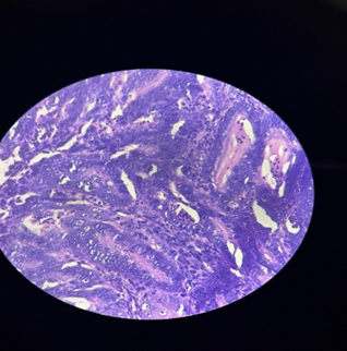
Figure 1: Endometrial hyperplasia with atypia / endometrial intraepithelial neoplasia
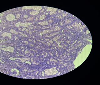
Figure 2: Endometrial hyperplasia without atypia
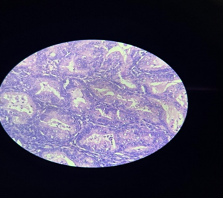
Figure 3: Endometrial hyperplasia without atypia
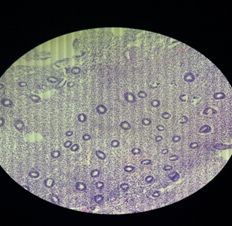
Figure 4: Proliferative endometrium
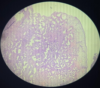
Figure 5: Endometrial polyp
DISCUSSION:
In the present study of 122 endometrial biopsy/curettage specimens, the mean age was 35.9 ± 9.8 years, with maximum cases in the 20-30 years age group (34.4%), followed by 31-40 years (31.1%). This indicates that endometrial sampling was more common in reproductive-age women in the present setting. In contrast, Hashmi et al. (2025)[1] reported maximum cases in 40-49 years (35.6%) with mean age 43.15 ± 11.70 years, while another study reported predominance in 41-50 years (55.4%)[2]. Menorrhagia was the most common complaint in the present study (46.7%), followed by dysmenorrhea (34.4%). Similar findings were noted by Hashmi et al. (2025)[1], who reported menorrhagia as the most prevalent presenting symptom, and by studies in which heavy menstrual bleeding was the main indication for endometrial biopsy.
In the present study, dysfunctional uterine bleeding was the most common clinical/radiological diagnosis (54.1%), followed by endometrial hyperplasia (23.0%). This was comparable with clinicopathological studies of AUB where functional causes formed the major group and organic lesions such as hyperplasia and polyp constituted important secondary causes. Vijayaraghavan et al. (2022)[3] emphasized that histopathological examination of endometrium remains essential because clinical symptoms alone cannot reliably distinguish functional endometrial changes from hyperplasia or malignancy. Alshdaifat et al. (2022)[4] also observed that normal cyclical endometrial patterns formed the majority of biopsy findings, supporting the diagnostic value of endometrial sampling in AUB.
The most common histopathological finding in the present study was proliferative endometrium (39.3%), followed by simple hyperplasia without atypia (26.2%), secretory endometrium (9.8%), and benign endometrial polyp (8.2%). These findings were consistent with GeethaMala et al. (2025)[5], who reported proliferative endometrium as the commonest finding (36.7%), followed by hyperplasia without atypia (25.6%) and endometrial polyps (8.9%). Similarly, Hashmi et al. (2025)[1] reported proliferative endometrium as the most common pattern (27.2%), followed by disordered proliferative endometrium (19.2%), hyperplasia (17.6%), and secretory endometrium (16.4%). Another study reported proliferative endometrium in 35.4% and secretory endometrium in 19.2%, closely supporting the predominance of functional endometrial patterns in AUB.
In the present study, premalignant lesions were found in 27.0% and malignancy in 0.8% of cases. The proportion of premalignant lesions was highest in the 41-50 years group (35.5%), while malignancy was seen only in women aged >50 years. This age-related increase in clinically significant lesions is similar to the findings of Hashmi et al. (2025)[1], who observed that hyperplasia and carcinoma were more common in older age groups. GeethaMala et al. (2025)[5] also reported carcinoma mainly among postmenopausal women. These findings support the importance of endometrial biopsy in perimenopausal and postmenopausal women presenting with AUB or postmenopausal bleeding.
The present study showed significant correlation between clinical/radiological diagnosis and histopathological findings (χ²=294.66, p<0.001). Endometrial hyperplasia was confirmed histologically in 89.3% of clinically/radiologically suspected cases, while endometrial polyp was confirmed in 83.3% of suspected cases. The single clinically suspected malignancy was confirmed as carcinoma. This highlights that although clinical and radiological assessment provides useful preliminary information, histopathology remains confirmatory. Similar conclusions were drawn by Singh et al. (2025)[6] and Yadav et al. (2025)[7], who emphasized histopathological evaluation for accurate differentiation of functional, benign, premalignant, and malignant endometrial lesions.
Overall, the findings of the present study were in agreement with recent literature showing that proliferative endometrium is the most frequent histopathological pattern in endometrial biopsies, while hyperplasia, polyps, endometritis, atrophic endometrium, and carcinoma form important pathological diagnoses. The statistically significant age-wise association in the present study indicates that premalignant and malignant lesions increase with advancing age, especially in perimenopausal and postmenopausal women. Therefore, endometrial biopsy remains a simple, reliable, and essential diagnostic tool for evaluation of AUB and for early detection of premalignant and malignant endometrial pathology.
CONCLUSION:
The present study demonstrated a wide histopathological spectrum of endometrial patterns and lesions among 122 endometrial biopsy and curettage specimens received at a multispeciality hospital in Amravati, Maharashtra. Proliferative endometrium was the most common histopathological finding, followed by simple endometrial hyperplasia without atypia, secretory endometrium, benign endometrial polyps, disordered proliferative endometrium, and progestin-associated changes. Inflammatory, atrophic, atypical hyperplastic, and malignant lesions were relatively uncommon. Benign lesions constituted the majority of cases; however, the proportion of premalignant lesions increased with advancing age, and the malignant lesion was observed in the older age group. A statistically significant association was observed between age and the histopathological category of endometrial lesions. There was also a significant correlation between clinical/radiological diagnoses and final histopathological findings, particularly for endometrial polyps, hyperplasia, and carcinoma. The findings emphasize that histopathological examination of endometrial biopsy and curettage specimens remains an essential diagnostic method for identifying functional changes and differentiating benign lesions from premalignant and malignant endometrial pathology. Appropriate endometrial sampling and timely histopathological evaluation, particularly among perimenopausal and postmenopausal women with abnormal uterine bleeding, may facilitate early diagnosis and appropriate clinical management.
LIMITATIONS OF THE STUDY
The present study had certain limitations. It was a retrospective, single-centre study conducted at a multispeciality hospital; therefore, the findings may not be generalizable to the wider population. The relatively small sample size of 122 specimens and the study duration of one year limited the evaluation of uncommon endometrial lesions, particularly atypical hyperplasia and endometrial carcinoma. As the study was based on previously recorded clinical and laboratory data, complete information regarding menstrual history, parity, body mass index, comorbidities, hormonal therapy, contraceptive use, and other potential risk factors was not available for all patients. Selection bias could have occurred because only women who underwent endometrial biopsy or curettage were included. Histopathological interpretation was based predominantly on Hematoxylin and Eosin-stained sections, and ancillary techniques such as immunohistochemistry and molecular testing were not routinely performed. Long-term follow-up information was unavailable; therefore, the subsequent progression, recurrence, treatment response, and clinical outcomes of patients with premalignant and malignant lesions could not be evaluated. Larger multicentric prospective studies with detailed clinical correlation, standardized histopathological classification, and long-term follow-up are recommended to validate the findings.
REFERENCES:
1. Hashmi ZF, et al. To study the endometrial histopathological spectrum in patients with abnormal uterine bleeding. 2025.
2. Shivakumar M, et al. Pattern of bleeding with histopathological correlation in patients presenting with abnormal uterine bleeding. J Med Sci Health. 2018.
3. Vijayaraghavan A, et al. A histopathological study of endometrial biopsy samples in abnormal uterine bleeding. Cureus. 2022;14(11):e31555.
4. Alshdaifat EH, et al. Histopathological pattern of endometrial biopsies in patients with abnormal uterine bleeding. Ann Saudi Med. 2022;42(3):204-213.
5. GeethaMala K, et al. Study of histopathological array of endometrium in abnormal uterine bleeding: our hospital experience. 2025.
6. Singh MN, et al. Histopathological spectrum of endometrium in abnormal uterine bleeding. 2025.
7. Yadav S, et al. Histopathological spectrum of endometrial lesions with correlation to hormonal status in abnormal uterine bleeding. 2025.
8. Karuna J, et al. Spectrum of pathologies observed in endometrial biopsies of cases with abnormal uterine bleeding. 2025.
9. Pathak M, et al. Histopathology of endometrium in cases of abnormal uterine bleeding. J Adv Med Pharm Sci. 2023.
10. Brahmaiah J, et al. Histopathological spectrum of endometrium in abnormal uterine bleeding. 2022.
11. Kumari SR, et al. Histopathological study of endometrium in abnormal uterine bleeding. 2017.
12. Shukla M, et al. Spectrum of endometrial lesions observed on histopathological examination in abnormal uterine bleeding. Int J Res Med Sci. 2019.
13. Munro MG, Critchley HOD, Fraser IS. FIGO systems for normal and abnormal uterine bleeding symptoms and classification of causes of AUB: 2018 revisions. Int J Gynaecol Obstet. 2018;143(3):393-408.