Comparison of USG Guided vs Landmark Technique for Easy Access of Spinal Anaesthesia in Elderly Patients.
- Dr. Arunraj , Assistant Professor, Department of ENT, Dhanalakshmi Srinivasan Medical College and Hospital, Siruvachur, Perambalur, Tamil Nadu, India.
- Dr. Dilipan Muthaiya , Senior Resident, Department of Anaesthesiology, Dhanalakshmi Srinivasan Medical College and Hospital, Siruvachur, Perambalur, Tamil Nadu, India.
- Dr. Geetha J. , Professor, Department of Anaesthesia, Dhanalakshmi Srinivasan Medical College and Hospital, Siruvachur, Perambalur, Tamil Nadu, India.
Article Information:
Abstract:
Background: Spinal anaesthesia is commonly used for lower abdominal, pelvic and lower-limb surgeries. However, age-related degenerative changes in the lumbar spine can make identification of surface anatomical landmarks difficult, potentially increasing needle passes, procedural time and patient discomfort. Preprocedural ultrasound may improve the accuracy of spinal needle placement. AIM: To compare the ease of access for spinal anaesthesia using ultrasound-assisted and landmark-guided techniques in elderly patients. METHODS: This prospective comparative clinical study was conducted in the Department of Anaesthesiology at Dhanalakshmi Srinivasan Medical College and Hospital, Siruvachur, Perambalur, over one year between 2023 and 2024. Seventy patients aged over 60 years undergoing elective surgery under spinal anaesthesia were randomly allocated into two groups: landmark-guided (Group L, n = 35) and ultrasound-assisted (Group U, n = 35). The primary outcome was the number of needle passes required for successful dural puncture. Secondary outcomes included first-pass success, procedure time, patient discomfort and haemodynamic parameters. RESULTS: The mean number of needle passes was 1.34 in Group L and 1.06 in Group U. First-pass success was 80% (28/35) in Group L and 91.4% (32/35) in Group U. The mean procedure time was 32.6 seconds in Group L compared with 19.9 seconds in Group U. The mean patient discomfort score was lower in Group U (2.60 ± 1.35) than in Group L (5.03 ± 1.65). Preoperative and postoperative pulse rates and blood pressure measurements were reported as comparable between the groups. CONCLUSION: In this study, ultrasound-assisted spinal anaesthesia was associated with fewer needle passes, higher first-pass success, shorter procedure time and lower patient discomfort than the landmark-guided technique. Preprocedural ultrasound may be a useful adjunct for facilitating spinal access in elderly patients. Further analysis with complete between-group statistical testing is needed to establish the statistical significance of these differences.
Keywords:
Article :
INTRODUCTION:
Spinal anaesthesia is a well-established regional anaesthetic technique commonly used for lower abdominal, pelvic and lower-extremity surgery. Its advantages include rapid onset, effective sensory and motor blockade, reduced intraoperative blood loss and avoidance of airway manipulation. These features are particularly relevant in older patients with multiple comorbidities.
Successful spinal anaesthesia depends on accurate identification of the intervertebral space and precise needle placement into the subarachnoid space. Age-related changes-including reduced intervertebral spaces, osteophyte formation, ligament calcification, reduced spinal mobility and altered alignment-can make surface landmarks difficult to identify. Obesity and spinal deformity may further obscure palpable landmarks, potentially increasing needle passes, redirections, procedure time and patient discomfort.
The conventional surface landmark-guided technique uses palpation of the iliac crests, spinous processes and interspinous spaces to estimate the L3–L4 or L4–L5 interspace. Preprocedural ultrasound can identify the midline and intervertebral space and estimate the depth to the dura, allowing the operator to mark an insertion site and plan the needle trajectory. This study compared ultrasound-assisted and landmark-guided spinal anaesthesia in elderly patients, with emphasis on the number of needle passes required for successful dural puncture.
Aim
To compare and assess the effectiveness of ultrasound-guided versus surface landmark-based methods of spinal anaesthesia in elderly patients based on the number of needle passes necessary for successful dural puncture.
Objectives
· Primary objective: To ascertain whether ultrasound guidance decreases the number of attempts required to perform successful spinal anaesthesia compared with the conventional landmark-guided technique.
· To compare successful dural puncture on the initial attempt between the two approaches.
· To compare the number of needle redirections during the procedure.
· To determine and compare the time taken for spinal anaesthesia.
· To record patient-perceived discomfort during the procedure using a visual analogue scale (VAS).
· To compare preoperative and postoperative pulse rate and blood pressure.
MATERIALS AND METHODS:
Study Design and Setting
This was a prospective comparative clinical study conducted in the Department of Anaesthesiology, Dhanalakshmi Srinivasan Medical College and Hospital, Siruvachur, Perambalur, following Institutional Ethics Committee approval. The study duration was one year, between 2023 and 2024.
Participants
Patients older than 60 years scheduled for elective lower abdominal, pelvic or lower-limb procedures requiring spinal anaesthesia were eligible.
Inclusion Criteria
· Age above 60 years.
· American Society of Anesthesiologists (ASA) physical status I–III.
· Elective surgery under spinal anaesthesia.
· Willingness to provide written informed consent.
Exclusion Criteria
· Bleeding disorders or anticoagulant therapy.
· Local infection at the lumbar puncture site.
· Previous spinal surgery or significant spinal abnormalities, such as severe kyphosis or scoliosis.
· Hypersensitivity to local anaesthetics.
· Unwillingness to participate.
Sample Size and Randomization
The dissertation describes a sample of 35 patients per group. The calculation was based on a first-attempt dural-puncture success proportion of 9.1% in the landmark group from Park et al., with 95% confidence and 7% precision. The formula used was N = Z² × p × (1−p) / d²; the document reports N = 64.85 and a total required sample of 65. The study groups comprised 35 patients each (70 total). Patients were randomly assigned using a computer-generated sequence to Group L (landmark-guided) or Group U (ultrasound-assisted).
Study Procedure
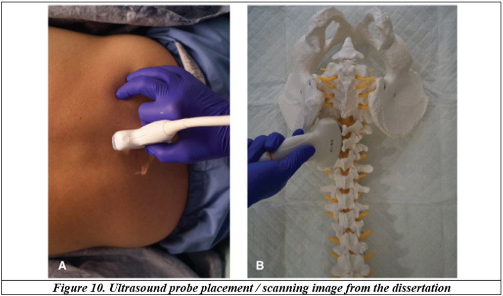
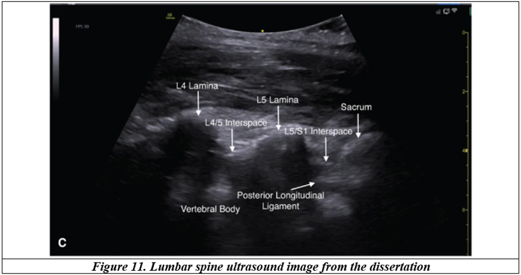
All patients were placed in the sitting position and a midline approach was used. In Group L, Tuffier’s (intercristal) line was palpated to estimate the L3–L4 or L4–L5 interspace. A 25-gauge Quincke spinal needle was used. In Group U, a low-frequency 2–5 MHz curved-array transducer was used for preprocedural scanning. The midline, intervertebral space and depth to the dura were identified and marked one hour before surgery in the preoperative holding area. Spinal anaesthesia was then performed using the same type of needle at the marked site.
Preoperative Preparation and Monitoring
All participants underwent pre-anaesthetic evaluation one day before surgery, including medical history, physical examination, airway and spinal assessment, and baseline vital signs. Routine investigations were reviewed and written informed consent was obtained. Standard aseptic precautions and ASA monitoring were used, including non-invasive blood pressure, ECG, pulse oximetry, heart rate and respiratory rate. Sensory block was assessed by pinprick and motor block by the Modified Bromage Scale. Haemodynamic parameters were recorded at baseline, every two minutes for the first ten minutes after the block, and then every five minutes until the end of the procedure.
Outcome Measures and Statistical Analysis
The primary outcome was the total number of needle passes required for successful dural puncture. Secondary outcomes included first-attempt success, needle redirections, time from needle insertion to CSF return, patient discomfort and haemodynamic parameters. An independent observer recorded procedural details using a standardized form. Continuous variables were presented as mean ± standard deviation and analysed using Student’s t-test. Categorical variables were compared using the chi-square or Fisher’s exact test. A p-value below 0.05 was considered statistically significant.
RESULTS:
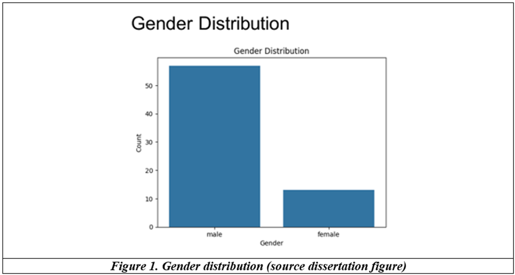
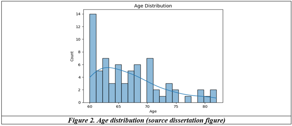
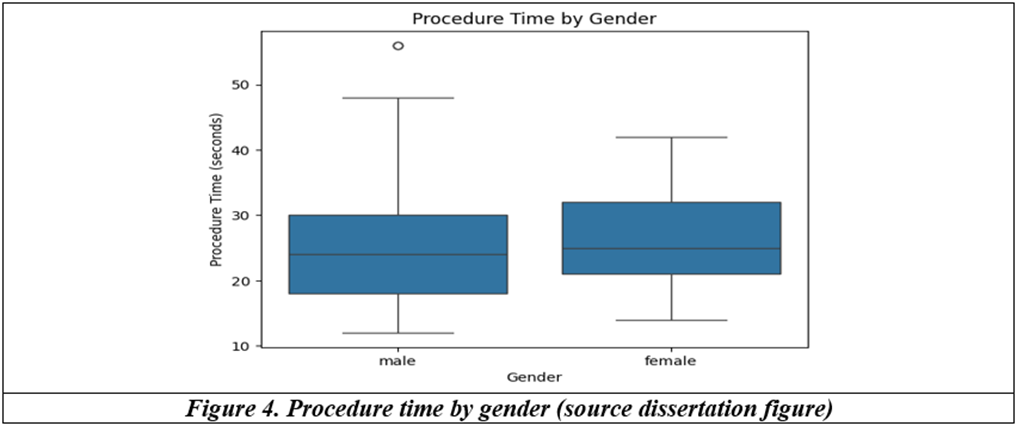
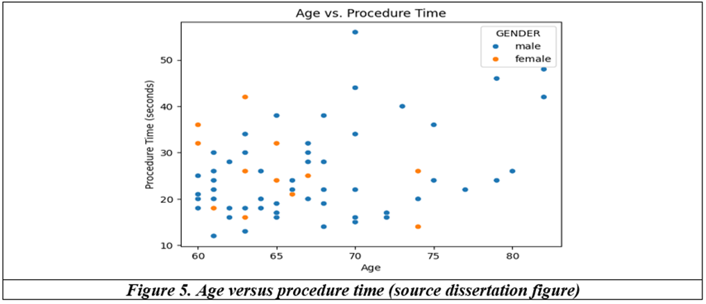
The study included 70 patients, 35 in each group. Participants were 60–82 years old; the dissertation reports 57 males and 13 females. The procedures included inguinal hernia surgery, vaginal hysterectomy, haemorrhoidectomy and fissurectomy. The source reports 17.8% inguinal hernia cases and nine vaginal hysterectomies.
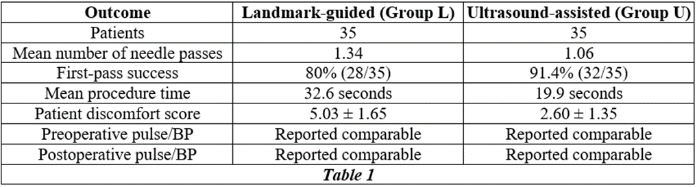
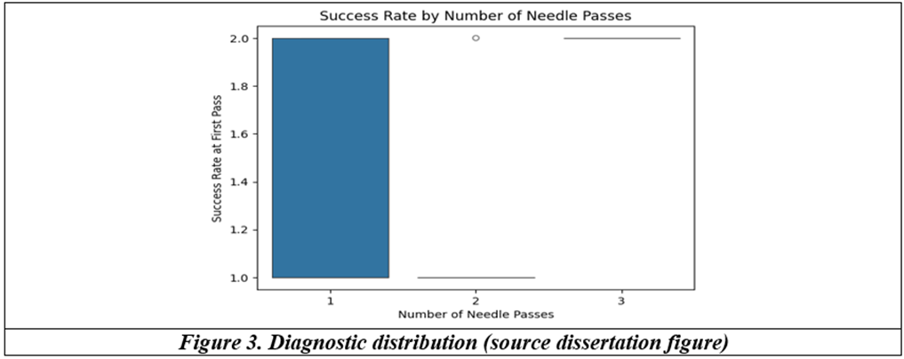
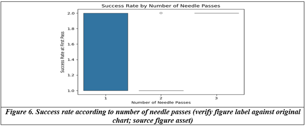
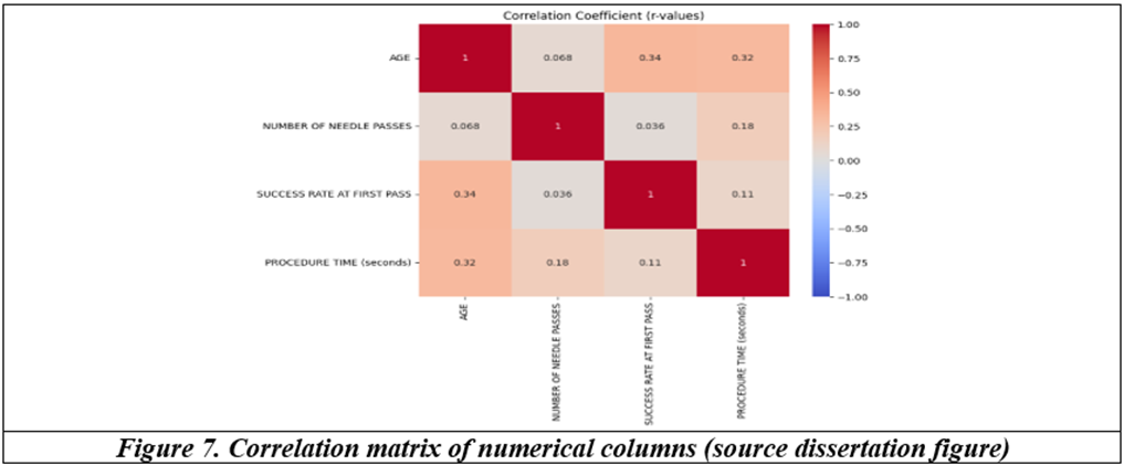
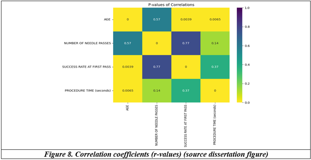
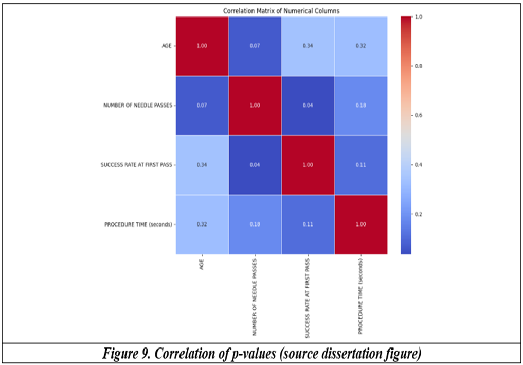
The dissertation reports p = 0.37 for the relationship between first-pass success and procedure time, and p = 0.14 for the relationship between number of needle passes and time to lumbar puncture. It states that correlations between age, number of needle passes, first-pass success and procedure time were not significant. Complete between-group p-values for the principal group comparisons are not consistently supplied in the source.
Preoperative pulse rate, systolic blood pressure and diastolic blood pressure were described as comparable between groups. Postoperative pulse rate and blood pressure were also reported as showing no significant difference. The dissertation does not provide a complete numerical group-wise haemodynamic table in the extracted results text.
DISCUSSION:
In this study, ultrasound-assisted spinal anaesthesia was associated with a lower mean number of needle passes than the landmark-guided technique (1.06 versus 1.34). First-pass success was reported as 91.4% in the ultrasound group and 80% in the landmark group. These findings are consistent with the proposed value of preprocedural ultrasound: identification of the midline and interspace and estimation of depth before needle insertion may help plan the needle trajectory in patients with age-related anatomical changes.
The reported mean procedure time was shorter in the ultrasound-assisted group (19.9 seconds) than in the landmark-guided group (32.6 seconds). Although ultrasound requires an initial scanning and marking step, the dissertation’s procedure-time measure is described as the interval from needle insertion to CSF return. The reported times should therefore be interpreted according to this definition and not as total preparation time.
Patient discomfort scores were lower in the ultrasound-assisted group (2.60 ± 1.35) than in the landmark-guided group (5.03 ± 1.65). Fewer needle insertions and shorter puncture time may explain this difference, although the source does not provide complete inferential statistics for this comparison. Preoperative and postoperative haemodynamic parameters were described as comparable, suggesting that the access technique did not materially alter the systemic haemodynamic effects of spinal anaesthesia in the reported observations.
The dissertation’s literature review discusses prior work by Broadbent et al., Grau et al., Chin et al., Park et al. and Goyal et al. reporting improved landmark identification or first-pass success with ultrasound in selected populations. The present study’s direction of findings is broadly consistent with that rationale. However, the source results contain limitations: complete between-group p-values are not provided for all outcomes, some graph labels and narrative interpretations are not fully aligned, and numerical haemodynamic results are not presented comprehensively. These issues should be checked against the original master chart and statistical output before journal submission.
Spinal anaesthesia is a core regional technique with great advantages for surgery of the lower abdomen, pelvis and lower limbs. However, the performance of a lumbar subarachnoid block could be challenging, especially in elderly patients. With advancing age, the spine undergoes anatomical changes such as calcification of the interspinous ligament and ligamentum flavum, reduced intervertebral spaces, reduced mobility of vertebral joints, development of osteophytes and spinal deformities with kyphoscoliosis, fat redistribution and muscle atrophy.
Traditionally, palpable anatomical landmarks serve to aid the location of the appropriate interspinous space, level and anticipated direction of the lumbar puncture needle. Tuffier's line-an imaginary line between the highest points of the iliac crests-is aligned with the L4 spinous process, thus determining the placement of the lumbar puncture needle at the L3–L4 or L4–L5 spaces.Broadbent et al. used MRI to compare estimated and actual vertebral levels, finding incorrect identification in about 70% of cases.[1] They compared the accuracy of identification of intervertebral spaces using surface landmarks and MRI and demonstrated that numerous needle insertions were one level higher than desired. This study accentuated the shortcomings of relying solely on surface anatomy for needle placement, particularly in patients with irregular spinal structures.
Several randomised trials were performed by Grau et al. on obstetric patients and concluded that ultrasound guidance helped to significantly improve the first-attempt success rate of spinal anaesthesia. Approximately 80–85% of patients in the ultrasound group had first-attempt success compared to 40–52% in the landmark-guided group.[2] The ultrasound group also had fewer needle passes and complications, such as traumatic dural puncture. Bartusseck F. and Comodi R. et al., along with Grau, conducted preliminary studies while training students for neuraxial anaesthesia and concluded that ultrasound imaging improved learning curves in obstetric epidural anaesthesia.
Chin et al. tested the usefulness of ultrasound (76%) over the standard technique (42%). They observed higher first-pass attempt success with the USG group, which was almost one and a half times more advantageous than conventional methods.[3] The study also recorded fewer needle redirections and reduced patient discomfort.
Elderly patients have anticipated difficult spinal anatomy, and hence this study validated the utility of ultrasound in difficult anatomical situations.[4] Park et al. documented a dramatic difference in first-pass success in elderly patients when ultrasound was used to locate the optimal and appropriate interspinous space. Although ultrasound required more time to scan and locate the appropriate space, it proved valuable in elective cases where it significantly reduced the number of attempts and complications, leading to improved procedural outcomes.[5]
Following increased interest in ultrasound usage to locate the interspinous space, various studies evaluated the sonoanatomy of the lumbar spine and better visualisation of the ligamentum flavum and dura mater. Karmakar et al.[6] demonstrated that the depth to the subarachnoid space estimated by ultrasound closely matched the actual insertion depth in more than 90% of cases, emphasising its accuracy in preprocedural planning. Geriatric patients have variable indices of frailty, either physical dependency or emotional instability.
These may be exacerbated in the event of physical or mental trauma. The variable metabolic response to injury or stress also needs to be considered. Hence, improved patient tolerance for a lumbar puncture and the ease of acceptance of the procedure are of prime importance in patient satisfaction with regard to elective surgeries. In spite of anatomical restraints in the elderly, as described earlier, various studies have focused attention on ultrasound to locate the appropriate space for first-shot success. Goyal et al. noted fewer attempts in the elderly when ultrasound-guided spinal anaesthesia was administered (1:1 vs. 2:3), with higher first-attempt success (84% vs. 48%) when compared with the traditional landmark technique. These findings further support the advantages of ultrasound guidance in geriatric populations with anticipated spinal difficulty.[7]
Benoist et al. (2003) describe spinal degeneration as a gradual, multifactorial process that typically starts in the second decade of life, beginning with biochemical and structural changes in the intervertebral discs, such as reduced proteoglycans, disc height loss, and matrix breakdown. These initial disc changes lead to secondary damage in nearby structures like the facet joints, causing arthritis, joint misalignment, and bone spur formation, which can narrow the spinal canal and reduce stability. Aging also affects the ligaments and muscles of the spine-thickening of the ligamentum flavum and weakening of postural muscles contribute to deformities like scoliosis and spondylolisthesis. Meanwhile, vertebral bodies may undergo osteoporotic changes, making them more prone to remodeling and misalignment. Pain in older adults is often due to the presence of nerve fibers in degenerated discs and joints, while nerve root compression from stenosis can cause claudication and radiculopathy. Altogether, aging of spinal tissues-driven by genetic, mechanical, and nutritional factors-creates a self-perpetuating cycle of degeneration that highlights the importance of prevention and early management (Benoist et al., 2003).[8]
Kasai et al. (2009) examined 14,250 pairs of anterior lumbar vertebral osteophytes in individuals aged over 60, identifying characteristic patterns of osteophyte development at various lumbar spine levels. They noted that in the upper lumbar regions (L1–L2 and L2–L3), osteophytes typically grew toward the adjacent disc, whereas in the lower segments (L3–S1), the growth was directed away from the disc-indicating greater degenerative changes and segmental instability. The study also distinguished between claw-shaped osteophytes, which tend to bridge the disc space, and traction osteophytes, which extend outward and are linked to instability. These formations were more prevalent with age and appeared to result from age-related mechanical stress and spinal alignment changes. This understanding is crucial for anticipating anatomical challenges during spinal procedures like neuraxial blocks in elderly patients.[9]
Sacralisation is a common irregularity of spine where fifth lumbar vertebra is fused with the sacrum. Estimates of the incidence of sacralization in the general population are very widely from 4 to 36%. The wide range of estimates is attributed to differences of diagnostic methods and definition. Among people under 30, an estimated 18.5 percent have sacralization.[10] Most of the elderly have maximum changes in the axial skeleton due to the weight-bearing action and due to the adaptational modifications towards varied occupation and environmental factors.
Popularly known as bamboo spine, ankylosing spondylitis is an inflammatory disorder that affects the axial skeleton, where inflammation of the vertebral joints with subsequent bridging of spinal segments results in reduced mobility of the spine and a stiff back. Zhu H et al. have conducted extensive studies on ankylosing spondylitis.[11] It is quite challenging when such patients necessitate a neuraxial blockade for surgery. Ultrasonogram provides valuable assistance in locating the appropriate interspinous space.
Castellvi et al. classified lumbosacral transitional vertebrae (LSTV) into four types based on the morphology and degree of articulation or fusion of the transverse process, with types II to IV being more clinically significant. Sacralization is a congenital structural variation in which the fifth lumbar vertebra is partially or completely fused to the sacrum. Its reported prevalence varies considerably depending on population characteristics and diagnostic criteria. An established system of classification separates sacralization into four types depending on the degree of articulation or co-ossification of the transverse processes, and some types have been determined to be more likely to be associated with clinical symptoms.
While the condition was originally reported in association with low back pain more than a century ago, research has yielded inconsistent results-some finding high prevalence within chronic back pain patients and others showing no statistical correlation. Anatomically, sacralization changes the biomechanical stress patterns of the lower spine, commonly raising stress at the level above the transitional vertebra, especially the L4–L5 segment. This can lead to expedited degenerative alterations, such as disc degeneration and facet joint arthropathy. Some subtypes of sacralization also carry an increased risk of herniated intervertebral disc. In symptomatic presentations, the application of focused conservative management like physiotherapy and spinal manipulative therapy has been found to yield positive results, highlighting the significance of diagnosing this anomaly in patients with longstanding or unexplained lower back pain.[12]
Technique Pictures from the Source Dissertation.
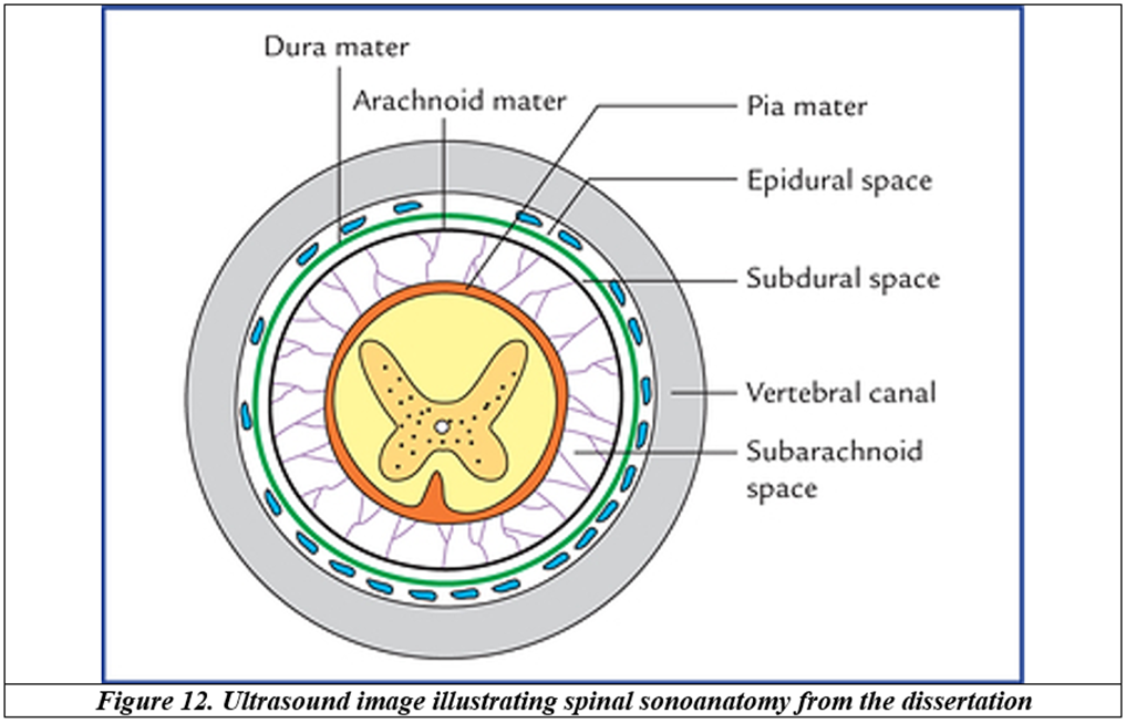
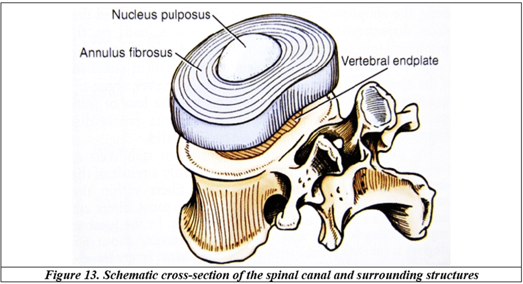
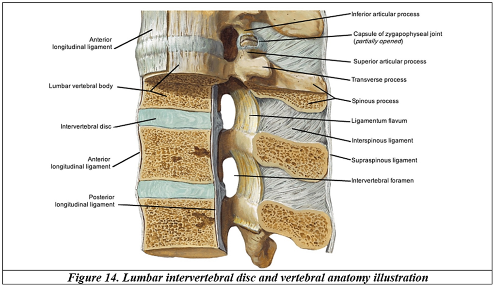
CONCLUSION:
In this prospective comparative study of elderly patients undergoing elective surgery under spinal anaesthesia, the ultrasound-assisted technique was associated with fewer needle passes, a higher first-pass success rate, shorter reported needle-insertion-to-CSF time and lower patient discomfort scores than the landmark-guided technique. Preoperative and postoperative haemodynamic parameters were reported as comparable between groups. Preprocedural ultrasound may be a useful adjunct for planning spinal access in elderly patients, particularly when surface landmarks are difficult to identify. The findings should be interpreted cautiously because complete between-group statistical results are not consistently reported in the dissertation.
REFERENCES:
1. Broadbent CR, Maxwell WB, Ferrie R, et al. Ability of anaesthetists to identify a marked lumbar interspace. Anaesthesia 2000;55(11):1122-6.
2. Grau T, Leipold RW, Horter J, et al. Paramedian access to the epidural space: the optimum procedure for obese patients. Anesth Analg 2001;93(5):1177-81.
3. Grau T, Bartusseck E, Conradi R, et al. Ultrasound imaging improves learning curves in obstetric epidural anesthesia: a preliminary study. Can J Anesth 2003;50(10):1047-50.
4. Chin KJ, Perlas A, Chan V, et al. Ultrasound imaging facilitates spinal anesthesia in adults with difficult surface anatomic landmarks. Anesthesiology 2011;115(1):94-101.
5. Park SK, Lee HJ, Cho YW, et al. Ultrasound-assisted spinal anesthesia in elderly patients: a randomized controlled trial. Anesthesia and Pain Medicine 2021;16(2):170-6.
6. Karmakar MK, Li X, Ho AM, et al. Sonoanatomy of the lumbar spine in a normal adult population using two-dimensional ultrasound imaging. Anesth Analg 2009;108(5):1851-7.
7. Goyal A, Sharma JP, Sethi N, et al. Comparative evaluation of ultrasound-guided and landmark-based spinal anesthesia in elderly. J Anaesthesiol Clin Pharmacol 2018;34(2):205-9.
8. Benoist M. Natural history of the aging spine. European Spine Journal 2003;12(Suppl 2):S86-9.
9. Kasai Y, Kawakita E, Sakakibara T, et al. Direction of the formation of anterior lumbar vertebral osteophytes. BMC Musculoskeletal Disorders 2009;10:4.
10. Hecht M. Sacralization: symptoms, causes, treatment, and outlook [Internet]. Healthline 2018 Mar 21 [cited 2025 Jun 30].
11. Zhu W, He X, Cheng K, et al. Ankylosing spondylitis: etiology, pathogenesis, and treatments. Bone Research 2019;7:22.
12. Castellvi AE, Goldstein LA, Chan DP. Lumbosacral transitional vertebrae and their relationship with lumbar extradural defects. Spine 1984;9(5):493-5.