Damage Control in Crush Injuries of Both Lower Limbs - A Case Report.
- Dr. Geetha J. , Professor, Department of Anaesthesia, Dhanalakshmi Srinivasan Medical College and Hospital, Siruvachur, Perambalur, Tamil Nadu, India.
- Dr. Meiyappan S , Assistant Professor, Department of Anaesthesia, Dhanalakshmi Srinivasan Medical College and Hospital, Siruvachur, Perambalur, Tamil Nadu, India.
- Dr. Tharun Balaji , Postgraduate in Anaesthesia, Department of Anaesthesia, Dhanalakshmi Srinivasan Medical College and Hospital, Siruvachur, Perambalur, Tamil Nadu, India.
- Dr. Tanya Anna Thomas , Postgraduate in Anaesthesia, Department of Anaesthesia, Dhanalakshmi Srinivasan Medical College and Hospital, Siruvachur, Perambalur, Tamil Nadu, India.
Article Information:
Abstract:
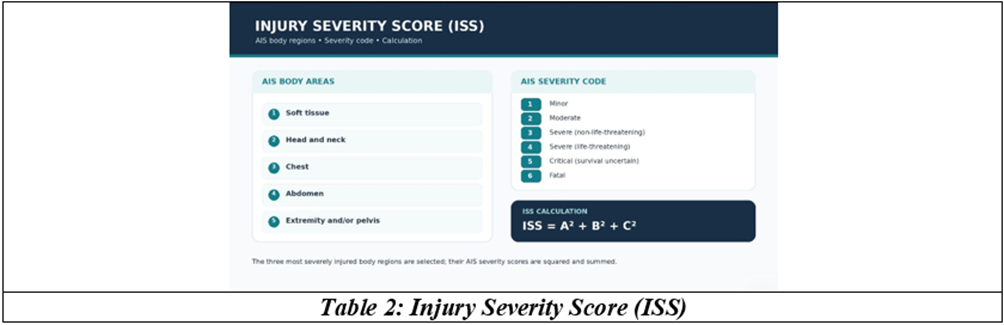
Introduction: Polytrauma activates the systemic inflammatory response syndrome (SIRS). In a patient with unstable haemodynamics, aggressive surgical intervention could be more damaging and is often called a second hit (trauma). Several studies performed widely all over the world have proved excellent outcomes when damage control strategies have been adopted in decision making, resuscitation and surgery. Case Report: This case report involved a severe degloving injury with fractures of both lower limbs, where the outcome improved with damage control surgery. General Examination: The patient was conscious, oriented and obeying commands. Heart rate was 136 beats/min, blood pressure was 110/70 mmHg and oxygen saturation (SpO2) was 98%. Systemic examination was within normal limits. The left lower limb was degloved below the knee, with impaired muscle, exposed tendons and active bleeding. There were fractures of both bones of the left leg at the mid-shaft level, with partial auto-amputation. The right lower limb had distal fractures of both bones, followed by dislocation, with partial auto-amputation. Investigations: Haemoglobin: 11.9 g/dL; prothrombin time (PT): 15.9 seconds; international normalised ratio (INR): 1.6; activated partial thromboplastin time (aPTT): 54.4 seconds; white blood cell count: 28,300/mm3; urea: 45 mg/dL; creatinine: 0.9 mg/dL; estimated glomerular filtration rate (eGFR): 114 mL/min/1.73 m2; sodium: 134 mmol/L; potassium: 3.2 mmol/L; and chloride: 103 mmol/L. Challenges: • Grossly degloved and partially amputated limbs. • Impending haemorrhagic shock. • Impending crush injury syndrome. • Impending disseminated intravascular coagulation (DIC). Plan of Anaesthesia: Endotracheal general anaesthesia (ET-GA) with controlled ventilation was planned. The presentation listed ketamine, propofol and rocuronium for general anaesthesia, with oxygen:nitrous oxide, intermittent positive-pressure ventilation (IPPV), and maintenance with rocuronium. The recorded vital signs were pulse rate 110/min, blood pressure 100/70 mmHg and SpO2 98%. Conduct of Anaesthesia: Preoxygenation was performed, followed by modified rapid sequence induction (RSI) using ketamine, rocuronium and fentanyl. The surgical procedures were left above-knee (AK) amputation with tourniquet and right below-knee (BK) amputation. Estimated blood loss was 2.5 L. Blood transfusion included 2 units of packed red blood cells (PRBC) and 2 units of fresh frozen plasma (FFP). Urine output was clear and adequate. Postoperatively, mechanical ventilation was continued overnight in the intensive care unit (ICU), and the patient was extubated the following morning. Conclusion: This case report indicated an Injury Severity Score (ISS) of 25/75. The lethal triad of hypothermia, acidosis and coagulopathy has to be constantly anticipated and monitored for aggressive intervention, against a background of damage control measures. Haemostatic resuscitation and permissive hypotension in a stable patient have proved to improve survival. Early hospitalisation and initiation of resuscitative measures prevent impending haemorrhagic shock, rhabdomyolysis and renal failure. In this era of advanced prosthesis facilities, a wise decision of amputation has saved the patient from crush trauma syndrome and prolonged multiple procedures with an indefinite outcome.
Keywords:
Article :
INTRODUCTION:
Injury is a leading cause of death and disability in people under fifty years of age worldwide. Urgent management of open fractures reduces the risk of infection, provides pain relief, promotes early mobility, and helps prevent pulmonary, thrombotic, and other complications. Damage-control surgery and related strategies are appropriate for achieving provisional bodily stability and avoiding haemorrhage and systemic deterioration.[1] Initial assessment should prioritise life-threatening and time-critical conditions. This case report describes a young male with crush injuries to both lower limbs. Complications were avoided through decisive damage-control management.
CASE REPORT:
History
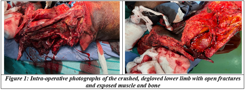
A 27-year-old male was brought to the casualty department with crush injuries and bleeding from exposed bones and muscles in both lower limbs. The patient had been pinned under a truck, with both legs extending outwards, and a mini-truck ran over his legs. He was transported to the hospital within thirty minutes of the incident. On arrival, his Glasgow Coma Scale (GCS) score was 15/15. Both lower limbs were severely injured and bleeding profusely. He was tachycardic, with a heart rate of 136 beats/min, indicating a high risk of impending hemorrhagic shock.
Clinical Features and Examination
The patient was pale. Oxygen saturation (SpO2) was 98%, pulse rate was 136 beats/min, and blood pressure was 110/60 mmHg. Systemic examination was reported to be within normal limits. The left lower limb was degloved below the knee, with muscles and tendons exposed and profuse bleeding. There were fractures of both the tibia and fibula at the mid-shaft level, with partial traumatic amputation. The right lower limb had fractures of the distal tibia and fibula, exposed tissues, and dislocation of the foot. The notes record an Abbreviated Injury Scale (AIS) score of 3 for soft-tissue injury and 5 for the extremity.
Secondary Survey and Initial Management
The secondary survey showed no head, chest, or pelvic injuries. Estimated blood loss was more than 2.5 litres. Blood samples were collected, and two 16-gauge intravenous cannulae were inserted. Two litres of crystalloids and two units of packed red blood cells were transfused.
Investigations
Investigations revealed a haemoglobin of 11 g/dL, a total white blood cell count of 28,800/mm3, a prothrombin time (PT) of 15.9 seconds, an activated partial thromboplastin time (aPTT) of 54.4 seconds, an international normalised ratio (INR) of 1.6, urea of 45 mg/dL, creatinine of 0.9 mg/dL, an estimated glomerular filtration rate (eGFR) of 114 mL/min/1.73 m2, sodium of 134 mmol/L, potassium of 3.2 mmol/L and chloride of 103 mmol/L. The urine was clear on bladder catheterisation.
Challenges in Management
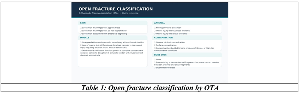
The challenges in management included: grossly degloved, fractured and bleeding limbs with contused muscles, exposed tendons and open fractures that indicate an impending hemorrhagic shock; the triad of coagulopathy, acidosis and hypothermia; an impending crush injury syndrome, constituting renal failure due to myoglobin and toxins insulting the proximal tubules of the nephrons; and a high risk of sepsis due to the automobile-type run-over on the road. According to the open fracture classification developed by the Orthopaedic Trauma Association (OTA),[2] the scores were 13/15, revealing the gravity of the injury.
Plan of Management
Oxygenation, two 16 G venous cannulations, blood products and activation of the massive transfusion protocol, bladder catheterisation, and blood samples for further investigations were all to progress simultaneously within minutes. A patient with such severe limb injuries can have a higher quality of life following a limb amputation rather than a heroic limb reconstruction.[3] After a life-changing limb injury, a well-performed rehabilitative amputation is a procedure that sets the patient on their own pathway of recovery to return to work, especially in this era of prosthesis. Amputation during the damage-control surgery phase may be necessary for a limb not amenable to reconstruction. The objectives at this phase are focussed on gaining haemorrhage control and removing gross contamination. The results of meticulous clinical examination should be documented as texts and images with date and time.
Anaesthesia
Regional anaesthesia was deferred due to haemodynamic instability. Endotracheal general anaesthesia with controlled ventilation was instituted with fentanyl 2 µg/kg, ketamine 2 mg/kg and rocuronium 1 mg/kg by the modified rapid sequence method of induction, and maintained with intermittent positive pressure ventilation with isoflurane at ½ MAC.
Monitors: pulse, mean arterial pressure by non-invasive blood pressure (NIBP), SpO2, EtCO2, ST segment monitoring, endotracheal cuff pressure (½ hourly), train-of-four (TOF) to guide muscle relaxant, continuous hourly urine output, and estimation of blood loss and tissue loss. Two units of fresh frozen plasma, two units of packed cells and two litres of crystalloids were administered.
Surgery
Right below-knee amputation and left above-knee amputation were performed, fashioning a rough end for closure. There was no major vascular bleed for a proximal vascular ligation. The estimated blood loss was about 2.5 litres and the patient compensated and tolerated it well, with no severe hypotension, probably due to his age and absence of comorbidities.
The urine output at the end of 90 minutes of the procedure was 400 ml and it was clear. His vitals were stable throughout the intra-operative period. The patient was electively ventilated overnight and extubation was smooth and uneventful the next morning. Pain relief was given by fentanyl infusion at the rate of 10 µg per hour. Psychological support and counselling were offered post-operatively and the patient was motivated for prosthesis.
The factors that averted the serious complications, namely shock, DIC, haemolysis, renal failure, embolism and ARDS, were hospitalisation within thirty minutes of injury, immediate replacement of blood products, quick decision for amputation, co-operation and consent from the patient’s relatives, young age and absence of comorbidities, aggressive resuscitation and prophylactic mechanical ventilation.
DISCUSSION:
The elaboration of the salient features of this case report is discussed under the following headings: crush syndrome, myoglobinuria, the lethal triad of polytrauma, damage control procedures and haemorrhagic shock.
Crush Syndrome
Crush injury or crush trauma occurs when soft tissues experience the effect of direct physical trauma due to external force. This results in extensive muscle and nerve damage and systemic ischaemia, tissue necrosis and systemic effects. Systemic manifestations that are induced by crush injury are referred to as crush syndrome.[4] Severe systemic complications of crush syndrome lead to muscle and end-organ injuries of rhabdomyolysis (myolysis), electrolyte imbalances, acute kidney injury, disseminated intravascular coagulation, dysrhythmias and sepsis. Once developed, the mortality rate in patients with crush injury is approximately 18%. Almost 10% of patients with crush injury develop crush syndrome (CS), while others may have an uneventful recovery. CS involves 74% of lower extremity trauma. The organs that are involved late are associated with the highest mortality rate.[5] The management of CS remains challenging due to the complexity of systemic manifestations that arise from muscle cell destruction. The prolonged pressure on soft tissues leads to ischaemia, reperfusion injury and release of toxic metabolites. These in turn lead to rhabdomyolysis, electrolyte imbalance, acute kidney injury and potential multiorgan failure.
History of Crush Syndrome
The phenomenon was first documented during the First World War as Meyer-Betz syndrome and later as Bywaters syndrome,[6] according to its description during the Second World War by Bywaters and Beall.
Pathophysiology
CS develops due to prolonged muscle compression, leading to ischaemia, hypoxia and muscle necrosis.
Stretching of skeletal muscle membranes opens calcium channels. The calcium influx triggers ATP consumption for ion balance, but excessive calcium accumulation weakens mitochondrial function and activates proteases and phospholipases. This cascade depletes cellular energy and disrupts sodium gradients, resulting in oedema. Muscles located distal to pressure sites become ischaemic. Complete ischaemia leads to oedema and lysosome degranulation within 30 minutes, and irreversible necrosis in 4–6 hours. In incomplete ischaemia, limited collateral circulation maintains minimal energy production, but ATP depletion still causes oedema. When the compressive force is released, reperfusion injury occurs, which could cause ischaemic damage through interaction between leukocytes and endothelial cells. The resulting superoxide radicals from ischaemic damage attack free fatty acids, leading to cellular oedema, cell death and necrosis.[7] The tissue damage is further exacerbated by an impaired sodium-potassium ATP pump that exchanges sodium for calcium, further disrupting intracellular metabolism. Direct pressure on the muscles during crush trauma results in release of potassium, purines, lactic acid, phosphate, myoglobin, urate, creatinine and thromboplastin. These lead to disruption of homeostasis, hyperkalaemia, metabolic acidosis, raised CPK levels and systemic inflammation. Myoglobin leads to tubular obstruction, oxidative stress and renal vasoconstriction. Phosphate accumulation, hyperuricaemia from breakdown of purines, and sodium imbalances all contribute to renal dysfunction. Mortality within the first 3 days after crush trauma is often recognised as hyperkalaemia-related deaths. Acute renal failure is the most significant cause of death within the first two weeks, and late deaths are due to multiorgan failure. Severe muscle breakdown is indicated by tea-coloured urine, which signifies myoglobinuria and potential renal failure.[8]
Rhabdomyolysis
The release of toxic intracellular material into the systemic circulation following damage to skeletal muscle results in the syndromic events of rhabdomyolysis. The pathogenesis is based on an increase in free ionised calcium in the cytoplasm. The main anticipated complications include: (a) acute renal failure triggered by renal vasoconstriction and ischaemia; (b) myoglobin cast formation in distal convoluted tubules; and (c) direct renal toxic effect of myoglobin on the epithelial cells of proximal convoluted tubules.[9] Hyperkalaemia from tissue injury causes cardiac arrhythmias, while metabolic acidosis, hyperphosphataemia, early hypocalcaemia and late hypercalcaemia are other metabolic derangements. Compartment syndrome and disseminated intravascular coagulopathy also emerge along with myoglobinuria.The management of myoglobinuric acute renal failure includes aggressive fluid administration to restore hypovolaemia and urine alkalinization.[10] Aggressive detection and management of anticipated concomitant electrolyte and metabolic disorders are essential to prevent the crush trauma sequelae.
The Lethal Triad in Critical Care
The lethal triad constitutes the trinity of hypothermia, acidosis and coagulopathy, directly related to a high risk of mortality.[11] The triad is a vicious cycle, each component potentiating the others, and is proportional to the severity of injury. It also relates to the resuscitative interventions received during the course of surgery or general management.[12] In a hypovolaemic trauma patient, these events occur rapidly in quick progression. The damage control measure entails damage control surgery, haemostatic resuscitation and permissive hypotension, and has been associated with improved survival in trauma patients.[13]
Hypothermia
Hypothermia in trauma is multifactorial in aetiology. Cold environment, exposure of tissues/organs leading to body heat loss, co-morbidities such as thyroid illness, shock, resuscitation with cold fluids, burns and central causes of traumatic brain injury all lead to hypothermia with a core temperature of less than 35 °C (95 °F). Hypothermia may lead to acidosis and coagulopathy. The hypothermia component of the lethal triad is associated with poor prognosis in trauma patients.[14] When patients are matched for injury severity, hypothermia has been shown to increase mortality.[15] Active and passive warming strategies should be utilised early to allay heat losses during resuscitation. By attending individually to every component of the lethal triad, the battered physiology may be restored to maintain the internal milieu.[16] Various studies were conducted defining hypothermia as temperature less than 35.5 °C, acidosis as pH less than or equal to 7.25, and coagulopathy as INR more than 1.2. It was concluded that metabolic acidosis setting in a trauma scenario was the most lethal of this triad. Patients with significant haemorrhage have added irreversible recovery in the presence of this lethal triad, according to Mitra et al, 2012. The cause of acidosis is usually due to inadequate tissue perfusion due to hypotension, anaemia and hypoxia, leading to anaerobic respiration and metabolic acidosis. Traumatised tissues activate the extrinsic pathway and induce fibrinolysis, leading on to consumption coagulopathy or disseminated intravascular coagulation. INR is an indicator of clotting efficiency. According to Hodgetts et al (2007), INR more than 1.5 suggests coagulopathy and the need for replacement by plasma. Damage control resuscitation combines permissive hypotension and haemostatic resuscitation with damage control surgery. Due to the role played by hypocalcaemia, the lethal triad has been modified to the lethal diamond, with four factors entering a vicious cycle indicating poor prognosis.
Case Summary
This patient was a young man who had crush injury of both lower limbs but escaped from impending haemorrhagic shock, rhabdomyolysis and renal failure. He had several positive factors to avert entering the lethal triad. He reached hospital within 30 minutes after injury and aggressive volume resuscitation was initiated. Borderline renal function and deterioration of urine output guided decision making towards amputation and avoidance of limb handling measures. Damage control procedures avoided the crush trauma syndrome.
CONCLUSION:
Multiple long bone fractures may lead to life-threatening haemorrhage with mortality up to 25%.[17] Degloved wounds lead to the lethal triad of mortality, heat loss causing hypothermia followed by acidosis and coagulopathy. Compression of soft tissues may eventuate myoglobinuria and its sequelae. Anticipating these grave and long drawn recovery situations and careful decision making focussing on damage control surgery and damage control resuscitation would greatly influence the lifestyle after injury for the victims.
REFERENCES:
1. Pape HC, Giannoudis PV, Krettek C. The timing of fracture treatment in polytrauma patients: relevance to damage control orthopaedic surgery. Am J Surg 2002;183(6):622-9.
2. Orthopaedic Trauma Association: Open Fracture Study Group. A new classification scheme for open fractures. J Orthop Trauma 2010;24(8):457-64.
3. Penn-Barwell JG, Kendrew TM, McCabe J, et al. Limb amputation after trauma. Chap- 21. In: Rockwood and Green’s Fractures in Adults. 10th edn. Wolters Kluwer 2025.
4. Akrivos VS, Koutalos A, Stefanou N, et al. Crush injury and crush syndrome: a comprehensive review. EFORT Open Rev 2025;10(6):424-30.
5. Long B, Liang SY, Gottlieb M. Crush injury and syndrome: a review for emergency clinicians. Am J Emerg Med 2023;69:180-7.
6. Bywaters EG. 50 years on: the crush syndrome. BMJ 1990;301(6766):1412-5.
7. Yokota J, Minei JP, Fantini GA, et al. Role of leukocytes in reperfusion injury of skeletal muscle after partial ischemia. Am J Physiol 1989;257:H1068-75.
8. Brattebø G, Fredriksen K, Wisborg T. Prevention of acute renal failure in muscular injuries. Tidsskr Nor Laegeforen 1992;112(18):2370-2.
9. Chatzizisis YS, Misirli G, Hatzitolios AI, et al. The syndrome of rhabdomyolysis: complications and treatment. Eur J Intern Med 2008;19(8):568-74.
10. Noji EK. Prophylaxis of acute renal failure in traumatic rhabdomyolysis. N Engl J Med 1990;323(8):550-1.
11. Mitra B, Tullio F, Cameron PA, et al. Trauma patients with the triad of death. Emerg Med J 2011;29(8):622-5.
12. Harris T, Davenport R, Hurst T, et al. Improving outcome in severe trauma: trauma systems and initial management. Postgrad Med J 2012;88(1044):588-94.
13. Ball CG. Damage control resuscitation: history, theory and technique. Can J Surg 2014;57(1):55-60.
14. McDermott FT, Cordner SM, Cooper DJ, et al. Management deficiencies and death preventability of road traffic fatalities before and after a new trauma care system. J Trauma 2007;63(2):331-8.
15. Langhelle A, Lockey D, Harris T, et al. Body temperature of trauma patients on admission to hospital: a comparison of anaesthetised and non-anaesthetised patients. Emerg Med J 2010;29(3):239-42.
16. Muthukumar V, Karki D, Jatin B. Concept of lethal triad in critical care of severe burn injury. Indian J Crit Care Med 2019;23(5):206-9.
17. O'Toole RV, Castillo RC, Pollak AN, et al. Determinants of patient satisfaction after severe lower extremity injury. J Bone Joint Surg Am 2008;90(6):1206-11.