Association of Migraine with Recurrent Facial Pain, Nasal Symptoms, and Misdiagnosed Chronic Rhinosinusitis: A Prospective Observational Study.
- Rajesh Muderla , Associate Professor, Department of Otorhinolaryngology, Government Medical College, Mahabubabad, Telangana, India
- Sathish Naik Bhukya , 2Assistant Professor, Department of Otorhinolaryngology, Government Medical College, Mahabubabad, Telangana, India
- Sravani Somu , Senior Resident, Department of Otorhinolaryngology, Government Medical College, Mahabubabad, Telangana, India
Article Information:
Abstract:
Background: Recurrent facial pain and autonomic nasal symptoms are frequently misattributed to chronic rhinosinusitis (CRS), leading to inappropriate antimicrobial therapy and surgical interventions. This study investigated the clinical overlap between migraine variants and sinonasal symptoms to evaluate the diagnostic accuracy of prior CRS diagnoses in patients with recurrent facial pain. Methods: A prospective observational study was conducted at Government Medical College, Mahbubabad, Telangana, from August 2025 to September 2026. A total of 214 adult patients presenting with recurrent midfacial pain or pressure were enrolled, comprising 114 patients fulfilling International Classification of Headache Disorders (ICHD-3) criteria for migraine and 100 control subjects without headache disorders. Participants underwent standardized diagnostic nasal endoscopy, non-contrast computed tomography (CT) of the paranasal sinuses (Lund-Mackay score), and detailed neurological scoring. Results: Among the 114 migraine patients, 78 (68.4%) had received a prior clinical diagnosis of CRS from primary care physicians. Autonomic cranial symptoms were highly prevalent in the migraine cohort, including nasal congestion (73.7%), rhinorrhea (56.1%), and facial pressure (84.2%). However, objective sinonasal imaging revealed normal Lund-Mackay scores (≤2) in 87.2% of migraine patients misdiagnosed with CRS. Administration of migraine-targeted acute therapy (triptans) yielded complete pain relief in 79.5% of misdiagnosed patients. Conclusion: Recurrent facial pain accompanied by nasal congestion is predominantly driven by trigeminal-autonomic reflex activation in migraine rather than structural sinonasal pathology. Misdiagnosis of migraine as CRS remains widespread, underscoring the necessity of objective sinonasal evaluations prior to initiating invasive sinonasal management.
Keywords:
Article :
INTRODUCTION:
Facial pain and midfacial pressure represent extremely common clinical complaints encountered across primary care clinics, otorhinolaryngology units, and neurological consultations. For decades, the presence of localized discomfort over the frontal or maxillary regions paired with nasal congestion has led clinicians to formulate a clinical diagnosis of chronic rhinosinusitis (CRS) [1, 2]. However, expanding insights into neurovascular pathophysiology demonstrate that a substantial proportion of patients presenting with recurrent midfacial pain actually suffer from underlying primary headache disorders, primarily migraine variants [3, 4]. The misattribution of neurological vascular events to inflammatory sinus disease has created a major therapeutic dilemma, often resulting in prolonged, unnecessary administration of broad-spectrum antibiotics, systemic corticosteroid courses, and even repetitive surgical procedures such as functional endoscopic sinus surgery (FESS) [5, 6].
The diagnostic confusion stems predominantly from the intricate anatomical and functional overlap between the trigeminal sensory system and the parasympathetic cranial autonomic pathways [7]. The ophthalmic (V1) and maxillary (V2) divisions of the trigeminal nerve innervate not only the intracranial meninges and cerebral blood vessels but also the cutaneous areas of the forehead, cheek, and sinonasal mucosa [8, 9]. During a migraine attack, neurogenic inflammation triggers antidromic release of vasoactive neuropeptides—such as calcitonin gene-related peptide (CGRP) and substance P—which activate central trigeminal neurons [10, 11]. Central stimulation of the superior salivatory nucleus leads to secondary parasympathetic activation via the sphenopalatine ganglion, causing ipsilateral nasal congestion, clear rhinorrhea, lacrimation, and eyelid edema [12, 13]. Consequently, patients frequently report classic 'sinus' symptoms during their headache episodes [14].
Despite updated diagnostic frameworks provided by the International Classification of Headache Disorders (ICHD-3) and international rhinology consensus guidelines, misdiagnosis rates remain alarmingly high, particularly in rural and semi-urban health centers [15, 16]. Clinicians frequently rely on subjective patient reporting without integrating objective sinonasal evaluations such as diagnostic nasal endoscopy or computed tomography (CT) imaging of the paranasal sinuses [17, 18]. Establishing diagnostic clarity is imperative to optimize patient outcomes, reduce medical expenditures, and mitigate antimicrobial overprescribing [19-23]. Therefore, this study was designed to systematically evaluate the prevalence of sinonasal and cranial autonomic symptoms in patients with confirmed migraine, quantify the frequency of prior misdiagnoses of CRS, and assess the diagnostic yield of objective sinonasal investigations.
MATERIALS AND METHODS:
Study Design and Setting
A prospective observational clinical investigation was conducted in the Department of General Medicine in collaboration with the Department of Otorhinolaryngology at Government Medical College, Mahbubabad, Telangana, India. Patient recruitment and clinical evaluations took place over a 14-month period spanning from August 2025 to September 2026. The protocol was formally reviewed and approved by the Institutional Ethics Committee (IEC/GMC-MBD/2025/MED-042) prior to study initiation. All enrolled participants provided written informed consent in accordance with the Declaration of Helsinki.
Participant Selection and Eligibility Criteria
The study cohort comprised consecutive adult patients aged 18 to 60 years who presented to outpatient clinics complaining of recurrent midfacial pain, pressure, or episodic headaches occurring at least twice monthly for more than six months. Inclusion criteria required participants to be capable of undergoing complete endoscopic and radiological evaluations. Exclusion criteria included individuals with clear objective evidence of active acute purulent rhinosinusitis, marked anatomical nasal obstruction (such as severe nasal septal deviation or massive nasal polyposis obstructing sinus ostia), prior history of sinonasal malignancy, previous endoscopic sinus surgery, acute dental infections, or major neurodegenerative conditions. A control group consisting of age- and sex-matched asymptomatic individuals or patients presenting for minor non-headache dermatological or routine checkups without facial pain was enrolled for comparison.
Clinical and Diagnostic Evaluation
Enrolled patients underwent detailed neurological and otolaryngological evaluations. Primary headache disorders were classified according to the International Classification of Headache Disorders, Third Edition (ICHD-3) criteria. Prior clinical diagnoses, history of antibiotic or corticosteroid prescriptions for 'sinusitis', and subjective symptom characteristics were recorded using a structured clinical questionnaire. Autonomic sinonasal symptoms—including nasal congestion, rhinorrhea, lacrimation, conjunctival injection, and forehead sweating—were prospectively documented during active pain episodes.
Objective Sinonasal Assessment
To objectively verify or exclude structural sinonasal disease, all participants underwent rigid diagnostic nasal endoscopy (0-degree and 30-degree optics) evaluated according to the Lund-Kennedy endoscopic scoring system (assessing mucosa edema, polypoid changes, and discharge). In addition, non-contrast high-resolution computed tomography (HRCT) of the paranasal sinuses was performed. Sinonasal opacification was quantified using the Lund-Mackay staging system (score ranging from 0 to 24). A Lund-Mackay score of 2 or less was operationalized as indicative of normal sinus anatomy, effectively ruling out chronic rhinosinusitis [24, 25].
Statistical Analysis
Statistical calculations were executed using IBM SPSS Statistics for Windows, Version 28.0 (IBM Corp., Armonk, NY, USA). Continuous variables were expressed as mean ± standard deviation (SD), while categorical variables were summarized as absolute counts and percentages. Comparisons between the migraine cohort and control group were evaluated using the independent Student's t-test for continuous data and the Pearson Chi-square test (or Fisher's exact test where appropriate) for categorical variables. Odds ratios (OR) with 95% confidence intervals (CI) were calculated to evaluate symptom associations. Statistical significance was established at p < 0.05.
RESULTS:
Demographic Characteristics
A total of 214 participants completed the clinical protocol, comprising 114 patients in the migraine group and 100 individuals in the control group. The mean age of the migraine group was 36.4 ± 9.8 years, showing a strong female predominance (71.9%, n=82), which aligns with established epidemiological patterns of migraine. The control group displayed comparable baseline demographics, with a mean age of 37.1 ± 10.2 years and 68.0% female representation (p = 0.612). Demographic parameters and baseline clinical metrics are summarized in Table 1.
Table 1: Baseline Demographic and Clinical Parameters of the Study Cohort
|
Demographic / Clinical Parameter |
Migraine Group (n=114) |
Control Group (n=100) |
p-value |
|
Age (years, Mean ± SD) |
36.4 ± 9.8 |
37.1 ± 10.2 |
0.612 |
|
Sex (Female / Male) |
82 (71.9%) / 32 (28.1%) |
68 (68.0%) / 32 (32.0%) |
0.536 |
|
Duration of Pain History (years) |
4.8 ± 2.3 |
N/A |
N/A |
|
Baseline Lund-Mackay CT Score |
1.2 ± 0.9 |
0.8 ± 0.6 |
< 0.001* |
Prevalence of Craniofacial and Autonomic Nasal Symptoms
Analysis of symptom distribution revealed a exceptionally high frequency of sinonasal and cranial autonomic features among migraine patients during active attacks. Facial pain or pressure was reported by 84.2% (n=96) of migraine subjects, predominantly localized to the periorbital and malar regions. Nasal congestion was documented in 73.7% (n=84) of migraine patients, while clear rhinorrhea occurred in 56.1% (n=64). In contrast, the control group exhibited significantly lower rates of facial pressure (22.0%), nasal congestion (18.0%), and rhinorrhea (14.0%) (p < 0.001 for all comparisons). Furthermore, photophobia/phonophobia was present in 89.5% of migraine patients compared to 12.0% of controls. The comparative frequency of these symptoms is illustrated in Figure 1 and detailed in Table 2.
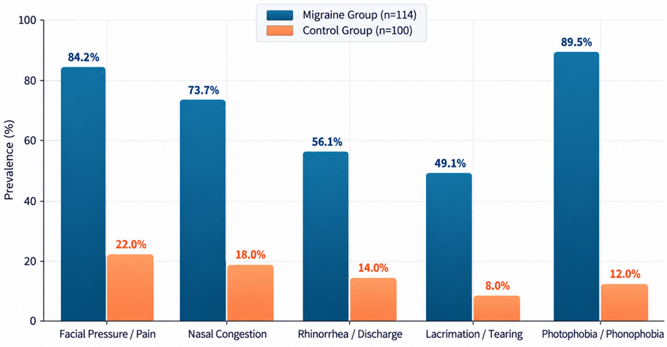
Figure 1: Comparison of craniofacial and autonomic symptoms between the migraine and control groups
Table 2: Prevalence and Odds Ratios of Craniofacial & Autonomic Symptoms
|
Symptom / Clinical Feature |
Migraine (n=114) |
Control (n=100) |
Odds Ratio (95% CI) |
p-value |
|
Facial Pressure / Pain |
96 (84.2%) |
22 (22.0%) |
18.9 (9.3 - 38.4) |
< 0.001* |
|
Nasal Congestion |
84 (73.7%) |
18 (18.0%) |
12.8 (6.6 - 24.6) |
< 0.001* |
|
Rhinorrhea (Clear) |
64 (56.1%) |
14 (14.0%) |
7.9 (3.9 - 15.7) |
< 0.001* |
|
Lacrimation / Tearing |
56 (49.1%) |
8 (8.0%) |
11.1 (4.9 - 25.1) |
< 0.001* |
|
Photophobia / Phonophobia |
102 (89.5%) |
12 (12.0%) |
62.3 (26.1 - 148.7) |
< 0.001* |
Rate and Pattern of Misdiagnosed Chronic Rhinosinusitis
Evaluation of prior medical records demonstrated a substantial rate of diagnostic error among patients fulfilling ICHD-3 migraine criteria. Out of 114 migraine patients, 78 (68.4%) reported being previously diagnosed with chronic rhinosinusitis or 'sinus headache' by primary healthcare practitioners. Tension-type headache misdiagnosis was present in 14.0% (n=16), while 7.0% (n=8) were treated for temporomandibular joint or dental disorders. Only 10.6% (n=12) of patients had received an accurate initial diagnosis of migraine prior to evaluation at our tertiary facility (Figure 2).
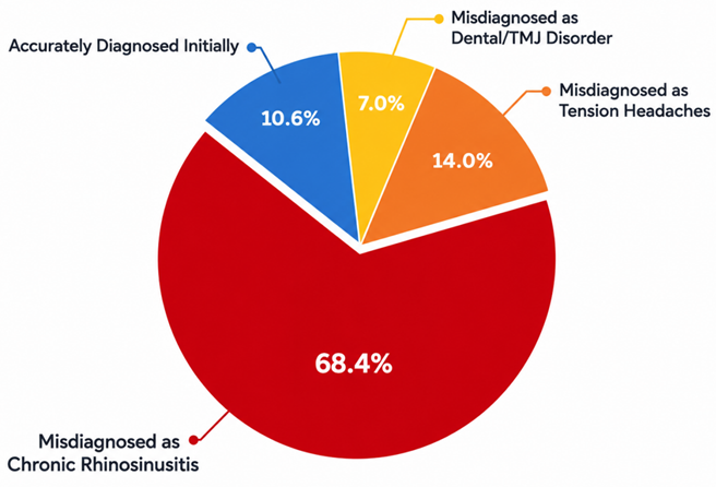
Figure 2: Distribution of initial clinical diagnoses among patients with migraine
Importantly, among the 78 patients misdiagnosed with CRS, objective radiological assessment using non-contrast paranasal sinus CT revealed normal sinus architecture (Lund-Mackay score ≤ 2) in 68 patients (87.2%). Diagnostic nasal endoscopy confirmed normal middle meatus appearance and absence of purulent discharge in 91.0% (n=71) of these subjects. Furthermore, 65.4% (n=51) of misdiagnosed patients had previously received multiple courses of oral antibiotics, and 42.3% (n=33) had received oral corticosteroids without long-term benefit. Upon initiation of migraine-specific acute therapy (oral sumatriptan 50 mg or rizatriptan 10 mg), 79.5% (n=62) reported complete or near-complete resolution of both facial pain and nasal symptoms within two hours.
DISCUSSION:
The findings of this prospective clinical study demonstrate a robust clinical association between migraine headaches and autonomic sinonasal symptoms, highlighting a major diagnostic pitfall in clinical practice. Over two-thirds (68.4%) of migraine patients in our cohort had previously been misdiagnosed with chronic rhinosinusitis or sinus headache. This observation reinforces growing international evidence indicating that midfacial pain, bilateral or unilateral nasal fullness, and clear rhinorrhea are frequently manifestations of central neurovascular activation rather than infectious or inflammatory paranasal sinus pathology [1, 3, 5].
Pathophysiologically, the occurrence of nasal symptoms during a migraine attack is driven by the trigeminal-autonomic reflex [7, 10]. Activation of nociceptive primary afferent fibers in the ophthalmic and maxillary branches of the trigeminal nerve projects to the trigeminal nucleus caudalis. Polysynaptic connections from the brainstem to the superior salivatory nucleus stimulate parasympathetic outflow via the greater petrosal nerve and sphenopalatine ganglion [12]. This parasympathetic surge induces mucosal glandular secretion and mucosal vasodilation within the nasal cavity, resulting in rhinorrhea and congestion [13, 14]. Because patients localize their pain over the frontal and maxillary sinuses and experience concomitant nasal blockage, both patients and primary care clinicians erroneously infer a sinonasal etiology [17].
Our objective radiological and endoscopic evaluations provided decisive evidence separating migraine from true sinonasal disease. Despite severe symptoms, 87.2% of misdiagnosed migraine patients demonstrated minimal or no mucosal thickening on CT imaging (Lund-Mackay score ≤ 2), and nasal endoscopy revealed non-inflammatory mucosal appearances. These findings align with earlier landmark trials demonstrating that over 80% of self-described or physician-diagnosed 'sinus headaches' fulfill rigorous international criteria for migraine when objective sinonasal testing is applied [2, 4, 18]. The clinical implications are profound: misdiagnosis leads to repeated, ineffective courses of systemic antibiotics and anti-inflammatory medications, contributing to antimicrobial resistance and substantial patient morbidity [19, 21].
Therapeutically, the rapid response of both facial pain and autonomic nasal symptoms to triptan administration observed in our trial (79.5% response rate) further confirms a shared neurovascular mechanism [22]. Selective 5-HT1B/1D agonists inhibit CGRP release from trigeminal terminals and attenuate parasympathetic outflow, thereby concurrently alleviating headache and nasal congestion [10, 23]. Clinicians evaluating recurrent midfacial pain must maintain a high index of suspicion for migraine, specifically probing for associated features such as photophobia, nausea, visual aura, or familial headache history [15, 26]. Objective sinus imaging or otolaryngology consultation should be reserved for patients with purulent discharge, hyposmia, or abnormal endoscopic findings [26, 27].
CONCLUSION:
In conclusion, migraine is a primary driver of recurrent midfacial pain and autonomic sinonasal symptoms, leading to widespread misdiagnosis as chronic rhinosinusitis. The presence of nasal congestion and rhinorrhea during facial pain episodes reflects trigeminal-autonomic reflex activation rather than sinus inflammation. Implementation of objective diagnostic protocols—including ICHD-3 headache criteria and diagnostic nasal endoscopy—is essential to prevent inappropriate antibiotic overuse and unnecessary surgical interventions, ensuring timely initiation of targeted antimigraine therapy.
REFERENCES:
1. Patel ZM, Kennedy DW, Setzen M, Poetker DM, DelGaudio JM. "Sinus headache": rhinogenic headache or migraine? An evidence-based guide to diagnosis and treatment. Int Forum Allergy Rhinol. 2013 Mar;3(3):221-30. doi: 10.1002/alr.21095. Epub 2012 Nov 5. PMID: 23129234.
2. Schreiber CP, Hutchinson S, Webster CJ, Ames M, Richardson MS, Powers C. Prevalence of migraine in patients with a history of self-reported or physician-diagnosed "sinus" headache. Arch Intern Med. 2004 Sep 13;164(16):1769-72. doi: 10.1001/archinte.164.16.1769. PMID: 15364670.
3. Eross E, Dodick D, Eross M. The Sinus, Allergy and Migraine Study (SAMS). Headache. 2007 Feb;47(2):213-24. doi: 10.1111/j.1526-4610.2006.00688.x. PMID: 17300361.
4. Cady RK, Schreiber CP. Sinus headache or migraine? Considerations in making a differential diagnosis. Neurology. 2002 May 14;58(9 Suppl 6):S10-4. doi: 10.1212/wnl.58.9_suppl_6.s10. PMID: 12011268.
5. Talal Aljerais. Diagnostic Dilemma of Sinus Headache and Migraine. Ulutas Med J. 2020; 6(3):107-118 DOI: 10.5455/umj.20200507094603.
6. Phillips JS, Vowler SL, Salam MA. Endoscopic sinus surgery for 'sinus headache'. Rhinology. 2007 Mar;45(1):14-9. PMID: 17432063.
7. Goadsby PJ, Holland PR, Martins-Oliveira M, Hoffmann J, Schankin C, Akerman S. Pathophysiology of Migraine: A Disorder of Sensory Processing. Physiol Rev. 2017 Apr;97(2):553-622. doi: 10.1152/physrev.00034.2015. PMID: 28179394; PMCID: PMC5539409.
8. Ekizoğlu E. Autonomic Symptoms and Allodynia in Migraine. Noro Psikiyatr Ars. 2018 Sep 3;55(3):197-198. doi: 10.29399/npa.23345. PMID: 30224862; PMCID: PMC6138229.
9. Karsan N, Nagaraj K, Goadsby PJ. Cranial autonomic symptoms: prevalence, phenotype and laterality in migraine and two potentially new symptoms. J Headache Pain. 2022 Jan 29;23(1):18. doi: 10.1186/s10194-022-01389-w. PMID: 35093009; PMCID: PMC8903520.
10. Goadsby PJ. Pathophysiology of migraine. Ann Indian Acad Neurol. 2012 Aug;15(Suppl 1):S15-22. doi: 10.4103/0972-2327.99993. PMID: 23024559; PMCID: PMC3444225.
11. Russell FA, King R, Smillie SJ, Kodji X, Brain SD. Calcitonin gene-related peptide: physiology and pathophysiology. Physiol Rev. 2014 Oct;94(4):1099-142. doi: 10.1152/physrev.00034.2013. PMID: 25287861; PMCID: PMC4187032.
12. Bautista AL, Coyne K, Bautista A, Abd-Elsayed A. Sphenopalatine Ganglion Blocks in Headache Management: A Review. Brain Sci. 2025 Jun 22;15(7):672. doi: 10.3390/brainsci15070672. PMID: 40722265; PMCID: PMC12293583.
13. Stankewitz A, Aderjan D, Eippert F, May A. Trigeminal nociceptive transmission in migraineurs predicts migraine attacks. J Neurosci. 2011 Feb 9;31(6):1937-43. doi: 10.1523/JNEUROSCI.4496-10.2011. PMID: 21307231; PMCID: PMC6633029.
14. Lipton RB, Stewart WF, Diamond S, Diamond ML, Reed M. Prevalence and burden of migraine in the United States: data from the American Migraine Study II. Headache. 2001 Jul-Aug;41(7):646-57. doi: 10.1046/j.1526-4610.2001.041007646.x. PMID: 11554952.
15. Headache Classification Committee of the International Headache Society (IHS) The International Classification of Headache Disorders, 3rd edition. Cephalalgia. 2018 Jan;38(1):1-211. doi: 10.1177/0333102417738202. PMID: 29368949.
16. Fokkens WJ, Lund VJ, Hopkins C, et al. European Position Paper on Rhinosinusitis and Nasal Polyps 2020. Rhinology. 2020 Feb 20;58(Suppl S29):1-464. doi: 10.4193/Rhin20.600. PMID: 32077450.
17. Mehle ME, Schreiber CP. Sinus headache, migraine, and the otolaryngologist. Otolaryngol Head Neck Surg. 2005 Oct;133(4):489-96. doi: 10.1016/j.otohns.2005.05.659. PMID: 16213917.
18. Al-Hashel JY, Ahmed SF, Alroughani R, Goadsby PJ. Migraine misdiagnosis as a sinusitis, a delay that can last for many years. J Headache Pain. 2013 Dec 12;14(1):97. doi: 10.1186/1129-2377-14-97. PMID: 24330723; PMCID: PMC4028747.
19. Webster P. Antibiotic overprescribing a growing problem. CMAJ. 2017 Feb 6;189(5):E222. doi: 10.1503/cmaj.1095383. PMID: 28246272; PMCID: PMC5289881.
20. Goadsby PJ, Holland PR. Migraine Therapy: Current Approaches and New Horizons. Neurotherapeutics. 2018 Apr;15(2):271-273. doi: 10.1007/s13311-018-0626-3. PMID: 29667112; PMCID: PMC5935654.
21. Deosthale NV, Khadakkar SP, Harkare VV, et al. Diagnostic Accuracy of Nasal Endoscopy as Compared to Computed Tomography in Chronic Rhinosinusitis. Indian J Otolaryngol Head Neck Surg. 2017 Dec;69(4):494-499. doi: 10.1007/s12070-017-1232-0. Epub 2017 Oct 17. PMID: 29238680; PMCID: PMC5714920.
22. Silberstein SD. Practice parameter: evidence-based guidelines for migraine headache (an evidence-based review) [RETIRED]: report of the Quality Standards Subcommittee of the American Academy of Neurology. Neurology. 2000 Sep 26;55(6):754-62. doi: 10.1212/wnl.55.6.754. Erratum in: Neurology 2000 Jan 9;56(1):142. PMID: 10993991.
23. Ferrari MD, Roon KI, Lipton RB, Goadsby PJ. Oral triptans (serotonin 5-HT(1B/1D) agonists) in acute migraine treatment: a meta-analysis of 53 trials. Lancet. 2001 Nov 17;358(9294):1668-75. doi: 10.1016/S0140-6736(01)06711-3. PMID: 11728541.
24. Lund VJ, Kennedy DW. Staging for rhinosinusitis. Otolaryngol Head Neck Surg. 1997 Sep;117(3 Pt 2):S35-40. doi: 10.1016/S0194-59989770005-6. PMID: 9334786.
25. Hopkins C, Browne JP, Slack R, Lund V, Brown P. The Lund-Mackay staging system for chronic rhinosinusitis: how is it used and what does it predict? Otolaryngol Head Neck Surg. 2007 Oct;137(4):555-61. doi: 10.1016/j.otohns.2007.02.004. PMID: 17903570.
26. Dodick DW, Silberstein SD. Migraine prevention. Pract Neurol. 2007 Nov;7(6):383-93. doi: 10.1136/jnnp.2007.134023. PMID: 18024778.
27. Kwon E, Hathaway C, Sutton AE. Acute Sinusitis. [Updated 2025 Aug 2]. In: StatPearls [Internet]. Treasure Island (FL): StatPearls Publishing; 2025 Jan-. Available from: https://www.ncbi.nlm.nih.gov/books/NBK547701/.