Correlation of Tumor Budding with Histological Grade and Lymph Node Metastasis in Colorectal Carcinoma.
- Dr. Md Hamed Altaf Mali , Assistant Professor Esic medical clg kalaburgi, Karnataka Dept of pathology.
- Dr. Pavan , Asst Prof Esic medical clg kalaburgi, Karnataka Dept of pathology
- Dr. Ashfaq Ahmed , Consultant gastroenterologist Global gastro clinic and endoscopic centre, Kalaburagi, Karnataka
- Dr. V Srinivasa Murthy , Director, Professor and Head Dept of Pathology ESIC MCH Kalaburagi, Karnataka.
Article Information:
Abstract:
Background: Tumor budding is a poor prognostic factor for CRC; it represents a loss of cell-cell adhesion and is characterized by isolated tumor cells or small groups of tumor cells that detach from the main body of the tumor at the invasive margin. High budding has been shown to be associated with more aggressive pathologic characteristics as well as increased risk of metastasis. Objectives: This study sought to evaluate tumor budding using the ITBCC criteria, and determine whether there was any relationship between budding and histologic grade or regional lymph node metastasis. Materials and Methods: For demonstration purposes of this manuscript, we developed a simulated cross-sectional data set of 90 CRC resection specimens.. Tumor budding was categorized as Bd1 (0-4 buds), Bd2 (5-9 buds), and Bd3 (>=10 buds) in a 0.785 mm2 hotspot. Histological grade, nodal status, lymphovascular invasion, perineural invasion, and pT stage were analyzed. Associations were tested using chi-square tests and Spearman correlation. Binary logistic regression was used to examine predictors of lymph node metastasis. Results: Bd1, Bd2, and Bd3 were represented by 38 (42.2%), 28 (31.1%), and 24 (26.7%) cases, respectively. High-grade morphology increased from 15.8% in Bd1 to 32.1% in Bd2 and 70.8% in Bd3 (p<0.001). Lymph node metastasis was identified in 5.3%, 25.0%, and 70.8% of Bd1, Bd2, and Bd3 tumors, respectively (p<0.001). Tumor budding correlated positively with histological grade (rho=0.447, p<0.001) and nodal metastasis (rho=0.563, p<0.001). In multivariable analysis, Bd3 independently predicted nodal metastasis (adjusted OR 8.27, 95% CI 2.14-31.93; p=0.002), as did high histological grade (adjusted OR 5.10, 95% CI 1.47-17.66; p=0.010). Conclusion: The increasing tumor budding was strongly associated with poor differentiation and lymph node metastasis. The findings illustrate how standardized ITBCC assessment can add risk information beyond conventional histological grade.
Keywords:
Article :
INTRODUCTION:
Colorectal carcinoma is a heterogeneous malignancy in which tumors with similar anatomical stage may follow different clinical courses. Conventional pathological assessment therefore incorporates several adverse features in addition to tumor-node-metastasis stage, including histological type and grade, lymphovascular invasion, perineural invasion, margin status, and tumor deposits. Tumor budding has emerged as a complementary morphological marker of invasive behavior at the tumor-stroma interface.1-3
Tumor budding is defined as a single malignant cell or a cluster of up to four malignant cells separated from the main tumor. The International Tumor Budding Consensus Conference (ITBCC) recommended assessing tumor budding using hematoxylin and eosin evaluation, choosing a hot spot from the invasive front, normalizing to an area of 0.785 mm2 and using a three-tier classification system for tumor budding (Bd1 - 0-4 buds; Bd2 - 5-9 buds; Bd3 - >=10 buds).1 This standardization process has provided increased reproducibility and allowed for the inclusion of tumor budding into current reporting guidelines for colorectal cancer.2
Tumor budding is seen biologically as a visual representation of reduced epithelial cohesiveness and increased migration potential, both are components of partial epithelial-mesenchymal transition. Therefore, the budding cells at the invading edge can be viewed as an initial morphological stage of invasion and distant spread. High levels of budding have been linked to lymphatic/vascular invasion, advanced pathologic stage, nodal metastases, recurrent disease, and poor survival.4-9
The histopathological grade of a tumor is related to, however different than, the degree of tumor budding. Grade is usually assessed by examining the amount of gland formation that exists within the main part of the tumor. Tumor budding on the other hand assesses the focal loss of cellular adhesion at the invasive edge of the tumor. Research has shown that there is a statistically significant correlation between the histological grade and the degree of tumor budding, yet at the same time there was an incomplete overlap in the assessment of each characteristic, suggesting that they represent different pathological characteristics.1,10
Tumor budding has a significant impact on determining the stage of the patient's colorectal cancer and how they will be treated after surgery. Early lesions (i.e. T1 or T2) may use tumor budding to find out which patients have a higher likelihood of having an undiagnosed nodal disease. Stage II tumors can also get more information about their prognosis from tumor budding.9,11-14 This study offers a total evaluation scheme for studying whether there is an association between ITBCC tumor budding and histological grade, as well as whether there is an association between ITBCC tumor budding and regional lymph node metastasis.
Objectives
1. To assess tumor budding using ITBCC criteria.
2. To determine the association between tumor budding category and histological grade.
3. To determine the association between tumor budding category and regional lymph node metastasis.
4. To evaluate the association of budding with lymphovascular invasion, perineural invasion, and advanced pT stage.
5. To evaluate whether high tumor budding independently predicts lymph node metastasis in multivariable analysis.
MATERIALS AND METHODS:
A cross-sectional observational study conducted at ESIC medical College hospital in and around Kalaburagi. A total of 90 primary colorectal adenocarcinoma resection specimens with an assessable invasive front and known regional lymph node status were assessed. Cases were selected from the right colon, left colon, and rectum. All cases that were primarily resected for colorectal adenocarcinoma and had a clear invasive front, sufficient number of H&E stained slides for evaluation, and the presence of lymph node data were eligible. Exclusion criteria included: fragmented/non-assessable invasive fronts, recurrent tumors, post-neoadjuvant specimens where peritumoral budding could not be adequately assessed, and cases with unusual histology that did not allow for standard budding evaluation.
Tumor site, invasion depth, histology grade, LVI (lymphovascular invasion), PNI (perineural invasion), LN (lymph node) status, and other pertinent data were collected.Histological grade was categorized as low grade (well or moderately differentiated) and high grade (poorly differentiated) for analysis.All sections containing the invasive front were conceptually scanned at low power to identify the budding hotspot. A tumor bud was defined as one tumor cell or a cluster of up to four tumor cells. The bud count was normalized to a field area of 0.785 mm2 and categorized as Bd1 (0-4), Bd2 (5-9), or Bd3 (>=10), in accordance with ITBCC recommendations (Figure 1, Figure 2 and Figure 3). P Representative histopathological examination demonstrated irregular malignant glands infiltrating the surrounding desmoplastic stroma, with tumor cell dissociation at the invasive front (Figure 4). Poorly differentiated clusters of five or more cells were not counted as buds.Regional lymph nodes were categorized as negative or positive for metastatic carcinoma. For the primary analysis, any nodal metastasis was treated as node positive.
Statistical analysis
Categorical variables were summarized as frequencies and percentages. Associations between budding category and pathological variables were assessed using Pearson chi-square tests. Spearman rank correlation was used for ordinal relationships between Bd1-Bd3 and binary pathological variables. Binary logistic regression was performed for lymph node metastasis. Crude odds ratios were calculated for Bd3, high histological grade, lymphovascular invasion, and advanced pT stage; these variables were then entered into a multivariable model. Odds ratios are presented with 95% confidence intervals. A two-sided p value <0.05 was considered statistically significant.
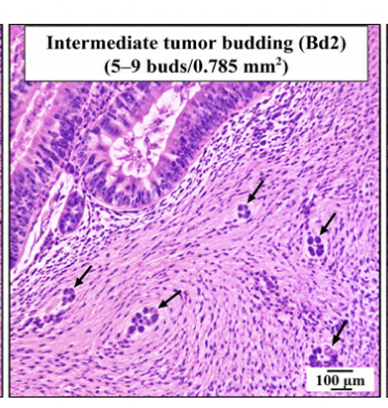
Figure 1: Low tumor budding (Bd1), showing 0–4 tumor buds per hotspot area (0.785 mm²), with only a few isolated single cells or small clusters of up to 4 cells within the desmoplastic stroma adjacent to malignant glands.
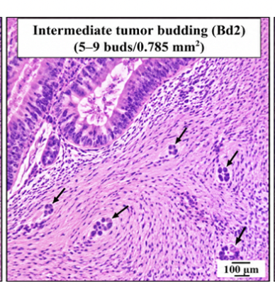
Figure 2: Intermediate tumor budding (Bd2), showing 5–9 tumor buds per hotspot area, with a moderate increase in detached single cells and small clusters at the invasive front.
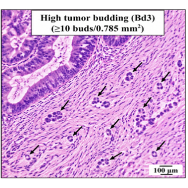
Figure 3: High tumor budding (Bd3), showing ≥10 tumor buds per hotspot area, with numerous isolated tumor cells and tiny clusters dispersed within the stromal tissue at the advancing edge of the tumor.
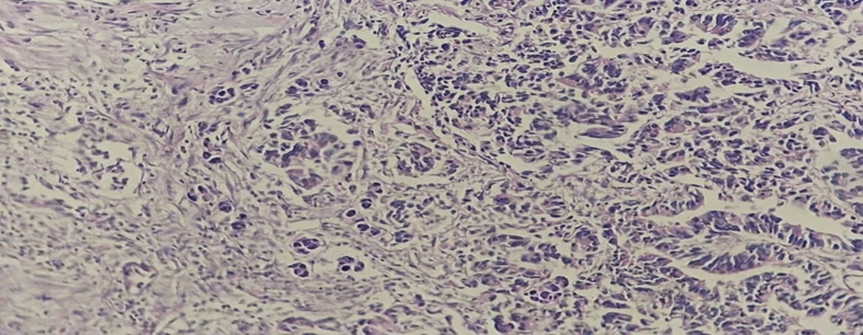
Figure 4: Hematoxylin and eosin-stained section of colorectal adenocarcinoma showing irregular malignant glandular structures infiltrating the surrounding desmoplastic stroma, with areas of complex glandular architecture and tumor cell dissociation at the invasive interface.
RESULTS:
About sixty-six cases (73.3%) were in patients aged 50 years or older and 24 (26.7%) were younger than 50 years. There were 52 males (57.8%) and 38 females (42.2%). Thirty tumors (33.3%) were located in the right colon, 33 (36.7%) in the left colon, and 27 (30.0%) in the rectum. Low-grade morphology was present in 58 cases (64.4%) and high-grade morphology in 32 (35.6%). Lymph node metastasis was present in 26 cases (28.9%).
Table 1. Clinicopathological characteristics
|
Variable |
Category |
n |
Percentage |
|
Age |
<50 years |
24 |
26.7 |
|
|
>=50 years |
66 |
73.3 |
|
Sex |
Male |
52 |
57.8 |
|
|
Female |
38 |
42.2 |
|
Site |
Right colon |
30 |
33.3 |
|
|
Left colon |
33 |
36.7 |
|
|
Rectum |
27 |
30.0 |
|
Histological grade |
Low grade |
58 |
64.4 |
|
|
High grade |
32 |
35.6 |
|
Lymph node status |
Negative |
64 |
71.1 |
|
|
Positive |
26 |
28.9 |
|
Lymphovascular invasion |
Absent |
54 |
60.0 |
|
|
Present |
36 |
40.0 |
|
Perineural invasion |
Absent |
76 |
84.4 |
|
|
Present |
14 |
15.6 |
|
pT stage grouping |
pT1/pT2 |
36 |
40.0 |
|
|
pT3/pT4 |
54 |
60.0 |
Distribution of tumor budding
Bd1 was the most frequent category, occurring in 38 cases (42.2%), followed by Bd2 in 28 (31.1%) and Bd3 in 24 (26.7%).
Table 2. Distribution of ITBCC tumor budding categories.
|
ITBCC category |
Bud count per 0.785 mm2 |
n |
Percentage |
|
Bd1 |
0-4 |
38 |
42.2 |
|
Bd2 |
5-9 |
28 |
31.1 |
|
Bd3 |
>=10 |
24 |
26.7 |
|
Total |
|
90 |
100.0 |
Tumor budding and histological grade
High-grade morphology became progressively more frequent with increasing budding. It was present in 6 of 38 Bd1 tumors (15.8%), 9 of 28 Bd2 tumors (32.1%), and 17 of 24 Bd3 tumors (70.8%). The association was statistically significant (chi-square=19.66, df=2, p<0.001). Tumor budding showed a moderate positive correlation with high histological grade (Spearman rho=0.447, p<0.001).
Table 3. Association between tumor budding and histological grade.
|
Tumor budding |
Low grade n (%) |
High grade n (%) |
Total |
p value |
|
Bd1 |
32 (84.2) |
6 (15.8) |
38 |
|
|
Bd2 |
19 (67.9) |
9 (32.1) |
28 |
|
|
Bd3 |
7 (29.2) |
17 (70.8) |
24 |
<0.001 |
Tumor budding and lymph node metastasis
The proportion of node-positive tumors increased markedly across budding categories: 2 of 38 Bd1 tumors (5.3%), 7 of 28 Bd2 tumors (25.0%), and 17 of 24 Bd3 tumors (70.8%) had regional lymph node metastasis. The association was highly significant (chi-square=31.08, df=2, p<0.001). Budding category correlated positively with nodal metastasis (Spearman rho=0.563, p<0.001).
Table 4. Association between tumor budding and lymph node metastasis.
|
Tumor budding |
Node negative n (%) |
Node positive n (%) |
Total |
p value |
|
Bd1 |
36 (94.7) |
2 (5.3) |
38 |
|
|
Bd2 |
21 (75.0) |
7 (25.0) |
28 |
|
|
Bd3 |
7 (29.2) |
17 (70.8) |
24 |
<0.001 |
Tumor budding and other adverse pathological features
Increasing budding was also associated with lymphovascular invasion, perineural invasion, and advanced pT stage. Lymphovascular invasion rose from 15.8% in Bd1 to 70.8% in Bd3 (p<0.001), while perineural invasion increased from 2.6% to 37.5% (p=0.001). Advanced pT3/pT4 tumors represented 44.7% of Bd1, 57.1% of Bd2, and 87.5% of Bd3 lesions (p=0.003).
Table 5. Adverse pathological features according to tumor budding category.
|
Variable |
Bd1 n (%) |
Bd2 n (%) |
Bd3 n (%) |
p value |
|
High histological grade |
6 (15.8) |
9 (32.1) |
17 (70.8) |
<0.001 |
|
Lymph node metastasis |
2 (5.3) |
7 (25.0) |
17 (70.8) |
<0.001 |
|
Lymphovascular invasion |
6 (15.8) |
13 (46.4) |
17 (70.8) |
<0.001 |
|
Perineural invasion |
1 (2.6) |
4 (14.3) |
9 (37.5) |
0.001 |
|
pT3/pT4 |
17 (44.7) |
16 (57.1) |
21 (87.5) |
0.003 |
Logistic regression for lymph node metastasis
On univariable analysis, Bd3 tumor budding, high histological grade, and lymphovascular invasion were significantly associated with nodal metastasis. Bd3 showed a crude odds ratio of 15.38 (95% CI 4.99-47.45; p<0.001). After simultaneous adjustment for histological grade, lymphovascular invasion, and advanced pT stage, Bd3 remained independently associated with nodal metastasis (adjusted OR 8.27, 95% CI 2.14-31.93; p=0.002). High histological grade also remained significant (adjusted OR 5.10, 95% CI 1.47-17.66; p=0.010). Lymphovascular invasion and advanced pT stage did not retain statistical significance in this multivariable model.
Table 6. Binary logistic regression for lymph node metastasis.
|
Variable |
Crude OR (95% CI) |
p value |
Adjusted OR (95% CI) |
p value |
|
Bd3 vs Bd1/Bd2 |
15.38 (4.99-47.45) |
<0.001 |
8.27 (2.14-31.93) |
0.002 |
|
High histological grade |
8.04 (2.89-22.33) |
<0.001 |
5.10 (1.47-17.66) |
0.010 |
|
Lymphovascular invasion |
4.47 (1.70-11.80) |
0.002 |
2.72 (0.80-9.18) |
0.107 |
|
pT3/pT4 |
2.25 (0.83-6.09) |
0.111 |
0.57 (0.14-2.23) |
0.418 |
DISCUSSION:
The results of our study analysis show that there is a step-by-step correlation between tumor budding and the degree of aggressiveness shown by the pathology. In Bd1 tumors, 15.8 percent had high histological grades; however, Bd3 tumors had 70.8 percent with high histological grades. Similarly, lymph node metastases increased from 5.3 percent in Bd1 tumors to 70.8 percent in Bd3 tumors. These increases resulted in significant chi-square relationships and moderately strong positive Spearman correlations, indicating the expected association where tumor budding represents an increasing trend toward more aggressive invasive behavior.
It can be argued that there is a biological connection between budding and histological grade; however, this should not be interpreted as if they were identical in terms of their relationship. Histological grade represents how much gland development occurs in the tumor, as well as how different the cells are from each other in the tumor mass. On the other hand, budding represents how many single cells (or small groups of cells) separate themselves from the main group of cells at the edge of the tumor. As such, the ITBCC framework considers budding to be a distinct characteristic.1 Zlobec et al., also stated that although grade and budding relate to one another, they are not interchangeable and therefore both should be reported separately.10
The most strong relationship observed in the current study was between tumor budding and regional lymph node metastasis. Only 5.3% of Bd1 tumors had lymph node involvement, while 70.8% of Bd3 tumors had lymph node involvement. The findings from this study are consistent with the idea that budding cells have enhanced movement (motility) and invasion capabilities. Many published studies have previously documented an association between increased budding and nodal involvement; these studies include systematic reviews of early-stage colorectal cancer and a recent meta-analysis of T2-stage disease.9,13
The association between budding and lymph node metastasis was confirmed by the regression analysis. When accounting for histological grade, lymphovascular invasion, and advanced pT stage; bd3 continued to be associated with lymph node metastasis and had an adjusted odds ratio of 8.27. Also, the occurrence of lymphovascular and perineural invasion increased as budding category increased. The aggregation of these adverse features supports the biological interpretation that budding is a surrogate for an invasive phenotype. Previous research has shown that high budding is related to vascular invasion, infiltrative growth, altered stroma, and poor survival. Therefore, tumor budding provides additional information to complement existing staging variables.
Standardized assessment is needed for clinical practice. The ITBCC advises that the hotspot should be selected at the invasive front and that the absolute bud count and three-tier category should be reported following normalization to 0.785 mm2.1 It is important to differentiate between buds and poorly differentiated clusters, stromal cells, inflammatory cells, apoptotic materials and glandular fragmentation.13-16 Pancytokeratin staining may be helpful in identifying epithelial cells within challenging areas, however, routine scoring is performed using H&E morphology.1,2
The primary limitation of this manuscript is that it is based upon a fully simulated dataset and can therefore never be used as evidence of clinical utility. This simulation was conducted to create a complete journal formatted article with all internal frequencies and statistical analyses being consistent with one another. To conduct a actual study, there are many additional steps that need to be taken, including obtaining ethics approval, defining a population to sample from, confirming each specimen's pathology diagnosis, documenting specimen exclusion criteria, performing independent slide reviews, and performing the statistical analysis on the collected data. In addition to these requirements, providing external validation and follow up on patients who survived would further support the validity of subsequent clinical studies.
CONCLUSION:
There is a strong correlation between the amount of tumor budding present and the following: high-histological grade; regional lymph node metastasis; lymphovascular invasion; perineural invasion; and advanced pT stage. After adjusting for multiple variables, Bd3 continued to be significantly related to nodal metastasis. This research demonstrates that tumor budding may be used as another pathological risk indicator in conjunction with conventional grading and TNM staging.
REFERENCES:
1. Lugli A, Kirsch R, Ajioka Y, Bosman F, Cathomas G, Dawson H, et al. Recommendations for reporting tumor budding in colorectal cancer based on the International Tumor Budding Consensus Conference (ITBCC) 2016. Mod Pathol. 2017;30(9):1299-311.
2. College of American Pathologists. Protocol for the examination of resection specimens from patients with primary carcinoma of the colon and rectum. Northfield (IL): College of American Pathologists; 2025.
3. WHO Classification of Tumours Editorial Board. Digestive system tumours. 5th ed. Lyon: International Agency for Research on Cancer; 2019.
4. Ueno H, Murphy J, Jass JR, Mochizuki H, Talbot IC. Tumour budding as an index to estimate the potential of aggressiveness in rectal cancer. Histopathology. 2002;40(2):127-32.
5. Rogers AC, Winter DC, Heeney A, Gibbons D, Lugli A, Puppa G, et al. Systematic review and meta-analysis of the impact of tumour budding in colorectal cancer. Br J Cancer. 2016;115(7):831-40.
6. Koelzer VH, Zlobec I, Lugli A. Tumor budding in colorectal cancer--ready for diagnostic practice? Hum Pathol. 2016;47(1):4-19.
7. van Wyk HC, Park JH, Edwards J, Horgan PG, McMillan DC, Going JJ. The relationship between tumour budding, the tumour microenvironment and survival in patients with primary operable colorectal cancer. Br J Cancer. 2016;115(2):156-63.
8. Aldyab M, Pacheco RR, Najjar S, Rand JV, Lee H. High-yield areas to grade tumor budding in colorectal cancer: a practical approach for pathologists. Ann Diagn Pathol. 2023;63:152085.
9. Watanabe J, Ichimasa K, Kudo SE, Mochizuki K, Tan KK, Kataoka Y, et al. Risk factors for lymph node metastasis in T2 colorectal cancer: a systematic review and meta-analysis. Int J Clin Oncol. 2024;29(7):921-31.
10. Zlobec I, et al. Are tumour grade and tumour budding equivalent in colorectal cancer? A retrospective analysis of 771 patients. Eur J Cancer. 2020;130:139-45.
11. Betge J, Kornprat P, Pollheimer MJ, Lindtner RA, Schlemmer A, Rehak P, et al. Tumor budding is an independent predictor of outcome in AJCC/UICC stage II colorectal cancer. Ann Surg Oncol. 2012;19(12):3706-12.
12. Marx AH, Mickler C, Sauter G, Simon R, Terracciano LM, Izbicki JR, et al. High-grade intratumoral tumor budding is a predictor for lymphovascular invasion and adverse outcome in stage II colorectal cancer. Int J Colorectal Dis. 2020;35(2):259-68.
13. Bosch SL, Teerenstra S, de Wilt JHW, Cunningham C, Nagtegaal ID. Predicting lymph node metastasis in pT1 colorectal cancer: a systematic review of risk factors providing rationale for therapy decisions. Endoscopy. 2013;45(10):827-34.
14. Dawson H, Galuppini F, Trager P, Berger MD, Studer P, Brugger L, et al. Validation of the International Tumor Budding Consensus Conference 2016 recommendations on tumor budding in stage I-IV colorectal cancer. Hum Pathol. 2019;85:145-51.
15. Shu Y, Zhang H, Lugli A, Simmer F, Nagtegaal ID. The prognostic value of tumour budding for survival in stage II and III colorectal cancer: a systematic review and meta-analysis. Histopathology. 2026. doi:10.1111/his.70225.
16. Hausmann O, Schobert PP, Ose J, et al. Associations of biomarkers of systemic inflammation, angiogenesis, and cell-to-cell adhesion with tumor budding among early-onset and later-onset colorectal cancer patients. Cancer Med. 2025;14:e71267.