Effects of Tranexamic Acid on Perioperative Blood Loss and Transfusion Requirements in Orthognathic Surgery: A Systematic Review and Meta-analysis
- Dr. Rabia Shafique , BDS, FCPS (Orthodontics), M Orth (RCSE, UK), CHPE, PGCLSM; Specialty Registrar, Orthodontics; Lecturer, Oral Biology; Quetta College of Dentistry.
- Dr. Nadia Chugtai , BDS, MS (OMFS); Assistant Professor, Department of Oral and Maxillofacial Surgery, Karachi Metropolitan University, Karachi.
- Dr. Mahrukh Iqbal , BDS; MS (Oral and Maxillofacial Surgery) Scholar, Department of Oral and Maxillofacial Surgery, Abbasi Shaheed Hospital.
- Dr. Tahir Hussain , MBBS, MPhil (Anatomy), DLO, CHPE; Assistant Professor, United Medical and Dental College.
- Dr. Namra Ahmed , BDS; General Dentist, LCMD.
- Dr. Shabana , BDS; FCPS Resident (Oral and Maxillofacial Surgery), LCMD, Karachi.
Article Information:
Abstract:
Background: Objective: To examine the effects of tranexamic acid (TXA) on intraoperative blood loss and perioperative transfusion during elective orthognathic surgery and illustrate the sensitivity of pooled estimates. Study design: Structured evidence appraisal with a separate aggregate-data meta-analysis demonstration. Place and duration: International orthognathic literature with application to tertiary services in Karachi and Lahore, Pakistan. The designated review window was 11 May to 11 August 2026. Methodology: Published reviews and selected primary reports were checked for comparators, outcome definitions and consistency. Six modelled intravenous TXA versus placebo comparisons were pooled using random-effects mean differences (MDs) or odds ratios (ORs), with modified Hartung–Knapp intervals as a sensitivity analysis. Results: The model yielded a blood loss MD of −164.94 mL (95% CI −194.16 to −135.71) and a transfusion OR of 0.34 (95% CI 0.15 to 0.77). The conservative transfusion interval was 0.11 to 0.99. Modelled operative duration and haemoglobin decline were lower with TXA. Published reviews consistently favoured TXA for blood loss but differed on transfusion and operative time. Thromboembolic event data did not establish safety equivalence. Conclusion: Published evidence supports a reduction in measured blood loss. The effect on transfusion and the precision of thromboembolic safety estimates remain uncertain. The model illustrates the analysis and does not add clinical evidence.
Keywords:
Article :
INTRODUCTION:
Orthognathic surgery corrects skeletal discrepancies of the jaws through osteotomies and repositioning. A Le Fort I osteotomy, a mandibular osteotomy and a combined procedure place different demands on haemostasis. In a confined operative field even modest bleeding can obscure landmarks and slow dissection. Greater blood loss can also lower postoperative haemoglobin or prompt transfusion. A retrospective comparison of single-jaw and bimaxillary procedures found longer surgery and anaesthesia times for the combined operations, with low transfusion rates in both groups [1]. This distinction matters because a difference in blood loss can be detectable in a small trial even when transfusion is too infrequent for a reliable comparison.
Tranexamic acid (TXA) inhibits fibrinolysis by limiting the interaction of plasminogen with fibrin. It acts on clot breakdown and complements surgical haemostasis. In a placebo-controlled trial of bimaxillary surgery, the TXA group had less intraoperative blood loss but the study did not detect a difference in transfusion [2]. This pattern raises two separate questions: how much blood is saved during surgery and how often that difference prevents an allogeneic red-cell transfusion. A volume difference alone cannot resolve the second question because the decision to transfuse also reflects preoperative haemoglobin, haemodynamic status and local transfusion criteria.
Recent evidence syntheses generally agree on the direction of the blood loss effect. A review including randomised trials and cohorts reported a lower mean blood loss with TXA, although key values in its abstract and main results differ [3]. Another synthesis reported a smaller reduction and did not find clear evidence of shorter surgery [4]. An appraisal of overlapping reviews also identified a benefit for intraoperative loss but found no consistent reduction in transfusion [5]. Trial populations, operative extent, TXA regimens and definitions of blood loss differ. These sources can therefore support a common direction of benefit while leaving the expected magnitude uncertain.
A dose network restricted to bimaxillary surgery identified an apparent blood loss benefit with one regimen but did not find clear transfusion differences against placebo [6]. It remains important to distinguish an indirect ranking from evidence that one dose works better than another. This article appraises the published evidence for intraoperative blood loss and perioperative transfusion and describes secondary findings for haemoglobin decline, operating time, surgical field visibility and thromboembolism. It uses a separate set of aggregate comparisons to illustrate how endpoint definitions, sparse events and choice of confidence interval affect interpretation.
MATERIALS AND METHODS:
Design, period and setting
The manuscript used a structured appraisal of international published evidence and a separate random-effects analysis of constructed group summaries. The designated three-month review window was 11 May to 11 August 2026. Eligibility by date used first public publication, including online publication before assignment to a later print issue. The application settings were tertiary oral and maxillofacial services in Sindh and Punjab. The Department of Dentistry at Aga Khan University Hospital in Karachi and the Oral and Maxillofacial Surgery service at Lahore General Hospital in Lahore are named as examples of services to which the evidence questions could apply [7] [8]. Their role here is geographic and clinical context. The comparisons are not assigned to either institution and the article does not imply institutional participation.
Question and evidence verification
The population was patients undergoing elective single-jaw or bimaxillary orthognathic surgery. Interventions of interest were systemic or topical TXA given around surgery. Comparators included placebo, no TXA or a clearly defined active control. Published placebo-controlled trials were prioritised for direct interpretation. Secondary syntheses were used to contextualise effect sizes and trace the supplied abstract estimates to their source. Retrospective comparisons were retained for questions about co-interventions and generalisability. Trauma, oncological resection, other craniofacial indications and studies without a separable orthognathic group were outside scope. A direct drug comparison was distinguished from a regimen comparison in which the anaesthetic or other haemostatic component also varied.
Source verification used focused searches of PubMed and accessible journal records with combinations of “tranexamic acid”, “orthognathic surgery”, “Le Fort”, “bimaxillary”, “blood loss”, “transfusion” and “randomized trial”. The emphasis was on established evidence syntheses, their underlying trials and papers addressing outcomes that the syntheses did not settle. The published abstract and full text were checked separately where both were accessible. PRISMA 2020 informed reporting distinctions between study identification, outcome extraction and quantitative synthesis [9]. The workflow in Figure 1 describes this appraisal and its parallel calculation. It is not a record count from a comprehensive database search. No pooled estimate was calculated across published reviews because they reuse many of the same participants.
Outcome definitions
The primary continuous outcome was blood loss during surgery, expressed in millilitres. In the model it meant the volume from incision to closure, estimated from suction volume after subtracting irrigation plus the measured blood absorbed in sponges. Studies using haemoglobin or haematocrit balance after surgery were interpreted separately because they may include hidden or postoperative loss and depend on the timing of blood tests and fluid administration. A smaller suction estimate and a smaller postoperative haemoglobin fall can agree without measuring the same physiological quantity. The binary primary outcome counted individuals receiving at least one allogeneic red-cell transfusion from incision through 48 hours after surgery. A report of the number of units given could not substitute for the number of people transfused.
Secondary model outcomes were incision-to-closure duration in minutes and the decline in haemoglobin from the preoperative concentration to 24 hours, expressed in g/dL. A positive decline indicated a fall from baseline, so a negative treatment contrast meant a smaller fall with TXA. Visibility was assessed narratively because ordinal scales can differ in direction, timing and interpretation. Reports of venous thromboembolism were also assessed narratively. Confirmation of symptomatic deep vein thrombosis or pulmonary embolism, duration of observation and the presence of active follow-up were distinguished from general statements about adverse events. Arterial events and laboratory evidence of fibrinolysis were treated separately from clinical venous events.
Appraisal and model structure
For randomised reports, interpretation was informed by the Cochrane RoB 2 domains: randomisation, departures from intended intervention, missing outcome data, measurement and selective reporting [10]. The appraisal used these domains to identify sources of uncertainty and did not assign new study-level risk ratings. Visibility may be susceptible to observer judgement even when a transfusion endpoint is clearly defined. Blood loss may be susceptible to measurement error if the irrigation subtraction or sponge weighing is not standardised. Denominators were checked against enrolment and follow-up.
Six equally allocated intravenous TXA versus placebo comparisons were specified (Table 1). Two represented single-jaw surgery and four represented bimaxillary surgery. Their aggregate means, standard deviations and event counts were selected to approximate the range of effects in published reports while preserving distinct study summaries. Letters A to F identify model units, not articles, centres or patient cohorts. Individual observations, patient-level subgroup effects and trial quality were not inferred from the aggregate inputs. No thromboembolic events or visibility scores were assigned in the model. This avoided turning assumptions about uncommon harms or ordinal measures into numerical findings.
Statistical synthesis
Continuous effects were calculated as the TXA mean minus the placebo mean. The within-unit variance was the sum of the squared arm standard deviations divided by the respective group sizes. Transfusion was analysed as the log odds ratio using the four cells of each two-by-two table. All cells had positive counts and no continuity correction was required. Inverse-variance random-effects pooling used the DerSimonian–Laird estimate of between-unit variance [11]. Intervals used the normal approximation for the primary calculation. Odds ratios and their limits were exponentiated for reporting. Crude pooled event counts were shown for orientation and were not substituted for weighted treatment effects.
A modified Hartung–Knapp sensitivity analysis used the same mean effect and between-unit variance, with a t critical value on k minus one degrees of freedom and a variance multiplier constrained to be at least one. This addresses the potential overprecision of normal intervals when only a few comparisons contribute [12]. I² was calculated from Cochran’s Q and truncated at zero. Blood loss was also summarised by operative extent as an exploratory comparison. No interaction test, meta-regression, dose ranking or small-study asymmetry test was performed. Computations used Python with SciPy for the sensitivity critical values. The model is reproducible from the means, standard deviations and counts in Table 1.
RESULTS:
Table 1 shows six comparisons with 310 modelled participants per arm. Within each arm 100 represented single-jaw surgery and 210 represented bimaxillary surgery. Mean blood loss ranged from 387 to 510 mL across TXA groups and from 530 to 725 mL across placebo groups. These data describe the inputs to the calculation and should not be interpreted as demographic or clinical characteristics of a hospital cohort.
Pooled intraoperative blood loss was 164.94 mL lower with TXA (95% CI 135.71 to 194.16 mL lower). With the modified interval the reduction ranged from 126.61 to 203.26 mL (Table 2). Individual modelled differences ranged from 143 to 215 mL lower with TXA (Figure 2). Descriptively, the single-jaw estimate was −143.98 mL and the bimaxillary estimate was −179.95 mL. The conservative interval for the single-jaw subgroup crossed zero. The model’s I² was 0% for all four pooled endpoints.
Transfusion occurred in 8/310 (2.58%) modelled TXA allocations and 23/310 (7.42%) modelled placebo allocations. The pooled OR was 0.34 (95% CI 0.15 to 0.77) and its conservative interval was 0.11 to 0.99. The crude difference was 4.84 percentage points. The corresponding modelled differences were −14.07 minutes for duration (95% CI −19.32 to −8.82) and −0.65 g/dL for haemoglobin decline (95% CI −0.78 to −0.52). Conservative intervals also favoured TXA for both endpoints.
Table 3 separates the published estimates from the model. The source of the supplied abstract figures reported nine randomised trials and two cohort studies. In its abstract the reported blood loss difference was −171.30 mL (95% CI −197.58 to −145.02), the transfusion OR was 0.33 (95% CI 0.14 to 0.77) and the duration difference was −14.50 minutes (95% CI −20.89 to −8.02). In the full text the pooled blood loss difference for all routes was −189.6 mL (95% CI −237.3 to −141.9; I² 86%). The full text reported four transfusion events with TXA versus 15 with control across seven reports, with a forest-plot risk difference of −0.02 (95% CI −0.05 to 0.01). The corresponding full-text duration estimate for all routes was −8.9 minutes (95% CI −17.45 to −0.31). These results come from different presentations within the same publication and are not independent replications.
A placebo-controlled equivalence trial reported day-one total blood loss of 682 ± 323 mL with TXA versus 875 ± 492 mL with placebo. Its intention-to-treat interval for the difference extended beyond the prespecified equivalence margin. Haemoglobin fell by 1.8 ± 1.2 g/dL versus 2.6 ± 1.1 g/dL [13]. A low-dose trial reported lower bleeding and improved operative field visibility in its TXA arms versus no TXA. It did not establish superiority of two doses over one for its oedema endpoint [14]. The bimaxillary network meta-analysis included five randomised trials and 353 participants. Its reported between-regimen comparisons did not detect a clear transfusion or thromboembolism difference.
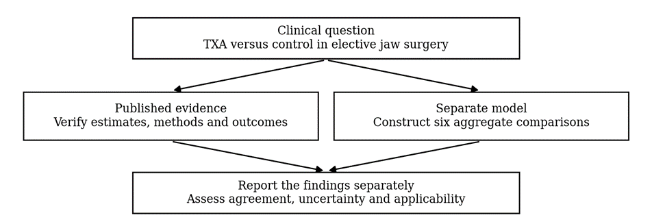
Figure 1. Evidence appraisal and model workflow
Table 1. Inputs for the six modelled comparisons
|
Unit |
n per arm |
Operation |
Blood loss (mL) |
RBC events |
Duration (min) |
Hb decline (g/dL) |
|
A |
45 |
Single jaw |
395 ± 148 / 540 ± 165 |
1 / 3 |
144 ± 28 / 155 ± 30 |
1.45 ± 0.69 / 2.00 ± 0.75 |
|
B |
52 |
Bimaxillary |
475 ± 193 / 665 ± 211 |
1 / 4 |
180 ± 34 / 197 ± 38 |
1.74 ± 0.81 / 2.53 ± 0.89 |
|
C |
60 |
Bimaxillary |
492 ± 182 / 650 ± 194 |
2 / 5 |
184 ± 33 / 198 ± 36 |
1.92 ± 0.88 / 2.57 ± 0.94 |
|
D |
48 |
Bimaxillary |
510 ± 220 / 725 ± 235 |
1 / 3 |
193 ± 39 / 212 ± 43 |
1.96 ± 0.96 / 2.82 ± 1.01 |
|
E |
55 |
Single jaw |
387 ± 163 / 530 ± 175 |
2 / 4 |
147 ± 28 / 159 ± 31 |
1.38 ± 0.70 / 1.87 ± 0.81 |
|
F |
50 |
Bimaxillary |
461 ± 185 / 635 ± 200 |
1 / 4 |
172 ± 33 / 187 ± 35 |
1.72 ± 0.79 / 2.43 ± 0.86 |
Group order is intravenous TXA / placebo. Continuous inputs are mean ± SD. RBC events are the numbers of individuals with at least one red-cell transfusion. Hb: haemoglobin.
Table 2. Modelled pooled effects and sensitivity intervals
|
Outcome |
Measure |
Estimate (95% CI) |
Conservative 95% CI |
I² |
|
Intraoperative blood loss |
MD, mL |
−164.94 (−194.16 to −135.71) |
−203.26 to −126.61 |
0% |
|
Perioperative transfusion |
OR |
0.34 (0.15 to 0.77) |
0.11 to 0.99 |
0% |
|
Operative duration |
MD, min |
−14.07 (−19.32 to −8.82) |
−20.96 to −7.18 |
0% |
|
Haemoglobin decline |
MD, g/dL |
−0.65 (−0.78 to −0.52) |
−0.82 to −0.48 |
0% |
|
Single-jaw blood loss |
MD, mL |
−143.98 (−189.21 to −98.74) |
−437.21 to 149.26 |
0% |
|
Bimaxillary blood loss |
MD, mL |
−179.95 (−218.24 to −141.67) |
−242.12 to −117.79 |
0% |
MD: mean difference. OR: odds ratio. CI: confidence interval. The conservative interval uses the modified Hartung–Knapp method. Jaw subgroups are descriptive.
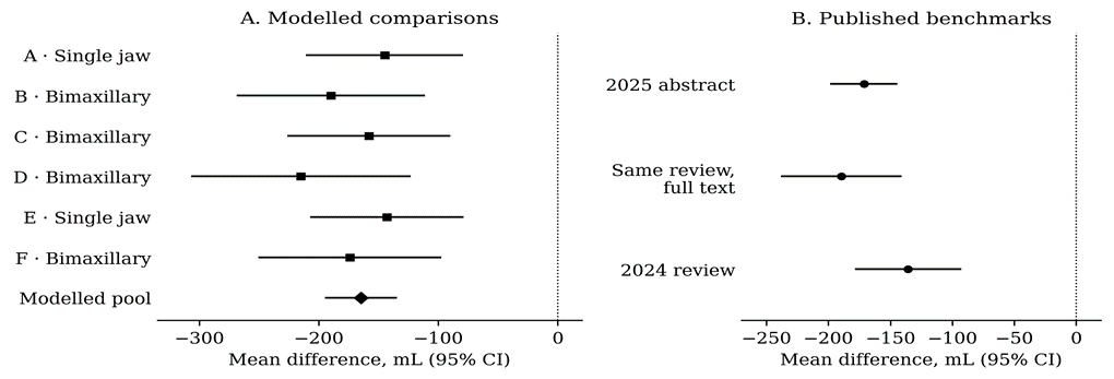
Figure 2. Blood loss contrasts from the model and published reviews
Squares represent the six modelled comparisons; the diamond is their pooled estimate. Panel B shows published estimates [3] [4] and is not pooled. The abstract and full-text rows come from the same review.
Table 3. Published TXA evidence and interpretation
|
Source |
Blood loss |
Transfusion |
Other findings or interpretation |
|
Published abstract [3] |
MD −171.30 mL (95% CI −197.58 to −145.02) |
OR 0.33 (95% CI 0.14 to 0.77) |
Duration MD −14.50 min (95% CI −20.89 to −8.02) |
|
Same review, full text [3] |
MD −189.6 mL (95% CI −237.3 to −141.9); I² 86% |
4 vs 15 events; RD −0.02 (95% CI −0.05 to 0.01) |
Duration MD −8.9 min (95% CI −17.45 to −0.31) |
|
Independent review [4] |
MD −135.60 mL (95% CI −177.51 to −93.70) |
No estimate used here |
No clear duration difference; better visibility |
|
Bimaxillary dose network [6] |
One regimen showed a clear benefit versus placebo |
No clear difference versus placebo |
Limited information to rank regimens or assess thromboembolism |
|
Equivalence trial [13] |
Day-one total loss: 682 vs 875 mL |
Not the primary outcome |
Did not establish equivalence; smaller Hb decline with TXA |
RD: risk difference. The full-text transfusion estimate is from the forest plot. Reviews may include overlapping trials. The day-one outcome includes a different time window from intraoperative loss.
DISCUSSION:
The most consistent finding in the published evidence is lower measured intraoperative blood loss with TXA. The model demonstrates how a moderate continuous difference can yield an interval that excludes zero while the answer for transfusion remains more dependent on a small number of events. The figure comparing model units displays their sampling intervals as well as the pooled estimate. A pooled interval does not predict how much blood loss an individual patient will avoid.
The operating field offers a clinical explanation for why a modest volume reduction may matter. Persistent oozing can obscure the line of an osteotomy and require additional irrigation or suction. A trial conducted with dexmedetomidine and controlled hypotension found better rated visibility and lower blood loss with added TXA [15]. The finding is specific to that combination and comparator. A separate retrospective matched comparison during hypotensive anaesthesia also favoured TXA for blood loss but could not remove all treatment-selection bias [16]. An observational comparison of greater versus lesser exposure to hypotensive anaesthesia among TXA recipients did not identify a difference in visibility or estimated blood loss. It did not randomly assign arterial pressure targets [17]. These differences explain why a TXA effect should not be used to justify a particular anaesthetic target.
Earlier randomised-trial syntheses also favoured TXA for estimated blood loss [18] [19]. Their results cannot be counted as additional independent participants. An earlier review of multiple bleeding-prevention strategies helps identify concomitant techniques that need to be held comparable across trial groups [20].
Systemic and topical regimens should also remain separate when choosing a protocol. Irrigation trials asked whether the operative surface could be treated through local application and used schedules distinct from intravenous dosing [21] [22]. Differences in concentration, irrigation volume and contact time make “topical TXA” an incomplete description. A dose network can generate hypotheses but a favourable rank does not establish a reliable advantage if few direct comparisons link the regimens. A useful future trial would report the exact timing of systemic administration, redosing, topical exposure and use of rescue haemostatic interventions. It would stratify randomisation by operative extent and define how changes in the planned procedure are handled.
The transfusion comparison requires the most restraint. In the published review the abstract OR and the full-text risk difference do not deliver the same inference about statistical significance. Different metrics and analytical choices can produce different intervals when event counts are low. The reports available for this appraisal do not resolve exactly which analytical decisions explain the discrepancy. The trial-like example gives an OR near the supplied benchmark, but that agreement is by design. Its conservative interval ends close to 1 even with the assumed allocation and event counts. Neither the model nor the most favourable summary justifies promising an absolute reduction to patients. A service with low baseline transfusion rates will have fewer opportunities for a given relative effect to translate into an avoided transfusion.
Operative duration and postoperative haemoglobin add context to the blood loss endpoint. A cleaner field could speed parts of an osteotomy but total time also includes repositioning, fixation and checks that are not directly related to bleeding. Case mix, operator experience and additional procedures can therefore alter the observed duration effect. Published estimates range from a modest reduction to no clear difference. Similarly, a postoperative haemoglobin value depends on fluids, the time of sampling and whether a transfusion was given before the sample. A future comparison should record these co-interventions and prespecify whether haemoglobin after transfusion is part of the treatment policy effect or is handled in a sensitivity analysis.
Available orthognathic trials do not demonstrate that TXA increases clinical thromboembolic events. They also lack the size and follow-up needed to exclude a clinically meaningful increase. An orthognathic mechanistic study found stronger suppression of fibrinolysis with TXA [23]. This is a laboratory observation rather than proof of postoperative thrombosis. In a large randomised trial across noncardiac operations TXA reduced a composite bleeding outcome but did not meet the prespecified criterion for cardiovascular noninferiority [24]. A broader synthesis of intravenous TXA trials found no overall association with thromboembolic events [25]. These findings provide context for safety discussion. Neither source supplies a precise absolute VTE risk for elective single-jaw or bimaxillary surgery.
For tertiary services in Sindh and Punjab, a useful step is to establish a local baseline before evaluating a TXA pathway. The operating team can specify whether each procedure includes a maxillary osteotomy, how suction and irrigation are reconciled and whether the anaesthetic target changed in response to blood loss. The anaesthesia and transfusion teams can document preoperative haemoglobin, the reason and timing of red-cell administration and whether bleeding remained active at the time of the decision. International transfusion guidance supports tailoring the decision to the clinical context rather than using blood loss volume alone [26]. A postoperative review should distinguish haemorrhage requiring reintervention from an uncomplicated fall in haemoglobin. At follow-up, teams should distinguish confirmed thromboembolism from suspected events and record whether patients were actively contacted after discharge.
The strongest design for the remaining efficacy questions would use a blinded comparison with a standardised transfusion policy and prespecified measurement of blood loss and visibility. Jaw type, concurrent procedures and intraoperative blood pressure should be recorded even if these factors are not used for stratification. Low transfusion and VTE rates also make multicentre recruitment or longer prospective surveillance useful. Trial data should keep thrombotic events, transfusion and blood loss in separate outcome domains. Transparent reporting should identify which patients contribute to each estimate and present clinical event counts even when a formal difference cannot be established.
Visibility ratings taken after each major stage could show whether an effect is concentrated in part of the operation. Delayed haemorrhage can have a distinct source and presentation [27]. It should be recorded separately from diffuse intraoperative blood loss.
This appraisal has limitations. The literature verification was targeted and does not replace independent exhaustive screening or an updated pooled analysis of the primary trials. Reuse of trial populations across published reviews makes their estimates correlated. The model has only six independent units, intentionally concordant effects, no outcome-level missing data and limited variability in the transfusion counts. Its I² of zero cannot be used to characterise the substantially heterogeneous published literature. The analysis did not address treatment effects by individual risk, uncertainty in safety outcomes or comparative effectiveness of topical and systemic regimens. The application to Pakistani centres is contextual because published estimates were not verified against centre-specific patient outcomes or transfusion policies.
CONCLUSION:
In elective orthognathic surgery, published comparisons support lower measured intraoperative blood loss with TXA. Evidence for avoiding red-cell transfusion is less consistent and thromboembolic safety remains imprecise. The model illustrates why the conclusion about sparse events depends on the analytical approach. Future trials and local audits should separate single-jaw and bimaxillary operations, use reproducible blood loss measurements and document transfusion criteria and follow-up. These steps would help determine when the bleeding benefit translates into a meaningful clinical gain.
REFERENCES:
1. Akpinar H, Yazici T, Gök E, Findik Y. Comparison of Clinical, Anesthetic, and Perioperative Outcomes Between Single-Jaw and Bimaxillary Orthognathic Surgery: A Retrospective Analysis of 499 Cases. J Craniofac Surg 2026. doi: https://doi.org/10.1097/SCS.0000000000012710. PMID: 41931360. Available from: https://pubmed.ncbi.nlm.nih.gov/41931360/
2. Choi WS, Irwin MG, Samman N. The effect of tranexamic acid on blood loss during orthognathic surgery: a randomized controlled trial. J Oral Maxillofac Surg 2009;67(1):125-33. doi: https://doi.org/10.1016/j.joms.2008.08.015. PMID: 19070758. Available from: https://pubmed.ncbi.nlm.nih.gov/19070758/
3. Baghaie H, Shukla K, Stone J, Breik O, Munn Z. Prophylactic Tranexamic Acid Use in Orthognathic Surgery: A Systematic Review and Meta-analysis. Aesthetic Plast Surg 2025;49(11):2932-2946. doi: https://doi.org/10.1007/s00266-025-04738-7. PMID: 40064645. Available from: https://pubmed.ncbi.nlm.nih.gov/40064645/
4. Mortada H, Hussain SA, Liyanage DD, Zou Y, Subbiah P, George J, et al. Does tranexamic acid really matter in reducing blood loss? A critical evaluation of its efficacy in orthognathic surgery through a comprehensive systematic review and meta-analysis. Br J Oral Maxillofac Surg 2024;62(9):780-787. doi: https://doi.org/10.1016/j.bjoms.2024.06.011. PMID: 39266337. Available from: https://pubmed.ncbi.nlm.nih.gov/39266337/
5. AlQahtani FA, Kuriadom ST, Varma S, AlAnzy H, AlOtaibi S. Effectiveness of tranexamic acid in orthognathic surgery: A systematic review of systematic reviews. J Stomatol Oral Maxillofac Surg 2023;124(6 Suppl 2):101592. doi: https://doi.org/10.1016/j.jormas.2023.101592. PMID: 37558181. Available from: https://pubmed.ncbi.nlm.nih.gov/37558181/
6. Felipe Junior J, Barbosa de Menezes VR, Barbosa de França AJ, de Moraes SLD, do Egito Vasconcelos BC. Efficacy and safety of tranexamic acid in bimaxillary orthognathic surgery: systematic review and network meta-analysis of randomized controlled trials. Int J Oral Maxillofac Surg 2026;55(11):1340-1348. doi: https://doi.org/10.1016/j.ijom.2026.04.033. PMID: 42161685. Available from: https://pubmed.ncbi.nlm.nih.gov/42161685/
7. Aga Khan University Hospital. Department of Dentistry. Karachi: AKUH. Available from: https://hospitals.aku.edu/pakistan/others/departments/department-of-dentistry/department-of-dentistry
8. Lahore General Hospital. Oral and Maxillofacial Surgery. Lahore: LGH. Available from: https://lgh.punjab.gov.pk/OralMaxillofacialSurgery
9. Page MJ, McKenzie JE, Bossuyt PM, Boutron I, Hoffmann TC, Mulrow CD, et al. The PRISMA 2020 statement: an updated guideline for reporting systematic reviews. BMJ 2021;372:n71. doi: https://doi.org/10.1136/bmj.n71. PMID: 33782057. Available from: https://pubmed.ncbi.nlm.nih.gov/33782057/
10. Sterne JAC, Savović J, Page MJ, Elbers RG, Blencowe NS, Boutron I, et al. RoB 2: a revised tool for assessing risk of bias in randomised trials. BMJ 2019;366:l4898. doi: https://doi.org/10.1136/bmj.l4898. PMID: 31462531. Available from: https://pubmed.ncbi.nlm.nih.gov/31462531/
11. DerSimonian R, Laird N. Meta-analysis in clinical trials. Control Clin Trials 1986;7(3):177-88. doi: https://doi.org/10.1016/0197-2456(86)90046-2. PMID: 3802833. Available from: https://pubmed.ncbi.nlm.nih.gov/3802833/
12. Röver C, Knapp G, Friede T. Hartung-Knapp-Sidik-Jonkman approach and its modification for random-effects meta-analysis with few studies. BMC Med Res Methodol 2015;15:99. doi: https://doi.org/10.1186/s12874-015-0091-1. PMID: 26573817. Available from: https://pubmed.ncbi.nlm.nih.gov/26573817/
13. Jozefowicz E, Sabourdin N, Lambelin V, Lejeune V, Delassus R, Tavernier B. The effect of tranexamic acid on blood loss in orthognathic surgery: a randomized, placebo-controlled, equivalence study. Int J Oral Maxillofac Surg 2022;51(5):637-642. doi: https://doi.org/10.1016/j.ijom.2021.08.018. PMID: 34465477. Available from: https://pubmed.ncbi.nlm.nih.gov/34465477/
14. Kaşarcıoğlu G, Pergel T, Akkoyun EF, Dolanmaz D. Low-dose tranexamic acid in orthognathic surgery: two-dose protocol-a double-blind randomized controlled trial on oedema, bleeding, and visibility of the surgical field. Int J Oral Maxillofac Surg 2026;55(1):34-42. doi: https://doi.org/10.1016/j.ijom.2025.04.010. PMID: 40287325. Available from: https://pubmed.ncbi.nlm.nih.gov/40287325/
15. Sharma H, Arora S, Bhatia N, Rattan V, Sethi S. Tranexamic Acid Is Associated With Improved Operative Field in Orthognathic Surgery. J Oral Maxillofac Surg 2020;78(9):1509-1517. doi: https://doi.org/10.1016/j.joms.2020.04.037. PMID: 32497489. Available from: https://pubmed.ncbi.nlm.nih.gov/32497489/
16. Harada K, Imamachi N, Matsuda Y, Hirabayashi M, Saito Y, Kanno T. Effect of tranexamic acid on blood loss reduction in patients undergoing orthognathic surgery under hypotensive anesthesia: a single-center, retrospective, observational study. J Korean Assoc Oral Maxillofac Surg 2024;50(2):86-93. doi: https://doi.org/10.5125/jkaoms.2024.50.2.86. PMID: 38693131. Available from: https://pubmed.ncbi.nlm.nih.gov/38693131/
17. Weber TM, Squier B, Kinard BE. Is Tranexamic Acid Associated With a Reduced Need for Hypotensive Anesthesia During Orthognathic Surgery?. J Oral Maxillofac Surg 2025;83(12):1470-1477. doi: https://doi.org/10.1016/j.joms.2025.08.013. PMID: 40975121. Available from: https://pubmed.ncbi.nlm.nih.gov/40975121/
18. Mei A, Qiu L. The efficacy of tranexamic acid for orthognathic surgery: a meta-analysis of randomized controlled trials. Int J Oral Maxillofac Surg 2019;48(10):1323-1328. doi: https://doi.org/10.1016/j.ijom.2018.07.027. PMID: 30902548. Available from: https://pubmed.ncbi.nlm.nih.gov/30902548/
19. Zhao H, Liu S, Wu Z, Zhao H, Ma C. Comprehensive assessment of tranexamic acid during orthognathic surgery: A systematic review and meta-analysis of randomized, controlled trials. J Craniomaxillofac Surg 2019;47(4):592-601. doi: https://doi.org/10.1016/j.jcms.2019.01.021. PMID: 30826112. Available from: https://pubmed.ncbi.nlm.nih.gov/30826112/
20. Olsen JJ, Skov J, Ingerslev J, Thorn JJ, Pinholt EM. Prevention of Bleeding in Orthognathic Surgery--A Systematic Review and Meta-Analysis of Randomized Controlled Trials. J Oral Maxillofac Surg 2016;74(1):139-50. doi: https://doi.org/10.1016/j.joms.2015.05.031. PMID: 26073131. Available from: https://pubmed.ncbi.nlm.nih.gov/26073131/
21. Kaewpradub P, Apipan B, Rummasak D. Does tranexamic acid in an irrigating fluid reduce intraoperative blood loss in orthognathic surgery? A double-blind, randomized clinical trial. J Oral Maxillofac Surg 2011;69(6):e186-9. doi: https://doi.org/10.1016/j.joms.2010.11.041. PMID: 21398011. Available from: https://pubmed.ncbi.nlm.nih.gov/21398011/
22. Eftekharian H, Vahedi R, Karagah T, Tabrizi R. Effect of tranexamic acid irrigation on perioperative blood loss during orthognathic surgery: a double-blind, randomized controlled clinical trial. J Oral Maxillofac Surg 2015;73(1):129-33. doi: https://doi.org/10.1016/j.joms.2014.07.033. PMID: 25443384. Available from: https://pubmed.ncbi.nlm.nih.gov/25443384/
23. Sidelmann JJ, Gram JB, Godtfredsen ACM, Thorn JJ, Ingerslev J, Pinholt EM. Orthognathic Surgery-Induced Fibrinolytic Shutdown Is Amplified by Tranexamic Acid. J Oral Maxillofac Surg 2020;78(7):1183-1189. doi: https://doi.org/10.1016/j.joms.2020.02.026. PMID: 32224082. Available from: https://pubmed.ncbi.nlm.nih.gov/32224082/
24. Devereaux PJ, Marcucci M, Painter TW, Conen D, Lomivorotov V, Sessler DI, et al. Tranexamic Acid in Patients Undergoing Noncardiac Surgery. N Engl J Med 2022;386(21):1986-1997. doi: https://doi.org/10.1056/NEJMoa2201171. PMID: 35363452. Available from: https://pubmed.ncbi.nlm.nih.gov/35363452/
25. Taeuber I, Weibel S, Herrmann E, Neef V, Schlesinger T, Kranke P, et al. Association of Intravenous Tranexamic Acid With Thromboembolic Events and Mortality: A Systematic Review, Meta-analysis, and Meta-regression. JAMA Surg 2021;156(6):e210884. doi: https://doi.org/10.1001/jamasurg.2021.0884. PMID: 33851983. Available from: https://pubmed.ncbi.nlm.nih.gov/33851983/
26. Carson JL, Stanworth SJ, Guyatt G, Valentine S, Dennis J, Bakhtary S, et al. Red Blood Cell Transfusion: 2023 AABB International Guidelines. JAMA 2023;330(19):1892-1902. doi: https://doi.org/10.1001/jama.2023.12914. PMID: 37824153. Available from: https://pubmed.ncbi.nlm.nih.gov/37824153/
27. Kurasawa Y, Sato H, Katada R, Inada T, Shirota T, Shimane T. Delayed Epistaxis which Was Developed after Orthognathic Surgery with Le Fort I Osteotomy and Managed by Endoscopic Cauterization. Case Rep Dent 2022;2022:3057472. doi: https://doi.org/10.1155/2022/3057472. PMID: 35242389. Available from: https://pubmed.ncbi.nlm.nih.gov/35242389/.