Clinical Applications of Pregnancy Induced Leucocytosis – an Analysis of 70 Cases.
- Dr. Geetha J , Professor, Department of Anaesthesia, Dhanalakshmi Srinivasan Medical College and Hospital, Siruvachur, Perambalur, Tamil Nadu, India
- Dr. Araviynd S , Postgraduate, Department of Anaesthesia, Dhanalakshmi Srinivasan Medical College and Hospital, Siruvachur, Perambalur, Tamil Nadu, India
- Dr. Mirthula S.S , Postgraduate, Department of Obstetrics & Gynaecology, Dhanalakshmi Srinivasan Medical College and Hospital, Siruvachur, Perambalur, Tamil Nadu, India
- Dr. Sreeharan A.R , CRMI, Department of Anaesthesia, Dhanalakshmi Srinivasan Medical College and Hospital, Siruvachur, Perambalur, Tamil Nadu, India
- Dr. H. Irfan Ahamed , CRMI, Department of Anaesthesia, Dhanalakshmi Srinivasan Medical College and Hospital, Siruvachur, Perambalur, Tamil Nadu, India.
Article Information:
Abstract:
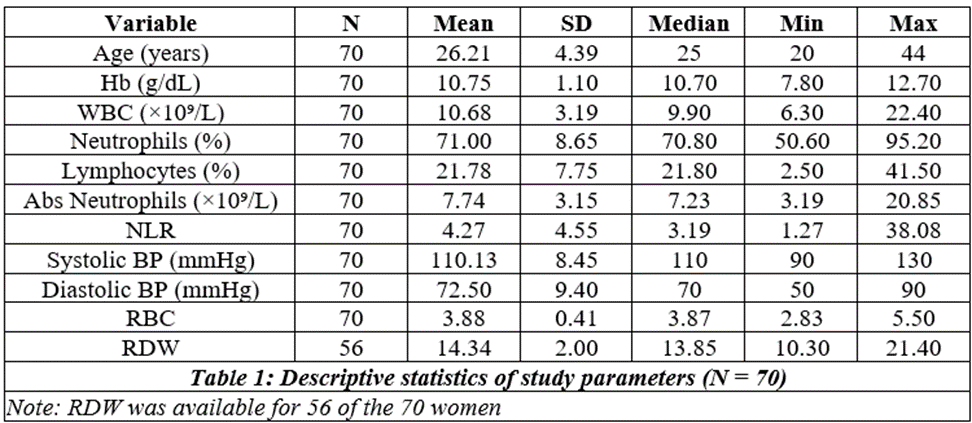
Introduction: Pregnancy induces adaptations in every system of a woman for the well-being of the developing fetus. Hematological changes focus on fetal oxygenation, adequate fetal tissue perfusion and immunity against fetal infections. Immunity and inflammatory triggers are activated in pregnancy. These create leucocytosis – a physiological response ranging from 9,000 to 11,000 cells per microlitre. Various studies have shown that a rise of the count from this level could be an early indication of infection, pregnancy induced hypertension and preterm labour. In this study, the medical undergraduates were directed to find the factors influencing leucocytosis in various trimesters of pregnancy and the granulocyte contribution to the leucocytosis. Materials And Methods: A prospective observational clinical study was conducted at DSMCH during the study period (Sep. 26 – Nov. 26) on 70 antenatal women attending the obstetric outpatient department. Patients with fever or any signs of infection were excluded. Age, Hb, trimester, systolic and diastolic BP, total and differential count, RBC count and RDW were recorded from case records and investigation reports, and the results were tabulated and analysed. Observations And Results: The mean age was 26.2 ± 4.4 years and the mean total count was 10.68 ± 3.19 × 10⁹/L; 20 women (28.6%) had a total count above 11 × 10⁹/L. The total count did not differ significantly between trimesters (p = 0.83). Total count correlated positively with neutrophil percentage (r = 0.55, p < 0.001) and negatively with lymphocyte percentage (r = −0.53, p < 0.001). Diastolic BP showed a weak positive correlation with total count (r = 0.32, p = 0.007), and women with a total count ≥ 15 × 10⁹/L had a higher mean diastolic BP (82.2 mmHg; p = 0.004). Systolic BP and Hb showed no significant correlation with total count. Hb fell with advancing pregnancy, and anaemia (Hb < 11 g/dL) was present in 57.1% of women overall and 78.6% in the third trimester (p < 0.001). Conclusion: Hematological indices are part of routine follow-up in antenatal mothers during the progress of pregnancy. A simple, common finding of an increase in white cell count from baseline could alert to the detection of any existing complications, so that management could be initiated earlier. Neutrophilia and the neutrophil–lymphocyte ratio could be valuable. However, larger studies are needed to substantiate the findings.
Keywords:
Article :
INTRODUCTION:
Pregnancy is a hyperdynamic, hypermetabolic and immunosuppressed state. Physiological changes occur in the mother, increasing demand and reducing her reserve to nourish the growth and well-being of the fetus. Anemia in pregnancy is attributed to hemodilution, nutritional deficiency and increased demand. Leucocytosis also occurs in pregnancy to combat immunosuppression. The total leucocyte count has been identified as an early marker of inflammatory conditions, infections and even pregnancy induced hypertension. In this study we intend to study the total leucocyte count across various trimesters, and its relationship to hemoglobin and blood pressure, in seventy patients who attended the department during the study period.
Aim
To evaluate the clinical applications of pregnancy induced leucocytosis.
Objectives
• To record the total and differential count of all antenatal women attending the obstetric outpatient department.
• To correlate with concomitant anemia by noting hemoglobin, RBC count and RDW.
• To associate the trimester of pregnancy and systolic and diastolic blood pressure to detect pregnancy induced hypertension.
MATERIALS AND METHODS:
Study Population
All antenatal patients attending the obstetric outpatient department.
Study Place
Dhanalakshmi Srinivasan Medical College and Hospital, Siruvachur, Perambalur, Tamil Nadu, India.
Study Design
Prospective observational clinical study.
Study Period
June 2026 – August 2026
Exclusion Criteria
Patients with fever or any signs of infection.
Methodology
Case records and investigation reports of 70 antenatal women were scrutinised and the following parameters were recorded:
• Age, Hb, Trimester, SBP, DBP, TC, DC, RBC and RDW were noted.
• The results were tabulated and analysed.
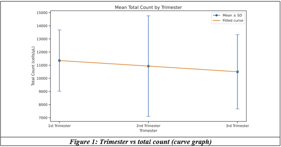
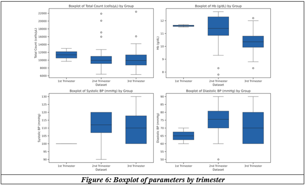
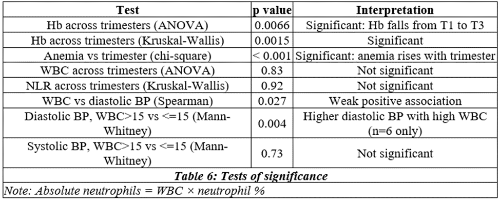
Mean total count fell slightly from the 1st (11,350) to the 2nd (10,931) and 3rd trimester (10,500 cells/µL). The difference was not statistically significant (Kruskal–Wallis p = 0.78).
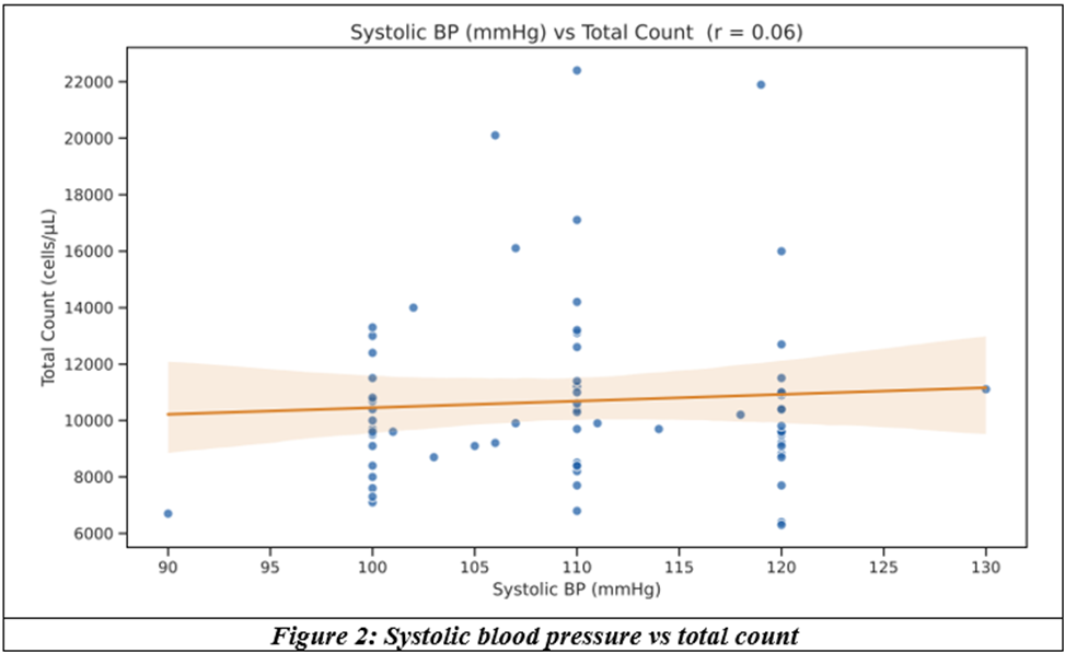
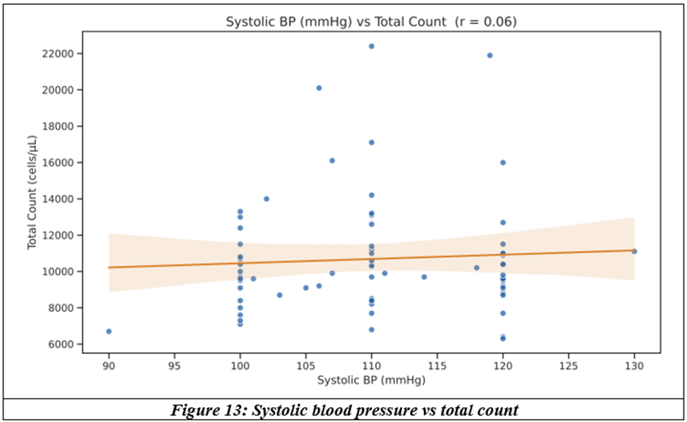
Systolic BP showed no meaningful correlation with total count (r = 0.06, p = 0.61).
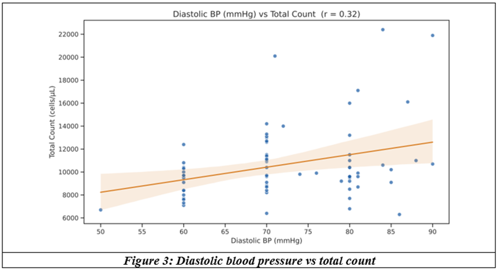
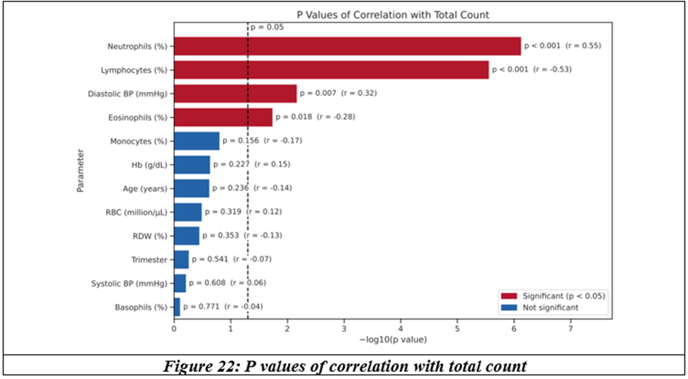
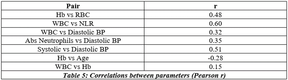
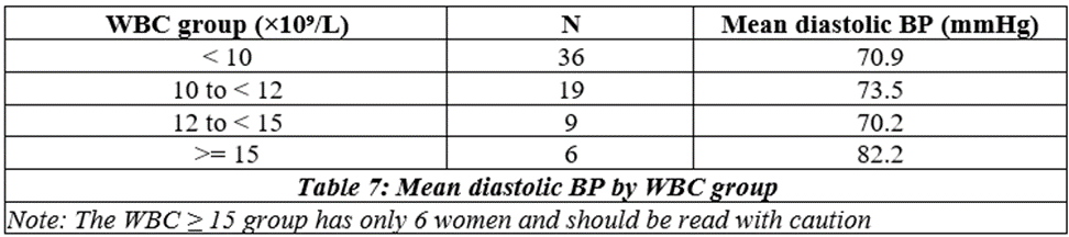
Diastolic BP showed a weak positive correlation with total count (r = 0.32, p = 0.007): higher diastolic pressure went with a higher total count.
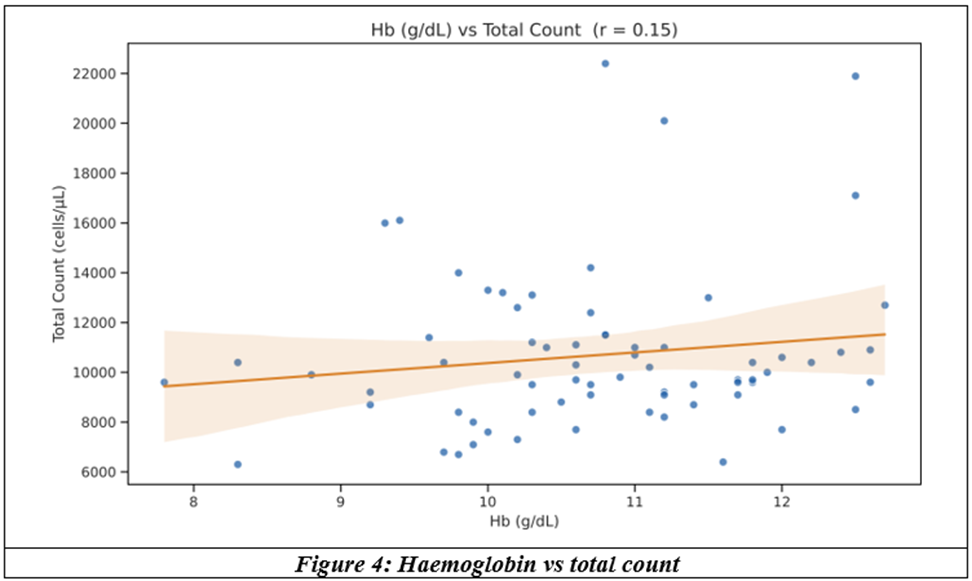
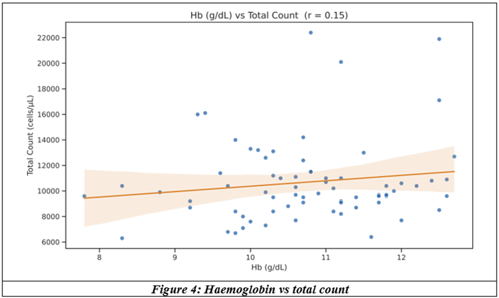
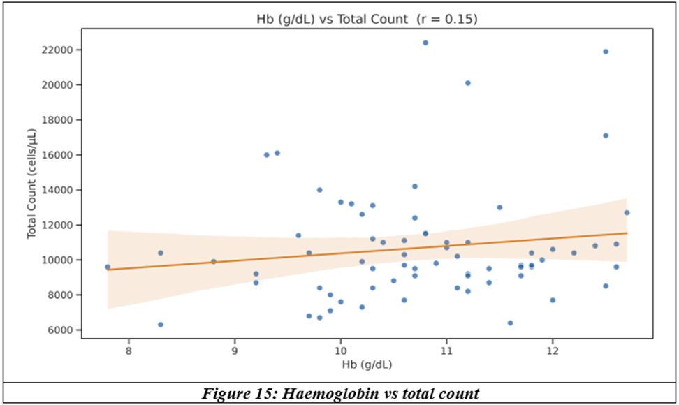
Haemoglobin showed no significant correlation with total count (r = 0.15, p = 0.23).
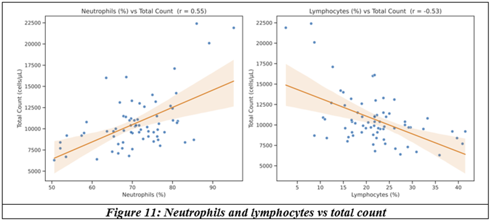
Total count correlated positively with neutrophils (0.55) and diastolic BP (0.32), and negatively with lymphocytes (−0.53) and eosinophils (−0.28). Hb correlated with RBC (0.48) and fell with advancing trimester (−0.37).
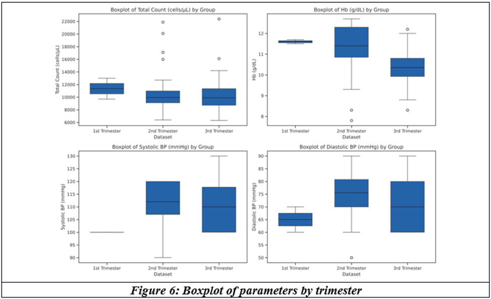
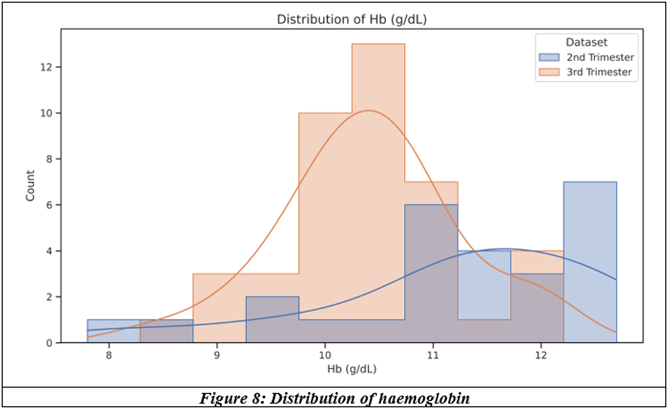
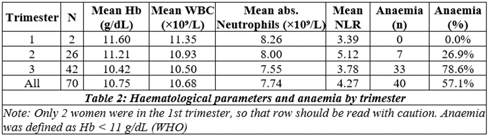
Hb was lower in the 3rd trimester (median 10.4 g/dL) than the 2nd (11.4 g/dL), p = 0.002. Total count, systolic and diastolic BP did not differ significantly between trimesters.
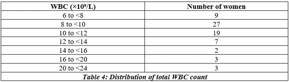
Most patients had a total count between 8,000 and 12,000 cells/µL in both trimesters, with a few high values above 16,000 cells/µL. 1st trimester (n = 2) not shown in distribution plots.
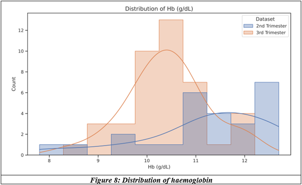
The 3rd trimester distribution is shifted to lower Hb values compared with the 2nd trimester. 1st trimester (n = 2) not shown in distribution plots.
Systolic BP clustered at 100, 110 and 120 mmHg in both trimesters. 1st trimester (n = 2) not shown in distribution plots.
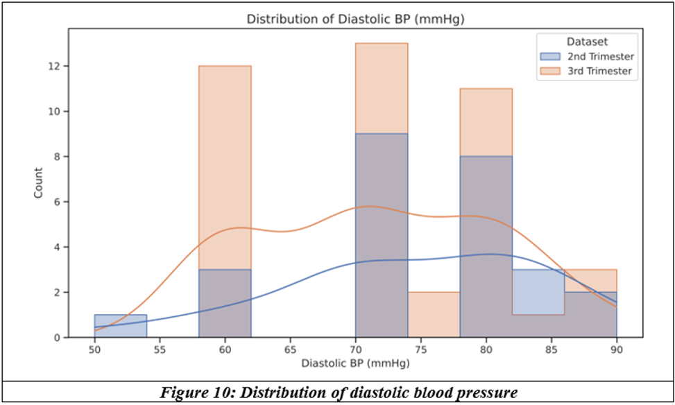
Diastolic BP clustered at 60, 70 and 80 mmHg, with a similar spread in both trimesters. 1st trimester (n = 2) not shown in distribution plots.
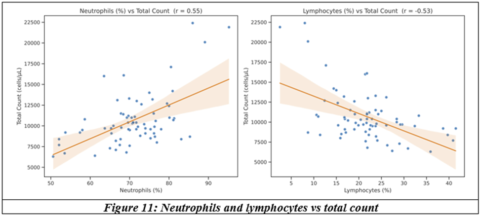
Total count rose with neutrophil percentage (r = 0.55) and fell with lymphocyte percentage (r = −0.53), both p < 0.001.
Mean total count fell slightly from the 1st (11,350) to the 2nd (10,931) and 3rd trimester (10,500 cells/µL). The difference was not statistically significant (Kruskal–Wallis p = 0.78).
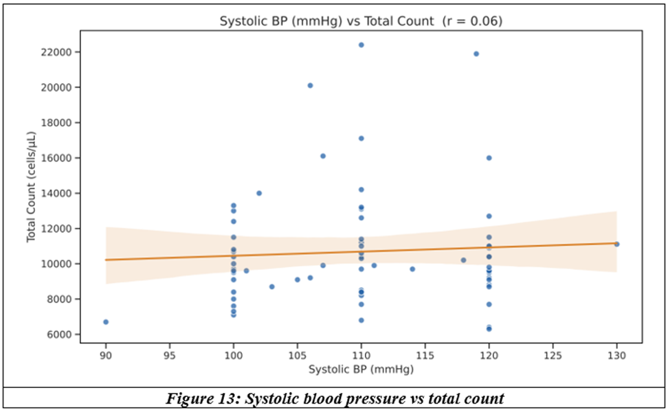
Systolic BP showed no meaningful correlation with total count (r = 0.06, p = 0.61).
Diastolic BP showed a weak positive correlation with total count (r = 0.32, p = 0.007): higher diastolic pressure went with a higher total count.
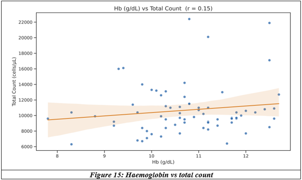
Haemoglobin showed no significant correlation with total count (r = 0.15, p = 0.23).
Total count correlated positively with neutrophils (0.55) and diastolic BP (0.32), and negatively with lymphocytes (−0.53) and eosinophils (−0.28). Hb correlated with RBC (0.48) and fell with advancing trimester (−0.37).
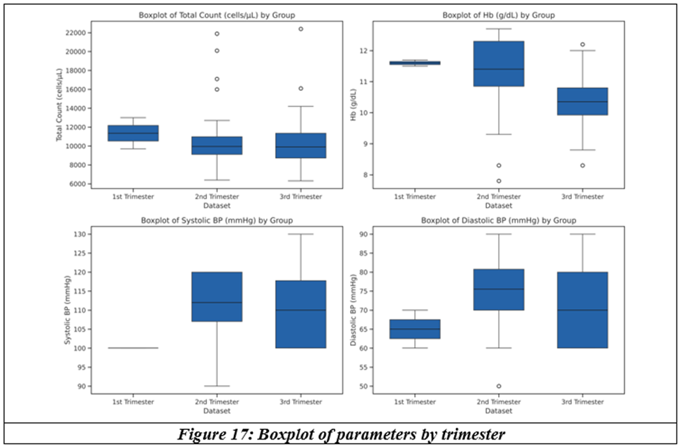
Hb was lower in the 3rd trimester (median 10.4 g/dL) than the 2nd (11.4 g/dL), p = 0.002. Total count, systolic and diastolic BP did not differ significantly between trimesters.
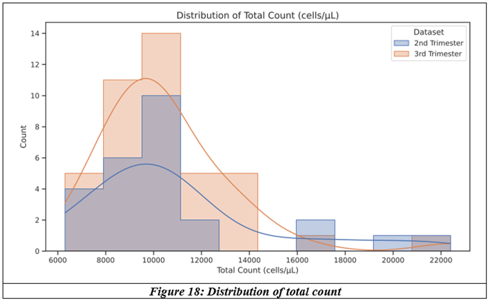
Most patients had a total count between 8,000 and 12,000 cells/µL in both trimesters, with a few high values above 16,000 cells/µL. 1st trimester (n = 2) not shown in distribution plots.
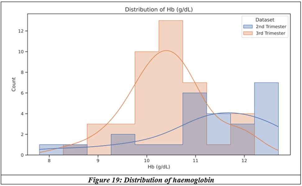
The 3rd trimester distribution is shifted to lower Hb values compared with the 2nd trimester. 1st trimester (n = 2) not shown in distribution plots.
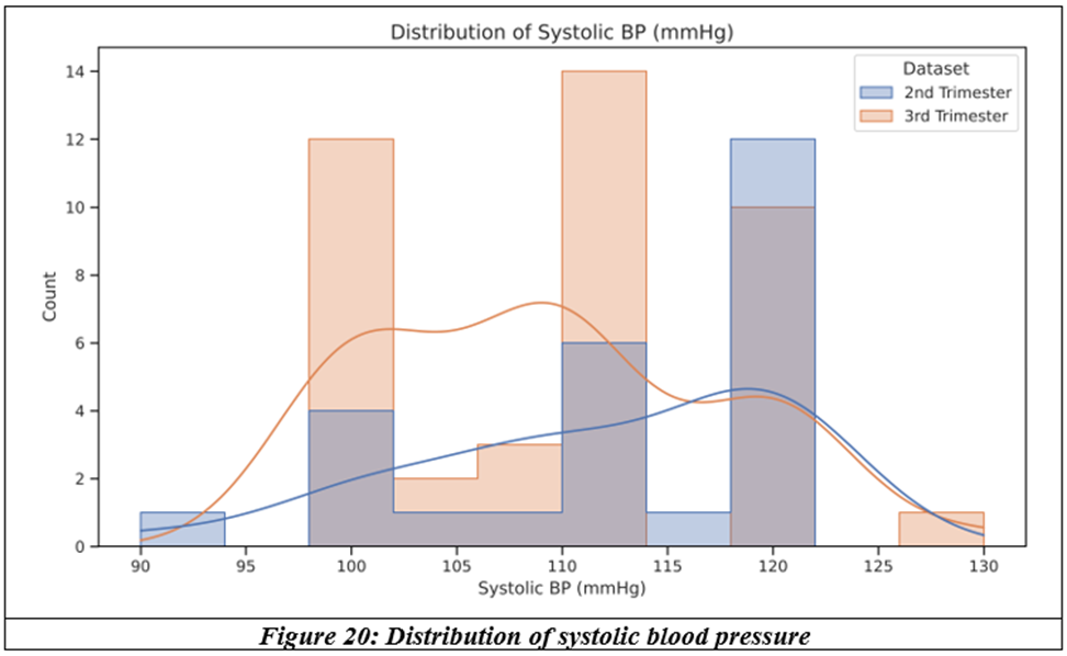
Systolic BP clustered at 100, 110 and 120 mmHg in both trimesters. 1st trimester (n = 2) not shown in distribution plots.
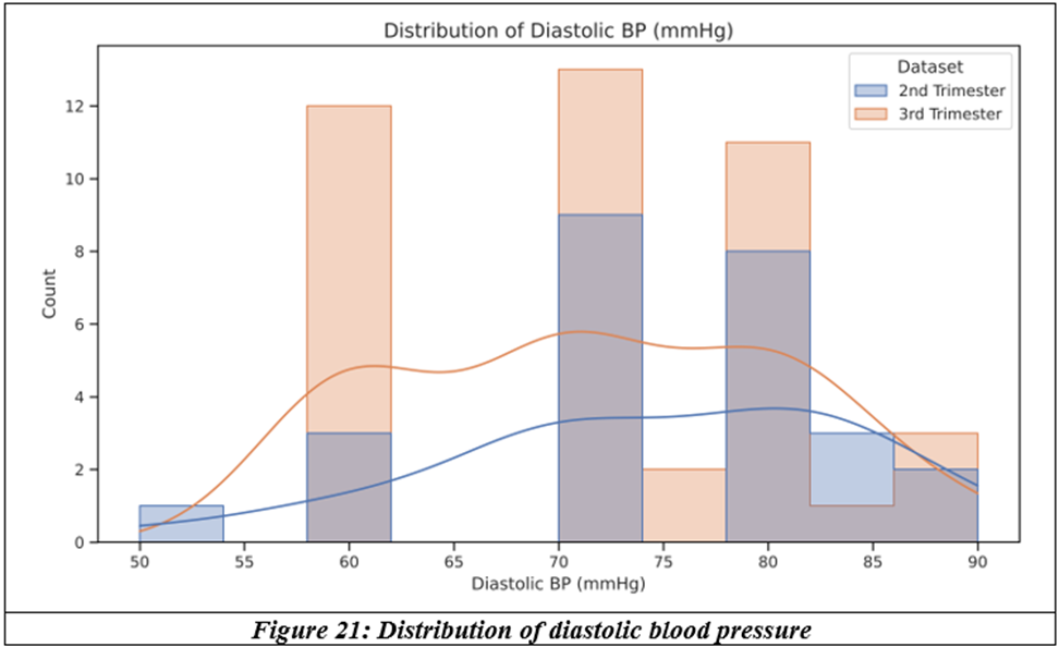
Diastolic BP clustered at 60, 70 and 80 mmHg, with a similar spread in both trimesters. 1st trimester (n = 2) not shown in distribution plots.
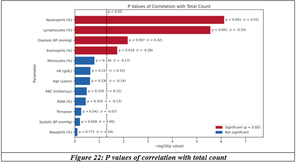
Neutrophils (p < 0.001), lymphocytes (p < 0.001), diastolic BP (p = 0.007) and eosinophils (p = 0.018) were significantly correlated with total count. Systolic BP (p = 0.608), Hb (p = 0.227) and trimester (p = 0.541) were not.
Pearson correlation p values for every pair of parameters. Dark cells are significant (p < 0.05); light cells are not.
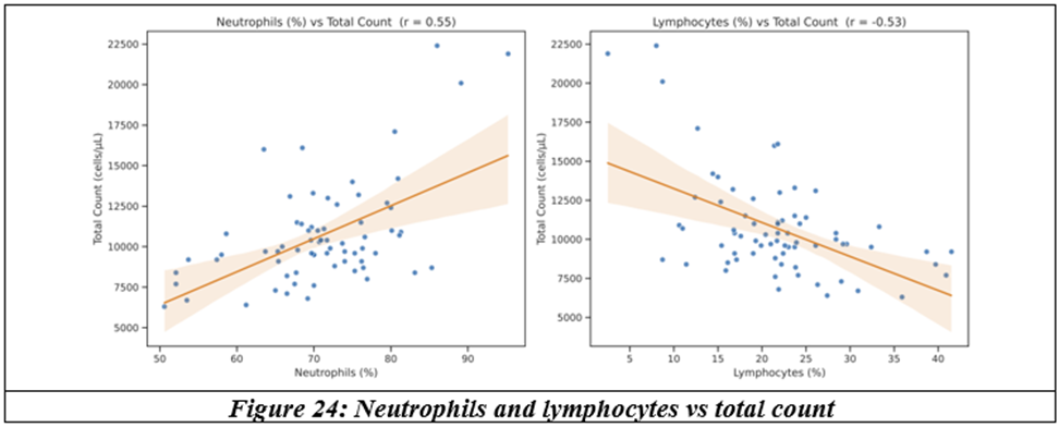
Total count rose with neutrophil percentage (r = 0.55) and fell with lymphocyte percentage (r = −0.53), both p < 0.001.
RESULTS:
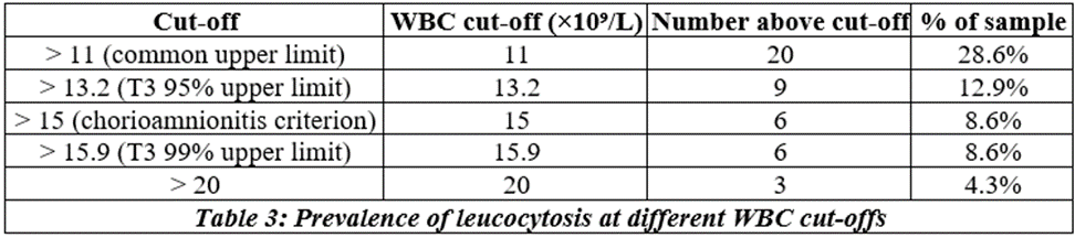
The summary statistics of the 70 antenatal women studied are shown in the tables below.


DISCUSSION:
The hematological system undergoes a series of adaptations in pregnancy in order to be prepared for fetal hematopoiesis and well-being. The increased plasma volume and red cell mass, increased RBC and WBC count, and raised fibrinogen levels all happen to increase circulatory volume in a hyperdynamic environment, compensate for anticipated blood loss and adapt for the immunosuppressed state of pregnancy. A pregnancy related leukocytosis with an increase in neutrophils has been observed since the second month of pregnancy, with an upward trend as pregnancy advances.[1] Lymphocyte count was decreased in the antenatal period. Several factors influence pregnancy, such as culture, environment, socioeconomic status and access to medical care. The hematological profile could affect pregnancy and its outcome. Akinlaja et al observed neutrophilia from the second month of pregnancy, gradually increasing with advancing pregnancy.
The increase in white cell counts through the trimesters was normal, and granulocytes progressively increased from the second trimester while platelets reduced.[2] The physiological leukocytosis was confined to neutrophils. The neutrophil count rises as early as the estrogen peak of a normal menstrual cycle, and if fertilization occurs, neutrophils continue to rise.[3] There is a subsequent rise to a peak at 30 weeks and a plateau during the third trimester. Several reviews by hematologists have confirmed the consistency of leucocytosis in pregnancy.[4]
Studies have shown that about 38.8% of pregnant women have leucocytosis in the first trimester, increasing to 52.1% in the third trimester. The rise in total white cells is due to a progressive increase of granulocytes from early to late pregnancy; the counts always remained high normal, from 9.7 to 10.4, at all stages of pregnancy. The mean lymphocyte count remains static at the non-pregnant level.
Dexamethasone is often administered in the antenatal period, especially in the third trimester, to pregnant women at risk of preterm labour to accelerate growth and surfactant production of the fetal lungs. The effect of intramuscular dexamethasone administration in late pregnancy on the maternal peripheral white cell count has been widely studied. The mean total count increased from a baseline of 11.3 × 10⁹/L to 16.2 × 10⁹/L on day one, normalising later.[5] This 43% increase represented composite changes in neutrophil and lymphocyte counts, which on average increased by 62% and decreased by 22% respectively. While steroid usage is generally considered immunosuppressive, the significant neutrophilia is useful information that may assist in monitoring infection. Similarly, studies quantifying the leucocytosis induced by betamethasone in late pregnancy have also been documented.[6]
As leucocytosis is a sensitive indicator of early infection, it is essential to understand the peak increase in white cell count beyond a physiological increase in the event of infection. The Faculty of Dental Medicine, Peru, and the Faculty of Dental Medicine, Romania, have conducted hematological studies in pregnant women with periodontitis and recorded a sharp and consistent increase in white cell count beyond 11,000 cells, especially more incidental in high-risk pregnancy and in women at risk of preterm labour.[7] Periodontitis is often triggered by gram-negative bacteria, namely Porphyromonas gingivalis, which disseminate into the peripheral circulation and cause inflammation in various areas of the body, and even form part of various systemic diseases.[8] The release of infiltrating leukocyte components such as lysosomal enzymes and reactive oxygen species leads to tissue destruction and an increased inflammatory response.
Thus, it is essential to differentiate physiological leucocytosis from pathological leucocytosis due to infection. Hypertensive disorders of pregnancy involve endothelial dysfunction and an exaggerated maternal inflammatory state.[9] During the inflammatory response, the leukocytes are activated with the release of cytokines, interleukin-8 and tumour necrosis factor-α, which lead to endothelial dysfunction. This predates the manifestation of preeclampsia. As there is a general leukocytosis, the platelet–lymphocyte ratio and neutrophil–lymphocyte ratio have recently been applied as systemic inflammatory response markers.[10] Aly et al detected superoxide production by maternal neutrophils in pregnant women with preeclampsia, proved by cell culture from syncytiotrophoblast microvillous membranes.[11] Several studies show that white cell count and neutrophilia tend to be higher in women with severe preeclampsia compared to those with mild PIH.
CONCLUSION:
This short study performed at the medical undergraduate level serves as an eye opener towards significant neutrophilic leucocytosis (p = 0.01) in pregnancy, with a higher increase in the second trimester. Abnormal ratios involving neutrophils, such as the neutrophil–leucocyte ratio and neutrophil–lymphocyte ratio, may offer sensitive detection of inflammatory responses. Diastolic blood pressure and a rise in total count (p = 0.007) have shown an association in this study of 70 pregnant women. An abnormally high total count of more than 11,000 cells may indicate an early phase of antenatal complications in hypertensive disorders, preeclampsia and preterm labour. We had around ten patients with a total count of more than 16,000, up to 22,000, in the second trimester of pregnancy with mild hypertension.
REFERENCES:
1. Azab AE, Albasha MO, Jbireal JM, et al. Hematological changes during pregnancy: insight into anemia, leukocytosis and thrombocytopenia. East African Scholars J Med Sci 2020;3(5):185-92.
2. Karim SA, Rizvi JH, Rizwana I, et al. Platelets and leucocyte counts in pregnancy. Journal of Pakistan Medical Association 1992;42:86.
3. Cruickshank JM, Morris R, Butt WR, et al. The relationship of total and differential leukocyte counts with urinary oestrogen and plasma cortisol levels. J Obstet Gynaecol Br Commonw 1970;77:634-9.
4. Letsky E. The hematological system. In: Hytten F, Chamberlain G. Clinical physiology in obstetrics. Oxford: Blackwell 1980:43-60.
5. Denison FC, Elliott CL, Wallace EM. Dexamethasone-induced leucocytosis in pregnancy. BJOG. 1997;104(7):851-3.
6. Wallace EM, Ekkel K, Cotter T, et al. Haematological effects of betamethasone treatment in late pregnancy. Aust N Z J Obstet Gynaecol 1998;38(4):396-8.
7. Camelia Fiera Maglaviceanu CF, Bechir ES, Ţuculină MJ, et al. The role of leucocytosis in the occurrence of pregnancy complications in women with periodontal disease. Rev Chim (Bucharest) 2019;70(11):3967-70.
8. Katz J, Chegini N, Shiverick KT, et al. Localization of P. gingivalis in preterm delivery placenta. Journal of Dental Research 2009;88(6):575-8.
9. Satish Korah Kuruvilla et al. Significance of leukocyte and platelet counts in hypertensive disorders of pregnancy. Int J Obstet Gynaecol Res 2024;11(3):398-402.
10. Yavuzcan A, Caglar M, Ustun Y, et al. Mean platelet volume, neutrophil-lymphocyte ratio and platelet-lymphocyte ratio in severe preeclampsia. Ginekologia Polska 2014;85(3):197-203.
11. Aly MA, Khandelwal MM, Zhao MJ, et al. Neutrophils are stimulated by syncytiotrophoblast microvillous membranes to generate superoxide radicals in women with preeclampsia. Am J Obstet Gynecol 2004;190(1):252-8.