Demographic Profile, Clinical Spectrum and Short-Term Outcomes of Mechanically Ventilated Children (1 Month to 14 Years) Admitted to the PICU of a Tertiary Care Hospital: A Prospective Observational Study.
- Dr. Sharanabasavaraj , Junior Resident, Department of Paediatrics, Ballari Medical College and Research Centre (BMCRC), Ballari, Karnataka, India.
- Dr. Krithika R , Assistant Professor, Department of Paediatrics, Ballari Medical College and Research Centre (BMCRC), Ballari, Karnataka, India.
- Dr. Riyaz Ahmed F.K. , Assistant Professor, Department of Paediatrics, Ballari Medical College and Research Centre (BMCRC), Ballari, Karnataka, India.
Article Information:
Abstract:
Background: Mechanical ventilation is a vital life-saving intervention in critically ill children admitted to the PICU (Pediatric Intensive Care Unit). However, it is associated with significant morbidity and mortality. Understanding the demographic profile, clinical spectrum, complications and outcomes of mechanically ventilated children helps improve pediatric critical care. Methods: This prospective observational study was conducted in the PICU of BMCRC, Ballari, over 18 months from May 2024 to November 2025. A total of 261 children aged 1 month to 14 years requiring invasive mechanical ventilation were included. Demographic characteristics, clinical presentation, indications for ventilation, ventilator parameters, complications and short-term outcomes were recorded and analysed using SPSS version 11.0. Results: Among 261 children, 37.5% were infants, with a mean age of 4.72 ± 4.48 years, and 54.8% were males. Respiratory failure (55.6%) was the leading indication for mechanical ventilation, followed by impending respiratory failure (41.4%). SIMV (Synchronized Intermittent Mandatory Ventilation) was the most frequently used mode (29.1%). The mean duration of ventilation was 2.98 ± 2.89 days. Laryngeal edema (77.4%) was the most common complication, followed by endotracheal tube blockage (62.5%) and ventilator-associated pneumonia (36%). Overall, 69% of children survived, while 29.5% succumbed and 1.5% were referred. Multivariate analysis identified multiorgan dysfunction syndrome (OR 7.21; p < 0.001) and ventilator-associated pneumonia (OR 2.67; p = 0.006) as significant predictors of mortality. Conclusion: Mechanical ventilation remains an essential life-saving intervention in critically ill children. However, the high complication rate and mortality highlight the importance of meticulous airway care, infection prevention, early identification of sepsis and prompt management of multiorgan dysfunction to improve outcomes.
Keywords:
Article :
INTRODUCTION:
Mechanical ventilation is one of the most important life-saving interventions in PICUs (Pediatric Intensive Care Units), providing essential respiratory support to critically ill children who are unable to maintain adequate spontaneous breathing due to respiratory failure, neurological impairment, shock, or multisystem organ dysfunction.[1] Despite significant advancements in pediatric critical care, mechanical ventilation continues to be associated with considerable morbidity and mortality, particularly in resource-limited healthcare settings.
The requirement for mechanical ventilation among critically ill children varies across healthcare settings and patient populations. Studies have reported that approximately 15–50% of children admitted to PICUs require invasive mechanical ventilation, depending on the severity of illness and clinical indications.[2,3] Respiratory disorders, neurological conditions, cardiac diseases, and postoperative complications constitute the major reasons for initiating ventilatory support. Acute lower respiratory tract infections, particularly pneumonia and bronchiolitis, contribute substantially to the burden of respiratory failure, especially among infants and young children.[4].
In developing countries, including India, infants and children below five years of age constitute a significant proportion of mechanically ventilated patients. Male predominance has also been observed in several studies. Pneumonia, sepsis, and central nervous system infections are among the leading underlying conditions requiring ventilatory support.[5,6] Additionally, demographic and nutritional factors, including age, gender, nutritional status, and socioeconomic background, may influence disease severity and clinical outcomes.[7]
The duration of mechanical ventilation depends on the underlying disease, severity of illness, and response to treatment. Previous studies have reported an average ventilation duration of approximately 4–7 days, although children with ARDS (Acute Respiratory Distress Syndrome) and multisystem organ dysfunction may require prolonged ventilatory support.[8,9] Prolonged mechanical ventilation increases the risk of complications such as VAP (Ventilator-Associated Pneumonia), barotrauma, and airway injuries, contributing to extended PICU stays and adverse clinical outcomes.[10,11] Among these complications, VAP remains a major concern, with reported incidence rates reaching up to 40% in certain pediatric intensive care settings.[12]
Aims and Objectives
The present study aims to assess the frequency of mechanical ventilation among children aged 1 month to 14 years admitted to the PICU (Pediatric Intensive Care Unit) and to evaluate the demographic profile, clinical spectrum, and short-term outcomes of mechanically ventilated patients during their PICU stay.
MATERIALS AND METHODS:
Study Design
The study was a prospective observational study conducted in the PICU (Pediatric Intensive Care Unit), Department of Pediatrics, BMCRC, Ballari, over a period of 18 months from 1st May 2024 to 30th November 2025. Children requiring invasive mechanical ventilation were observed from the initiation of ventilation until discharge or death, without any intervention or modification of standard treatment protocols. The study aimed to assess the demographic profile, clinical spectrum, and short-term outcomes of mechanically ventilated children admitted to the PICU.
Inclusion and Exclusion Criteria
Children aged more than 1 month and less than 14 years, admitted to the PICU and requiring invasive mechanical ventilation, with intubation and ventilation initiated within the study hospital, were included in the study. Children presenting with cardiopulmonary arrest at the time of admission and those referred from other hospitals with an endotracheal tube already in situ were excluded.
Sample Size Calculation
The sample size was calculated using the single proportion formula with absolute precision:
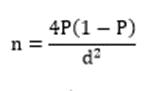
Where:
· P = Prevalence of admission to PICU = 19%
· 1 – P = 0.81
· d = Absolute precision = 0.05
Based on this formula, the minimum calculated sample size was 237. Considering a 10% non-response rate, an additional 24 participants were added. Thus, the final minimum sample size was 261 children. All eligible mechanically ventilated children admitted during the study period were included until this sample size was achieved.
Data Collection Procedure
Data were collected prospectively using a structured and pre-designed proforma for all eligible children admitted to the PICU who required invasive mechanical ventilation during the study period. Initially, demographic details such as age, sex, residence, and socioeconomic status were recorded. Clinical information, including presenting complaints, relevant past history, clinical examination findings, and system involvement, was documented at the time of admission.
The indication for mechanical ventilation, clinical condition at the time of intubation, and relevant investigations, including arterial blood gas analysis, were recorded. Details of mechanical ventilation, such as mode of ventilation, respiratory rate, tidal volume, peak inspiratory pressure, PEEP (Positive End-Expiratory Pressure), fraction of inspired oxygen, and inspiratory and expiratory times, were documented.
Patients were monitored regularly throughout their PICU stay, and daily records of ventilator settings, clinical progress, treatment administered, complications related to mechanical ventilation, and duration of ventilation were maintained. The duration of PICU stay and the clinical criteria for weaning from mechanical ventilation were also recorded. All patients were followed until their final hospital outcome, which was categorized as improvement and discharge, discharge against medical advice, or death. Post-discharge follow-up was advised after four weeks to assess the short-term clinical status of the patients. All collected information was entered into the study proforma and subsequently used for statistical analysis to evaluate the demographic profile, clinical spectrum, and short-term outcomes of mechanically ventilated children.
Statistical Analysis
The collected data were systematically entered into a database and analyzed using the SPSS (Statistical Package for Social Sciences), version 11.0 for Windows. Categorical variables were expressed as frequencies and percentages, while continuous variables were presented as mean and standard deviation. The Chi-square test was used to determine the association between categorical variables and clinical outcomes. A p-value of less than 0.05 was considered statistically significant.
RESULTS:
Table 1. Age Distribution of the Study Population (n = 261)
|
Age Group |
Frequency (n) |
Percentage (%) |
|
|
Less than 1 year |
98 |
37.5 |
|
|
1–5 years |
60 |
23.0 |
|
|
6–10 years |
62 |
23.8 |
|
|
11–14 years |
41 |
15.7 |
|
|
Total |
261 |
100 |
|
|
Age (years) |
Mean ± SD |
Minimum |
Maximum |
|
Age |
4.72 ± 4.48 |
0.08 |
14 |
Table 1 illustrates the age-wise distribution of the study population. The majority of children were below 1 year of age (37.5%), followed by the 6–10-year-old age group (23.8%) and 1–5-year-old age group (23.0%). Children aged 11–14 years constituted 15.7% of the study population. The mean age was 4.72 ± 4.48 years, with an age range of 0.08 to 14 years.
Table 2. Gender Distribution of the Study Population (n = 261)
|
Gender |
Frequency (n) |
Percentage (%) |
|
Male |
143 |
54.8 |
|
Female |
118 |
45.2 |
|
Total |
261 |
100 |
Table 2 demonstrates the gender distribution of the study participants. Males constituted the majority, accounting for 143 (54.8%) children, whereas females accounted for 118 (45.2%). A slight male predominance was observed among mechanically ventilated children.
(Modified B.G. Prasad Classification)
Table 3. Distribution of Patients According to Socioeconomic class (n = 261)
|
Socioeconomic Class |
Frequency (n) |
Percentage (%) |
|
Upper class |
9 |
3.4 |
|
Upper middle class |
32 |
12.3 |
|
Lower middle class |
74 |
28.4 |
|
Upper lower class |
82 |
31.4 |
|
Lower class |
64 |
24.5 |
|
Total |
261 |
100 |
Table 3 illustrates the socioeconomic distribution of the study population according to the Modified B.G. Prasad Classification. The majority of children belonged to the upper lower socioeconomic class (31.4%), followed by the lower middle class (28.4%) and lower class (24.5%). The upper middle class accounted for 12.3%, while only 3.4% belonged to the upper socioeconomic class.
Table 4. Geographical Distribution of the Study Population (n = 261)
|
Residence |
Frequency (n) |
Percentage (%) |
|
Rural |
118 |
45.2 |
|
Urban |
72 |
27.6 |
|
Semi-urban |
71 |
27.2 |
|
Total |
261 |
100 |
Table 4 illustrates the geographical distribution of the study participants. A higher proportion of children were from rural areas, accounting for 118 (45.2%) cases. Children from urban areas constituted 72 (27.6%), while 71 (27.2%) belonged to semi-urban areas. This indicates that the majority of mechanically ventilated children were from rural backgrounds.
Table 5. Distribution of Patients According to Immunization Status (n = 261)
|
Immunization Status |
Frequency (n) |
Percentage (%) |
|
Complete |
145 |
55.6 |
|
Partial |
105 |
40.2 |
|
None |
11 |
4.2 |
|
Total |
261 |
100 |
Table 5 demonstrates the immunization status of the study population. More than half of the children (55.6%) were completely immunized, whereas 105 (40.2%) were partially immunized. Only 11 (4.2%) children had received no immunization. These findings indicate that although the majority had completed immunization, a considerable proportion had incomplete or no immunization.
Table 6. Distribution of Patients According to Clinical Presentation (n = 261)
|
Clinical presentation |
Present n (%) |
Absent n (%) |
|
Respiratory failure |
152 (58.2) |
109 (41.8) |
|
Altered sensorium |
96 (36.8) |
165 (63.2) |
|
Status epilepticus |
68 (26.1) |
193 (73.9) |
|
Respiratory distress |
122 (46.7) |
139 (53.3) |
|
Shock |
70 (26.8) |
191 (73.2) |
Table 6 illustrates the clinical presentation of mechanically ventilated children at admission. Respiratory failure was the most common clinical presentation, observed in 152 (58.2%) children, followed by respiratory distress in 122 (46.7%). Altered sensorium was noted in 96 (36.8%) children, while shock and status epilepticus were observed in 26.8% and 26.1% of cases, respectively. These findings indicate that respiratory compromise was the predominant clinical feature, although neurological and cardiovascular manifestations were also observed.
Table 7. Distribution of Patients According to Organ System Involvement (n = 261)
|
Organ System |
Present n (%) |
Absent n (%) |
|
Central nervous system (CNS) |
83 (31.8) |
178 (68.2) |
|
Cardiovascular system (CVS) |
64 (24.5) |
197 (75.5) |
|
Respiratory system |
152 (58.2) |
109 (41.8) |
|
Gastrointestinal tract (GIT) |
53 (20.3) |
208 (79.7) |
|
Genitourinary tract (GUT) |
39 (14.9) |
222 (85.1) |
|
Sepsis |
78 (29.9) |
183 (70.1) |
|
Multiorgan dysfunction syndrome (MODS) |
45 (17.2) |
216 (82.8) |
Table 7 demonstrates the distribution of organ system involvement among the study participants. Respiratory system involvement was the most common, observed in 152 (58.2%) children, followed by central nervous system involvement in 83 (31.8%) and sepsis in 78 (29.9%) children. Cardiovascular system involvement was seen in 24.5%, while gastrointestinal and genitourinary involvement accounted for 20.3% and 14.9%, respectively. Multiorgan dysfunction syndrome was observed in 45 (17.2%) children. These findings highlight the predominance of respiratory involvement, with considerable contributions from neurological and systemic infections.
DISCUSSION:
Age Distribution
In the present study, infants below 1 year constituted the largest proportion of mechanically ventilated children (37.5%), followed by those aged 6–10 years (23.8%), 1–5 years (23.0%) and 11–14 years (15.7%). The mean age was 4.72 ± 4.48 years, indicating a greater burden of mechanical ventilation among younger children. Similarly, Linssen et al. reported an increasing PICU burden of RSV bronchiolitis, predominantly among infants aged ≤3 months, while Patra et al. reported a median age of 3.7 years.[13] Vidal et al. identified age below 4 years as a significant predictor of prolonged mechanical ventilation (OR 3.21; 95% CI 1.38–7.48; p = 0.007).[14] Berkenbosch et al. reported a slightly higher mean age of 5.7 ± 6.1 years.[15] These findings highlight the vulnerability of infants and younger children to severe illnesses requiring ventilatory support.
Gender Distribution
The present study showed a slight male predominance, with 143 (54.8%) males and 118 (45.2%) females. However, gender was not significantly associated with patient outcome (χ² = 1.514; p = 0.679). Similarly, Pereira et al. reported 60% male representation among children assessed following PICU discharge,[16] while Patra et al. observed a male-to-female ratio of 1.9:1 among intubated children.[13]
Socioeconomic Class and Residence
Most children in the present study belonged to the upper lower (31.4%), lower middle (28.4%) and lower (24.5%) socioeconomic classes. Rural children constituted the largest proportion (45.2%), followed by urban (27.6%) and semi-urban (27.2%) residents. These findings suggest a greater representation of children from socioeconomically disadvantaged and rural backgrounds among mechanically ventilated patients. Although the comparison studies did not directly assess these demographic variables, Pereira et al. reported a median Functional Status Scale score of 11.5 following PICU discharge, with poorer outcomes associated with prolonged ventilation and PICU stay.[16] Urrea et al. demonstrated significantly longer hospitalization among children with nosocomial infections (22.5 versus 9 days; p < 0.001).[17] These observations emphasize the potential influence of healthcare accessibility, delayed referral and socioeconomic vulnerability on paediatric critical illness.
Immunization Status
In the present study, 145 children (55.6%) were completely immunized, whereas 105 (40.2%) were partially immunized and 11 (4.2%) were unimmunized. Thus, 44.4% had incomplete or no immunization, indicating a considerable gap in preventive healthcare coverage. Although the comparative studies did not directly investigate immunization status, Linssen et al. highlighted the increasing PICU burden of severe RSV bronchiolitis.[18] Patra et al. reported nosocomial pneumonia in 30.5% of intubated children, with 47% of deaths directly attributable to pneumonia.[13] These findings reinforce the importance of infection prevention and timely immunization in reducing severe infectious illnesses during childhood.
Clinical Presentation and Organ System Involvement
Respiratory failure was the predominant clinical presentation in the present study, affecting 152 children (58.2%), followed by respiratory distress (46.7%), altered sensorium (36.8%), shock (26.8%) and status epilepticus (26.1%). Respiratory involvement was also the most frequently affected organ system (58.2%), followed by central nervous system involvement (31.8%), sepsis (29.9%), cardiovascular involvement (24.5%), gastrointestinal involvement (20.3%), multiorgan dysfunction syndrome (17.2%) and genitourinary involvement (14.9%). Similarly, Amigoni et al. reported respiratory failure in 33% and neurological impairment in 24% of PICU admissions.[19] Vidal et al. found that respiratory disease was significantly associated with prolonged mechanical ventilation (OR 4.94; 95% CI 1.51–16.10; p = 0.008).[14]
Indications for Intubation and Mechanical Ventilation
Respiratory failure was the leading indication for mechanical ventilation in the present study (55.6%), followed by impending respiratory failure (41.4%), acute meningitis (28.4%), status epilepticus (26.8%), severe asthma (24.1%), chronic respiratory failure (21.1%) and respiratory muscle paralysis (16.1%). These findings demonstrate that respiratory compromise was the predominant indication for invasive ventilation, although neurological and neuromuscular conditions also contributed considerably. Similarly, Amigoni et al. reported respiratory failure in 33% and neurological impairment in 24% of PICU admissions.[19] Linssen et al. documented an increasing burden of severe RSV bronchiolitis requiring PICU respiratory support.[18] Vidal et al. further demonstrated a significant association between respiratory disease and prolonged ventilation (OR 4.94).[14] Overall, the findings emphasize the importance of respiratory and neurological conditions as major indications for paediatric mechanical ventilation.
Mode of Ventilation and Ventilator Course
Synchronized intermittent mandatory ventilation was the most frequently used mode in the present study (29.1%), followed by volume support (24.9%), pressure support ventilation (24.5%) and controlled mechanical ventilation (21.5%). The mean duration of mechanical ventilation was 2.98 ± 2.89 days, ranging from 0.25 to 16 days. The mean maximum and minimum PIP values were 24.25 ± 3.54 and 15.51 ± 1.10 cm H₂O, respectively, while maximum and minimum PEEP values were 7.94 ± 1.43 and 4.50 ± 0.50, respectively. Randolph et al. reported extubation failure rates of 15%, 24% and 17% with pressure support ventilation, volume support ventilation and usual care, respectively, with median weaning durations of 1.6, 1.8 and 2.0 days.[20] In contrast, Vidal et al. reported prolonged mechanical ventilation of ≥7 days in 50.3% of children.[14] Maue et al. identified higher postoperative PIP as an independent predictor of prolonged invasive ventilation (adjusted OR 1.2; 95% CI 1.1–1.3).[21] The relatively shorter mean ventilation duration in the present study may reflect differences in disease severity and patient characteristics.
Complications of Mechanical Ventilation
Mechanical ventilation-related complications constituted a considerable burden in the present study. Laryngeal edema was the most common complication (77.4%), followed by endotracheal tube blockage (62.5%), ventilator-associated pneumonia (36.0%), endotracheal tube displacement (29.1%), reintubation (21.1%), pneumothorax (8.0%) and tension pneumothorax (3.1%). Similarly, Patra et al. reported nosocomial pneumonia in 30.5% of intubated children and identified reintubation as the only independent predictor of nosocomial pneumonia.[13] El-Rebigi et al. reported adverse events in 24.1% of ventilated children, with ventilator-associated pneumonia (13%), post-extubation stridor (7.9%) and pneumothorax (6%) being the commonly observed complications.[22] Urrea et al. also demonstrated that nosocomial infections significantly prolonged hospitalization.[17] The higher complication frequencies observed in the present study highlight the need for meticulous airway management, appropriate ventilator care and strict infection prevention practices.
Outcome of Patients
In the present study, 180 children (69.0%) survived, of whom 36 (13.8%) improved without complications and 144 (55.2%) improved with complications. Mortality was observed in 77 children (29.5%), while 4 (1.5%) were referred. These findings indicate that although survival was achieved in the majority, uncomplicated recovery was relatively uncommon. Pereira et al. reported moderate functional impairment following PICU discharge, with a median Functional Status Scale score of 11.5, particularly affecting motor and feeding domains.[16] Patra et al. reported a mortality rate of 21% among intubated children, with 47% of deaths directly attributable to nosocomial pneumonia.[13] El-Rebigi et al. reported a comparatively lower mortality rate of 9.8%, with adverse events occurring in 24.1% of patients.[22] Maue et al. reported mortality of only 1.8% in paediatric liver transplant recipients, representing a specialized postoperative population.[21] The differences in mortality across studies may reflect variations in patient characteristics, underlying diseases and severity of critical illness.
Factors Associated with Poor Outcome and Mortality
On multivariate logistic regression, MODS emerged as the strongest independent predictor of mortality (OR 7.21; p < 0.001), followed by ventilator-associated pneumonia (OR 2.67; p = 0.006). Duration of ventilation also remained statistically significant (OR 0.87; p = 0.031). Similarly, Amigoni et al. identified multiple organ failure as an independent predictor of PICU mortality (OR 4.83).[19] Vidal et al. demonstrated an association between organ dysfunction severity and prolonged ventilation,[14] while Patra et al. identified reintubation as an important predictor of nosocomial pneumonia.[13] These findings emphasize that systemic illness severity, organ dysfunction and ventilator-associated infections are important determinants of mortality in mechanically ventilated children.
Association between Sepsis and Outcome
Sepsis was present in 78 children (29.9%) and showed a statistically significant association with patient outcome (χ² = 9.957; p = 0.019). This indicates that sepsis contributed considerably to adverse outcomes among mechanically ventilated children. Vidal et al. reported that septic shock was significantly associated with prolonged mechanical ventilation (OR 4.66; 95% CI 1.10–19.65; p = 0.036).[14] Similarly, Amigoni et al. demonstrated that multiple organ failure independently increased PICU mortality (OR 4.83; 95% CI 1.66–15.71).[19] Urrea et al. reported significantly prolonged hospitalization among children with nosocomial infections compared with those without infections (22.5 versus 9 days; p < 0.001).[17]
Association between MODS and Outcome
MODS was observed in 45 children (17.2%) and demonstrated a highly significant association with patient outcome (χ² = 24.631; p < 0.001). Furthermore, multivariate analysis identified MODS as the strongest independent predictor of mortality, with an odds ratio of 7.21 (p < 0.001). Similarly, Vidal et al. reported that a PELOD score greater than 10 was associated with prolonged mechanical ventilation (OR 2.58; 95% CI 1.17–5.58; p = 0.018).[14] Amigoni et al. also identified multiple organ failure as an independent predictor of PICU mortality (OR 4.83; 95% CI 1.66–15.71).[19] These findings establish MODS as an important prognostic indicator and emphasize the need for early recognition of organ dysfunction and timely multisystem supportive management.
Limitations
The present study has certain limitations that should be acknowledged. Being a single-centre observational study, the findings may have limited generalizability to other PICUs with different patient populations, infrastructure and ventilation practices. The observational design also limits the establishment of causal relationships between clinical variables and outcomes. Furthermore, the study focused only on short-term PICU outcomes and did not evaluate long-term survival, neurodevelopmental status or post-discharge quality of life. Standardized paediatric severity scoring systems were not assessed in detail for all patients, and certain factors such as nutritional status, microbiological profiles, pre-referral treatment, sedation practices and weaning parameters were not explored comprehensively. Variations in clinical management and the unusually high frequencies of certain complications, particularly laryngeal edema and endotracheal tube blockage, may also have influenced the reported outcomes. Therefore, the findings should be interpreted cautiously, highlighting the need for further multicentre studies with long-term follow-up to validate these observations.
CONCLUSION:
The present study concludes that mechanical ventilation is a crucial life-saving intervention in critically ill children, particularly among infants, who constituted 37.5% of the study population. Respiratory failure (58.2%) was the predominant clinical presentation, although neurological involvement, sepsis (29.9%) and multiorgan dysfunction syndrome (17.2%) also contributed significantly to the need for ventilatory support. The mean duration of ventilation was 2.98 ± 2.89 days. Mechanical ventilation-related complications, particularly laryngeal edema (77.4%), endotracheal tube blockage (62.5%) and ventilator-associated pneumonia (36.0%), contributed considerably to morbidity. Although 69.0% of children survived, the mortality rate was 29.5%. Multiorgan dysfunction syndrome (OR 7.21; p < 0.001) emerged as the strongest independent predictor of mortality, followed by ventilator-associated pneumonia (OR 2.67; p = 0.006), highlighting the importance of disease severity and prevention of ventilator-related complications in determining outcomes. Therefore, early recognition of critical illness, timely initiation of ventilatory support, meticulous airway management, strict infection prevention, prompt treatment of sepsis and aggressive management of multiorgan dysfunction are essential to improving survival and reducing morbidity among mechanically ventilated children.
REFERENCES:
1. Blackwood B, Tume LN, Morris KP, et al. Effect of a sedation and ventilator liberation protocol vs usual care on duration of invasive mechanical ventilation in paediatric intensive care units: a randomized clinical trial. JAMA 2021;326(5):401-10.
2. Farias JA, Fernandez A, Monteverde E, et al. Mechanical ventilation in paediatric intensive care units during the season for acute lower respiratory infection: a multicentre study. Pediatr Crit Care Med 2012;13(2):158-64.
3. Egbuta C, Easley RB. Update on ventilation management in the Paediatric Intensive Care Unit. Paediatr Anaesth 2022;32(2):354-62.
4. Sood S, Ganatra HA, Perez Marques F, et al. Complications during mechanical ventilation-A paediatric intensive care perspective. Front Med 2023;10:1016316.
5. Cheifetz IM. Invasive and non-invasive paediatric mechanical ventilation. Respir Care 2003;48(4):442-58.
6. Bose SN, Defante A, Greenstein JL, et al. A data-driven model for early prediction of need for invasive mechanical ventilation in paediatric intensive care unit patients. PloS One 2023;18(8):e0289763.
7. Rowan CM, Gertz SJ, McArthur J, et al. Invasive mechanical ventilation and mortality in paediatric hematopoietic stem cell transplantation: a multicentre study. Pediatr Crit Care Med 2016;17(4):294-302.
8. Farias JA, Frutos F, Esteban A, et al. What is the daily practice of mechanical ventilation in paediatric intensive care units? A multicentre study. Intensive Care Med 2004;30(5):918-25.
9. Pisitcholakarn V, Sunkonkit K, Reungrongrat S. Incidence and factors associated with prolonged use of mechanical ventilation in paediatric intensive care unit in a single tertiary care hospital. PloS One 2024;19(11):e0311275.
10. Martins LD, Ferreira AR, Kakehasi FM. Adverse events related to mechanical ventilation in a paediatric intensive care unit. Rev Paul Pediatr 2021;39:e2019180.
11. Ma L, Yin M, Yang XL, et al. Risk factors for air leakage during invasive mechanical ventilation in paediatric intensive care units. Eur J Med Res 2022;27(1):218.
12. Newth CJ, Khemani RG, Jouvet PA, et al. Mechanical ventilation and decision support in paediatric intensive care. Pediatr Clin North Am 2017;64(5):1057-70.
13. Patra PK, Jayashree M, Singhi S, et al. Nosocomial pneumonia in a pediatric intensive care unit. Indian Paediatr 2007;44(7):511-8.
14. Vidal S, Pérez A, Eulmesekian P. Fluid balance and length of mechanical ventilation in children admitted to a single paediatric intensive care unit. Arch Argent Pediatr 2016;114(4):313-8.
15. Berkenbosch JW, Fichter CR, Tobias JD. The correlation of the bispectral index monitor with clinical sedation scores during mechanical ventilation in the paediatric intensive care unit. Anaesth Analg 2002;94(3):506-11.
16. Pereira GA, Schaan CW, Ferrari RS. Functional evaluation of paediatric patients after discharge from the intensive care unit using the Functional Status Scale. Rev Bras Ter Intensiva 2017;29(4):460-5.
17. Urrea M, Pons M, Serra M, et al. Prospective incidence study of nosocomial infections in a paediatric intensive care unit. Pediatr Infect Dis J 2003;22(6):490-4.
18. Linssen RS, Bem RA, Kapitein B, et al. Burden of respiratory syncytial virus bronchiolitis on the Dutch pediatric intensive care units. Eur J Pediatr 2021;180(10):3141-9.
19. Amigoni A, Boscato S, Mondardini MC, et al. Exploring risk factors for paediatric cancer patients admitted to the Paediatric Intensive Care Unit: insight from a multicentre observational study revealing no association with mechanical ventilation. J Anaesth Analg Crit Care 2025;5(1):63.
20. Randolph AG, Wypij D, Venkataraman ST, et al. Effect of mechanical ventilator weaning protocols on respiratory outcomes in infants and children: a randomized controlled trial. JAMA 2002;288(20):2561-8.
21. Maue DK, Martinez M, Alcamo A, et al. Critical care and mechanical ventilation practices surrounding liver transplantation in children: a multicentre collaborative. Pediatr Crit Care Med 2023;24(2):102-11.
22. El-Rebigi AM, Fekry AN, Elfaramawy MA, et al. Mechanical ventilation-associated complications and comorbidities in children admitted at paediatric intensive care unit: a cross-sectional retrospective study. Turk J Emerg Med 2025;25(3):230-8.