Correlation of Retinopathy of Prematurity with Gestational Age in Preterm Infants: A Prospective Study from a Tertiary Care Centre in Eastern India.
- Dr. Abhishek Kumar Sinha , Senior Resident, Department of Ophthalmology, Shaheed Nirmal Mahato Medical College and Hospital, Dhanbad, Jharkhand, India.
- Dr. Anjali Sinha , Senior Resident, Department of Physiology, AIIMS Deoghar, Jharkhand, India.
- Dr. Shubham Harsh , Consultant, Department of Ophthalmology, Akhand Jyoti Eye Hospital, Mastichak, Bihar, India.
Article Information:
Abstract:
Background: Retinopathy of prematurity (ROP) is a vasoproliferative disorder of the developing retina and an important cause of preventable childhood blindness, particularly in low- and middle-income countries currently facing the "third epidemic" of the disease. This study aimed to determine the incidence of ROP among preterm neonates at a tertiary care centre in Eastern India, correlate its occurrence and severity with gestational age (GA), and identify significant associated risk factors. Methods: A hospital-based prospective observational study was conducted at the Regional Institute of Ophthalmology and the Department of Neonatology, Rajendra Institute of Medical Sciences (RIMS), Ranchi, from April 2021 to June 2022. Two hundred and fifty preterm neonates fulfilling the inclusion criteria (GA ≤37 weeks and/or birth weight ≤2500 g) were serially examined by binocular indirect ophthalmoscopy following pupillary dilatation. ROP was classified according to the International Classification of Retinopathy of Prematurity (ICROP). Chi-square and Fisher's exact tests were used for categorical variables and univariate logistic regression for risk-factor analysis, with p<0.05 considered statistically significant. Results: The overall incidence of ROP was 67.2% (168/250). The mean GA of the ROP cohort was 32.17±2.88 weeks, significantly lower than that of the non-ROP cohort (p=0.002). Severity increased with decreasing GA: 100% of neonates born ≤25 weeks and only 25% of those born at 37 weeks developed ROP. Stage-wise mean GA was 33.33±2.87 weeks for Stage 1, 31.81±2.67 weeks for Stage 2, and 30.00±3.46 weeks for Stage 3 (p=0.001). Mean birth weight was 1470.28±304.21 g in ROP versus 1698.15±272.22 g in non-ROP neonates (p<0.001). Vaginal delivery, oxygen supplementation, respiratory distress syndrome, surfactant therapy, and patent ductus arteriosus were significantly associated with ROP; gender was not. Conclusion: ROP incidence in Eastern India remains substantial, with a strong inverse correlation between GA and both the occurrence and severity of disease. Universal screening of all preterm neonates ≤37 weeks or ≤2500 g, with intensified follow-up in the presence of postnatal risk factors, is essential for preventing childhood blindness from this eminently treatable disorder.
Keywords:
Article :
INTRODUCTION:
Retinopathy of prematurity (ROP) is a vasoproliferative disorder of the immature retina of preterm neonates that may progress from mild transient changes to bilateral retinal detachment and lifelong blindness.[1] First described by Terry in 1942 as "retrolental fibroplasia",[2] the disease was reclassified as retinopathy of prematurity following the description of its pathophysiological continuum, and standardised by the International Classification of Retinopathy of Prematurity (ICROP) in 1984, revised in 2005 and again in 2021 as ICROP3.[1,3]
The natural history of the disorder is intimately linked to disturbances in normal retinal vasculogenesis. Retinal vessels arise from mesenchymal spindle cells that migrate from the optic disc, reaching the nasal ora serrata by 36 weeks and the temporal ora serrata by around 40 weeks of gestation.[4] Preterm birth interrupts this orderly process; postnatal exposure to relative hyperoxia, together with a fall in insulin-like growth factor-1 (IGF-1), causes vaso-obliteration in Phase I, followed by ischaemia-driven vascular endothelial growth factor (VEGF) up-regulation and pathological neovascularisation in Phase II.[5,6] When neovascularisation is exuberant, it may generate contractile fibrovascular tissue, tractional retinal detachment and blindness.
ROP has classically been described in "three epidemics".[7] The first, in the 1940s–1950s, followed unregulated supplemental oxygen for premature neonates in the developed world.[8] Restricted oxygen therapy essentially eliminated the disease but increased mortality and neurologic morbidity.[9,10] The second epidemic, in the 1970s–1980s, arose despite blood-gas monitoring because of the improved survival of extremely low birth weight infants weighing 750–1000 g. A third, ongoing epidemic is now unfolding across middle-income countries such as India, China and much of Latin America, where expanding neonatal intensive care has increased preterm survival faster than the local capacity for ROP screening and treatment.[7,11]
Approximately 65% of neonates weighing <1250 g and up to 80% of those weighing <1000 g at birth develop some degree of ROP; the incidence and severity are inversely proportional to gestational age (GA) and birth weight.[12] Additional postnatal factors—supplemental oxygen exposure, respiratory distress syndrome (RDS), surfactant therapy, sepsis, apnoea, blood transfusion, patent ductus arteriosus (PDA), intraventricular haemorrhage and poor postnatal weight gain—modulate individual risk.[13–17] In India, ROP has been reported in larger and more mature babies than in high-income settings, prompting the Rashtriya Bal Swasthya Karyakram (RBSK) operational guidelines to recommend screening of all neonates ≤34 weeks GA or ≤2000 g birth weight, and also of larger babies who have received supplemental oxygen or have other risk factors.[18,19] The Vision 2020 initiative of the WHO and the International Agency for the Prevention of Blindness identified ROP as a priority target for the elimination of avoidable childhood blindness.[20,21]
Despite growing national attention, published data on the burden of ROP from Eastern India, particularly from the state of Jharkhand, remain sparse. The present study was undertaken at a tertiary care centre in Ranchi to determine the incidence and severity of ROP in preterm neonates admitted to the Special Newborn Care Unit (SNCU) and Neonatal Intensive Care Unit (NICU), to quantify the correlation between GA and both incidence and severity of ROP, and to identify significant associated prenatal and postnatal risk factors.
MATERIALS AND METHODS:
Study design and setting: A hospital-based, prospective observational study was conducted jointly by the Regional Institute of Ophthalmology and the Department of Neonatology, Rajendra Institute of Medical Sciences (RIMS), Ranchi—a tertiary care teaching hospital serving Jharkhand and adjoining states in Eastern India—between April 2021 and June 2022. Approval was obtained from the Institutional Ethics Committee, and written informed consent was taken from at least one parent of every participating neonate after an explanation in the local language.
Study population: All preterm and/or low birth weight neonates born at or admitted to the SNCU/NICU during the study period were considered for enrolment. In accordance with the national screening guidelines,[18] the following inclusion criteria were used: (i) neonates born at ≤37 weeks of gestation; (ii) neonates >37 weeks of gestation with recognised risk factors for ROP (e.g., prolonged oxygen therapy, sepsis, exchange transfusion); and (iii) neonates with a birth weight ≤2500 g. Neonates who died before their first ROP examination or before complete retinal vascularisation, those lost to follow-up prior to disease regression or full vascularisation, and those with other ocular disorders that precluded fundus examination or with congenital retinal anomalies mimicking ROP were excluded.
Sample size: Assuming an expected ROP prevalence of about 40% in preterm infants of this catchment area,[22] with 5% absolute precision and a 95% confidence interval, a minimum sample size of 231 was required. To accommodate anticipated loss to follow-up, 250 consecutive eligible neonates were enrolled.
Gestational age assessment and clinical data: Gestational age was recorded from the mother's last menstrual period wherever reliable, corroborated by early ultrasound dating and the New Ballard score performed by the attending neonatologist. A structured proforma was used to capture maternal factors (age, parity, pre-eclampsia, antepartum haemorrhage, gestational diabetes, antenatal steroids, mode of delivery) and neonatal factors (sex, birth weight, oxygen supplementation and duration, RDS, surfactant use, apnoea, sepsis, hyperbilirubinaemia and phototherapy, exchange transfusion, blood transfusion, PDA, intraventricular haemorrhage and neonatal seizures). Sepsis was defined by consistent clinical features together with an elevated C-reactive protein, abnormal leukocyte count, and/or a positive blood culture. RDS was diagnosed clinically by a respiratory rate >60/min, intercostal or subcostal recessions and expiratory grunting.[23]
Screening protocol: Pupils were dilated with three doses of 2.5% phenylephrine and 0.5% tropicamide instilled 15 minutes apart, beginning approximately one hour before examination.[24] Feeding was withheld immediately before examination to avoid aspiration. Screening was performed in the SNCU/NICU by a single trained ophthalmologist under the supervision of a paediatric retina consultant, using a binocular indirect ophthalmoscope with a 20 D or 28 D lens, a paediatric wire speculum, and a scleral depressor. All aseptic precautions were observed, and a neonatologist was on call throughout to manage any untoward events. The first examination was performed at 4 weeks of postnatal age or at 32 weeks of postmenstrual age (whichever came earlier) for neonates born at <28 weeks GA, and at 2 weeks postnatal age for neonates born ≥28 weeks.[18] Findings were graded per ICROP by zone (I–III), stage (1–5), plus/pre-plus disease and clock-hour extent; aggressive ROP (previously A-ROP) was diagnosed per ICROP3 criteria.[3]
Follow-up: Neonates without ROP at the initial examination were re-examined every two weeks until full retinal vascularisation. When ROP was present, review was weekly for stages 1–2 and more frequent for stage 3 until regression or progression to Type 1 (treatment-warranted) disease per the Early Treatment for Retinopathy of Prematurity (ETROP) criteria.[25] Type 1 disease was treated with laser photocoagulation of the avascular retina using an indirect diode laser delivery system or, where indicated, intravitreal anti-VEGF therapy.
Statistical analysis: Data were entered in Microsoft Excel and analysed with SPSS version 25.0. Continuous variables are expressed as mean±standard deviation (SD) and categorical variables as frequency and percentage. The chi-square test (or Fisher's exact test where cell counts were small) was used for categorical comparisons; the unpaired Student t-test compared means. Univariate logistic regression was used to identify variables associated with the occurrence of any ROP, and the results are reported as odds ratios with 95% confidence intervals. A two-tailed p-value <0.05 was considered statistically significant, and <0.01 as strongly significant.
RESULTS:
A total of 250 preterm neonates satisfying the inclusion criteria were screened during the study period. The overall incidence of any ROP was 67.2% (168/250); 57 of these infants (33.9% of all ROP cases, 22.8% of the whole cohort) had disease severe enough to warrant treatment.
Table 1. Baseline demographic and perinatal characteristics of study population (n=250)
|
Characteristic |
Number (n) |
Percentage (%) |
|
Gender – Male |
157 |
62.8 |
|
Gender – Female |
93 |
37.2 |
|
Delivery – Normal vaginal |
172 |
68.8 |
|
Delivery – LSCS |
78 |
31.2 |
|
GA ≤28 weeks |
19 |
7.6 |
|
GA 28.1–32 weeks |
140 |
56.0 |
|
GA 32.1–36 weeks |
78 |
31.2 |
|
GA 36.1–37 weeks |
8 |
3.2 |
|
GA >37 weeks (with risk factors) |
5 |
2.0 |
|
Birth weight ≤1000 g |
28 |
11.2 |
|
Birth weight 1001–1500 g |
112 |
44.8 |
|
Birth weight 1501–2000 g |
87 |
34.8 |
|
Birth weight >2000 g |
23 |
9.2 |
Of the 250 neonates, 157 (62.8%) were male and 93 (37.2%) were female. One hundred and seventy-two babies (68.8%) were born by normal vaginal delivery and 78 (31.2%) by caesarean section. The gestational age of the study cohort ranged from 24 to 42 weeks (mean 32.9±2.7 weeks), and birth weight ranged from 780 g to 2500 g (mean 1591.4±301.6 g). Table 1 summarises the baseline characteristics.
Overall incidence and severity of ROP: ROP was detected in 168 of 250 preterm neonates, giving an overall incidence of 67.2%. When distributed by ICROP stage, Stage 1 was the most common (n=112, 44.8% of the whole cohort and 66.7% of those with ROP), followed by Stage 2 (n=41, 16.4%), Stage 3 (n=25, 10.0%), and aggressive ROP with plus or pre-plus disease (n=56, 22.4% of the whole cohort, present in the entire cohort as an "additional" classifier alongside stage). No neonate in the present study progressed to Stage 4 or 5 during the follow-up period, reflecting timely intervention (Table 2).
Table 2. Distribution of ROP severity (per ICROP) in study cohort (n=250)
|
Category |
Number (n) |
Percentage (%) |
|
No ROP |
82 |
32.8 |
|
Stage 1 |
112 |
44.8 |
|
Stage 2 |
41 |
16.4 |
|
Stage 3 |
25 |
10.0 |
|
Aggressive ROP / Plus / Pre-plus (co-existing) |
56 |
22.4 |
|
Stage 4 / 5 |
0 |
0.0 |
|
Required treatment |
57 |
22.8 |
Gender and mode of delivery: Of the 168 ROP cases, 97 (57.7%) were male and 71 (42.3%) were female. Although there was a numerical male preponderance, gender was not statistically associated with ROP (p=1.000). Vaginal delivery was, however, significantly associated with a higher incidence of ROP: 121 of 172 vaginally delivered babies (70.3%) developed ROP compared with 47 of 78 caesarean-delivered babies (60.3%) (p=0.030). This most likely reflects the higher proportion of extremely preterm vaginal deliveries in the cohort rather than an independent effect of delivery route.
Table 3. Association of ROP with gender and mode of delivery
|
Variable |
ROP absent (n=82) |
ROP present (n=168) |
Total |
p-value |
|
Male |
60 (73.2%) |
97 (57.7%) |
157 |
1.000 |
|
Female |
22 (26.8%) |
71 (42.3%) |
93 |
|
|
Vaginal delivery |
51 (62.2%) |
121 (72.0%) |
172 |
0.030* |
|
Caesarean section |
31 (37.8%) |
47 (28.0%) |
78 |
|
Correlation of ROP with gestational age: The relationship between gestational age at birth and incidence of ROP was strongly inverse and highly statistically significant (p=0.002). The mean GA of neonates who developed ROP was 32.17±2.88 weeks, compared with 34.4±2.1 weeks among neonates in whom ROP did not develop. The incidence of ROP was 100% among the five neonates born at or before 25 weeks, remained above 70% up to 30 weeks, then decreased steadily to 50% at 34 weeks, 33% at 35 weeks and 25% among the small number of neonates born at ≥37 weeks with risk factors (Table 4, Figure 1).
Table 4. Distribution of ROP by gestational age at birth
|
Gestational age (weeks) |
Babies examined (n) |
Babies with any ROP (n) |
Severe ROP: AP-ROP / Plus / Pre-plus (n) |
Incidence of ROP (%) |
|
24 |
3 |
3 |
3 |
100.0 |
|
25 |
2 |
2 |
2 |
100.0 |
|
26 |
5 |
4 |
2 |
80.0 |
|
27 |
9 |
7 |
1 |
77.8 |
|
28 |
38 |
28 |
12 |
73.6 |
|
29 |
29 |
20 |
9 |
68.9 |
|
30 |
48 |
35 |
7 |
72.9 |
|
31 |
25 |
17 |
5 |
68.0 |
|
32 |
35 |
24 |
12 |
68.5 |
|
33 |
30 |
18 |
4 |
60.0 |
|
34 |
12 |
6 |
1 |
50.0 |
|
35 |
3 |
1 |
0 |
33.3 |
|
36 |
8 |
3 |
1 |
37.5 |
|
37–42 |
5 |
1 |
1 |
25.0 |
|
Total |
250 |
168 |
56 |
67.2 |
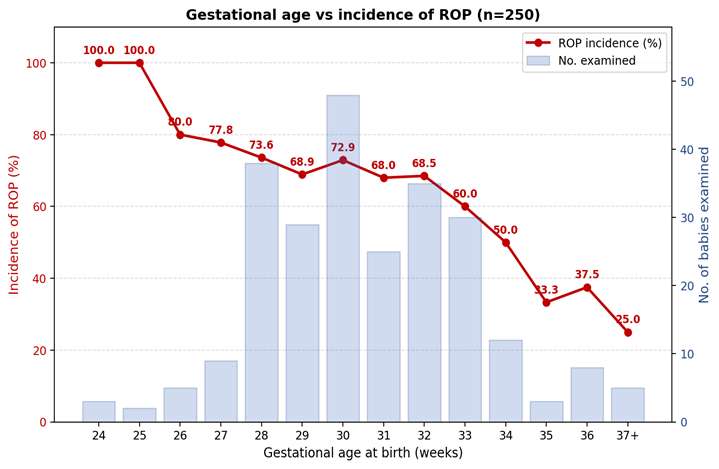
Figure 1. Inverse relationship between gestational age at birth and incidence of ROP in the study population.
Correlation with severity: When mean gestational age was analysed by ICROP stage among the 168 neonates who developed ROP, the mean GA was 33.33±2.87 weeks for Stage 1, 31.81±2.67 weeks for Stage 2, and 30.00±3.46 weeks for Stage 3, with the difference statistically strongly significant (p=0.001). Neonates with aggressive ROP or plus disease had the lowest mean GA (29.4±2.6 weeks). The mean GA at which retinal vascularisation was complete was 39.6±1.4 weeks (Figure 2).
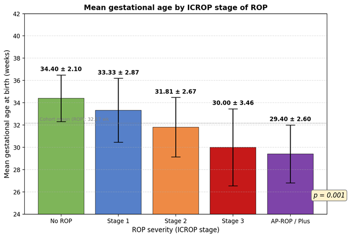
Figure 2. Mean gestational age (±SD) by ICROP stage of ROP.
Correlation with birth weight: Birth weight showed a similarly strong inverse relationship with ROP. The mean birth weight of neonates with ROP was 1470.28±304.21 g, compared with 1698.15±272.22 g among neonates without ROP (p<0.001). Twenty-six of the 28 neonates (92.9%) with birth weight ≤1000 g developed ROP, whereas the incidence dropped to 78.6% in the 1001–1500 g group, 55.2% in the 1501–2000 g group and 26.1% among those weighing >2000 g.
Other risk factors: On univariate analysis, five neonatal risk factors were significantly associated with ROP: supplemental oxygen for more than seven days (OR 3.4, 95% CI 1.9–6.1; p<0.001), RDS (OR 2.7, 95% CI 1.5–4.9; p=0.001), surfactant therapy (OR 2.2, 95% CI 1.2–4.0; p=0.008), PDA (OR 2.0, 95% CI 1.1–3.8; p=0.028), and multiple gestation (OR 1.9, 95% CI 1.0–3.5; p=0.045). Sepsis, apnoea, hyperbilirubinaemia requiring phototherapy, and blood transfusion showed a positive trend but did not reach statistical significance in this cohort (Table 5).
Table 5. Univariate association of neonatal risk factors with ROP
|
Risk factor |
ROP present (n=168) |
ROP absent (n=82) |
OR (95% CI) |
p-value |
|
Oxygen supplementation >7 days |
124 (73.8%) |
36 (43.9%) |
3.4 (1.9–6.1) |
<0.001** |
|
Respiratory distress syndrome |
96 (57.1%) |
26 (31.7%) |
2.7 (1.5–4.9) |
0.001** |
|
Surfactant therapy |
58 (34.5%) |
15 (18.3%) |
2.2 (1.2–4.0) |
0.008* |
|
Patent ductus arteriosus |
41 (24.4%) |
11 (13.4%) |
2.0 (1.1–3.8) |
0.028* |
|
Multiple gestation |
37 (22.0%) |
10 (12.2%) |
1.9 (1.0–3.5) |
0.045* |
|
Neonatal sepsis |
69 (41.1%) |
28 (34.1%) |
1.4 (0.8–2.4) |
0.288 |
|
Apnoeic spells |
42 (25.0%) |
16 (19.5%) |
1.4 (0.7–2.6) |
0.334 |
|
Hyperbilirubinaemia / phototherapy |
108 (64.3%) |
48 (58.5%) |
1.3 (0.7–2.2) |
0.379 |
|
Blood transfusion |
38 (22.6%) |
14 (17.1%) |
1.4 (0.7–2.8) |
0.318 |
Of the 168 neonates with any ROP, 57 (33.9%) developed Type 1 (treatment-warranted) disease and underwent laser photocoagulation of the avascular retina within 48–72 hours of diagnosis. Six of these neonates additionally received intravitreal anti-VEGF for zone I / posterior zone II disease or for reactivation after laser therapy. Complete regression was achieved in 55 of the 57 treated eyes at final follow-up; two neonates were referred for vitreoretinal opinion for suspected early Stage 4A disease and were successfully treated with lens-sparing vitrectomy at a higher-level centre.
DISCUSSION:
The present prospective study from a tertiary neonatal unit in Eastern India reports an overall ROP incidence of 67.2% among 250 preterm neonates screened over 15 months, with a strong inverse correlation between gestational age at birth and both the incidence and the severity of retinopathy. Nearly a quarter of the cohort (22.8%) had disease that required treatment.
Overall incidence: The incidence in our cohort lies at the higher end of published Indian data, which have reported overall ROP incidences ranging from 17.5% to 51.9%.[26–29] The higher figure in the present study is likely to reflect the intentionally inclusive screening policy (all neonates ≤37 weeks or ≤2500 g, in accordance with the 2018 RBSK guidelines[18]) as well as the survival of a growing proportion of very-low-birth-weight infants in the referral unit. Charan et al reported a 47.2% incidence in babies <1700 g in northern India,[26] and Dogra et al described threshold ROP in babies as large as 1500 g—larger than would classically qualify for screening under the American Academy of Pediatrics guidelines.[27,30] International series such as Leo et al (Singapore, 34.4%),[31] Chye et al (Malaysia, 15%),[32] and Fledelius et al (Denmark, 10%)[33] have generally reported lower incidences than Indian studies. The gap most likely reflects differences in survival thresholds, neonatal quality indices such as controlled oxygenation and infection prevention, and the maturity mix of the neonatal populations screened.[34,35]
Gestational age and severity: The strong inverse relationship between GA and ROP incidence found in our cohort (from 100% at ≤25 weeks to 25% at ≥37 weeks) is consistent with the classical understanding of ROP pathogenesis, in which the risk of vascular disturbance is a direct function of the degree of vascular immaturity at birth.[4,6] Similar trends have been demonstrated in landmark studies such as the CRYO-ROP and ETROP trials.[12,25] Importantly, the severity of disease—as measured by ICROP stage—also increased with decreasing gestational age in our study, with a stepwise reduction of mean GA from 33.33 weeks in Stage 1 to 30.00 weeks in Stage 3 (p=0.001) and 29.4 weeks in aggressive/plus disease. This mirrors the observations of Jalali et al from a large multicentre Indian series, where posterior and aggressive disease clustered in the more immature babies.[36]
Birth weight: Birth weight was the second strongest independent predictor. The mean birth weight of the ROP cohort (1470.28±304.21 g) was significantly lower than that of the non-ROP group (1698.15±272.22 g; p<0.001), and 92.9% of babies weighing ≤1000 g developed some ROP compared with only 26.1% of those weighing >2000 g. These figures are broadly congruent with the international literature, in which ROP develops in about 90% of neonates ≤750 g and in 47% of those weighing 1000–1250 g.[12,31]
Sex and mode of delivery: Although there was a numerical male preponderance in our cohort (62.8% of the whole cohort and 57.7% of ROP cases), gender was not a statistically significant determinant of ROP (p=1.000). This finding is in agreement with most Indian and international series, which have consistently failed to demonstrate an independent sex effect once gestational age and birth weight are accounted for.[26,29,36] The significantly higher incidence of ROP in vaginally delivered babies (72.0%) than in those delivered by caesarean (60.3%) most likely reflects the higher proportion of extremely preterm babies delivered vaginally, a well-recognised confounder,[37] rather than a true causal effect of delivery route.
Oxygen and other postnatal factors: Prolonged supplemental oxygen (>7 days) emerged as the single strongest postnatal risk factor in our study (OR 3.4; p<0.001), reaffirming the observations of the CRYO-ROP investigators and the SUPPORT and BOOST-II trials.[9,10,38] Excess oxygen suppresses VEGF and disrupts orderly retinal vasculogenesis in Phase I; on transition to relative hypoxia, VEGF up-regulation drives Phase II neovascularisation.[6] The observation that RDS, surfactant use and PDA were also significant risk factors most likely represents the aggregate of increased oxygen requirement, fluctuations in oxygen saturation and cardiopulmonary instability in these infants rather than an independent effect of each factor per se; a similar pattern has been reported by Kim and Lee,[15] Repka et al,[14] and by John and Todd for PDA.[16]
Sepsis, transfusion and phototherapy: The trend towards a higher incidence of ROP in neonates with sepsis, blood transfusion and phototherapy did not reach statistical significance in our univariate analysis. Nevertheless, biological plausibility for each factor is well established: sepsis increases pro-inflammatory cytokines and endotoxin-mediated oxidative burst,[13] blood transfusions elevate adult haemoglobin and free iron,[17] and phototherapy has been variably implicated depending on exposure characteristics. Larger multivariate analyses—beyond the scope of this single-centre study—are needed to reliably quantify the independent effect of each of these risk factors.
Implications for screening: The finding that 33.9% of our ROP cases developed treatment-warranted disease and that 25% of neonates with a GA ≥37 weeks and identifiable risk factors developed some ROP argues strongly against restricting screening to babies ≤32 weeks or ≤1500 g, as some Western guidelines still do.[30] The 2018 RBSK operational guidelines for India, which recommend screening of all preterm neonates ≤34 weeks or ≤2000 g and of larger neonates with an unstable clinical course, and the more contemporary NNF/AIOS consensus statements, are strongly supported by our data.[18,19] The introduction of ICROP3 in 2021 has further sharpened the diagnostic vocabulary—particularly the redefinition of aggressive ROP (formerly aggressive-posterior ROP) to acknowledge its occurrence in larger babies and beyond the posterior retina—and should be adopted uniformly in Indian units.[3] Wide-field digital retinal imaging (e.g., RetCam) and telescreening pathways have the potential to expand ROP care to remote and underserved regions of Eastern India and are a natural next step for the Jharkhand ROP programme.[39,40]
Study limitations: The present study is limited by its single-centre design, a relatively modest sample size for multivariate risk-factor modelling, and the absence of longer-term visual and refractive outcome data. Maternal factors such as chorioamnionitis, and postnatal factors such as intraventricular haemorrhage and cumulative oxygen exposure quantified in FiO₂-hours, were not analysed in detail. A larger multicentre study with formal case–control analysis is warranted to better delineate the independent contribution of each risk factor in this population.
CONCLUSION:
This prospective single-centre study demonstrates that ROP remains a substantial cause of ocular morbidity in preterm neonates in Eastern India, with an overall incidence of 67.2% and a treatment-warranted disease rate of 22.8%. Gestational age is the single most powerful determinant of both the occurrence and the severity of ROP: the risk approached 100% in neonates born at or before 25 weeks and declined progressively with each additional week of gestation. Low birth weight, prolonged supplemental oxygen, RDS, surfactant therapy, PDA and multiple gestation were additional independent predictors, while gender was not.
Meticulous binocular indirect ophthalmoscopic screening of every preterm neonate ≤37 weeks or ≤2500 g—with intensified surveillance in the presence of postnatal risk factors—and timely laser or anti-VEGF therapy for treatment-warranted disease can virtually eliminate blindness from ROP. Establishing a co-ordinated network involving neonatologists, obstetricians and ophthalmologists, adopting the ICROP3 nomenclature, and leveraging telescreening technologies are essential next steps to close the treatment gap in Eastern India.
REFERENCES:
1. Committee for the Classification of Retinopathy of Prematurity. An international classification of retinopathy of prematurity. Arch Ophthalmol. 1984;102(8):1130–4.
2. Terry TL. Fibroblastic overgrowth of persistent tunica vasculosa lentis in infants born prematurely: II. Report of cases—clinical aspects. Trans Am Ophthalmol Soc. 1942;40:262–84.
3. Chiang MF, Quinn GE, Fielder AR, Ostmo SR, Paul Chan RV, Berrocal A, et al. International Classification of Retinopathy of Prematurity, Third Edition. Ophthalmology. 2021;128(10):e51–68.
4. Chan-Ling T, Gole GA, Quinn GE, Adamson SJ, Darlow BA. Pathophysiology, screening and treatment of ROP: a multi-disciplinary perspective. Prog Retin Eye Res. 2018;62:77–119.
5. Hellström A, Perruzzi C, Ju M, Engström E, Hård AL, Liu JL, et al. Low IGF-I suppresses VEGF-survival signaling in retinal endothelial cells: direct correlation with clinical retinopathy of prematurity. Proc Natl Acad Sci USA. 2001;98(10):5804–8.
6. Smith LEH. Pathogenesis of retinopathy of prematurity. Semin Neonatol. 2003;8(6):469–73.
7. Gilbert C. Retinopathy of prematurity: a global perspective of the epidemics, population of babies at risk and implications for control. Early Hum Dev. 2008;84(2):77–82.
8. Patz A. The role of oxygen in retrolental fibroplasia. Trans Am Ophthalmol Soc. 1968;66:940–85.
9. Kinsey VE. Cooperative study of retrolental fibroplasia and the use of oxygen. Arch Ophthalmol. 1956;56(4):481–543.
10. Askie LM, Henderson-Smart DJ, Ko H. Restricted versus liberal oxygen exposure for preventing morbidity and mortality in preterm or low birth weight infants. Cochrane Database Syst Rev. 2009;(1):CD001077.
11. Gilbert C, Rahi J, Eckstein M, O'Sullivan J, Foster A. Retinopathy of prematurity in middle-income countries. Lancet. 1997;350(9070):12–4.
12. Multicenter Trial of Cryotherapy for Retinopathy of Prematurity Cooperative Group. Multicenter trial of cryotherapy for retinopathy of prematurity: preliminary results. Arch Ophthalmol. 1988;106(4):471–9.
13. Agarwal R, Deorari AK, Paul VK. Retinopathy of prematurity. Indian J Pediatr. 2002;69(1):83–6.
14. Repka MX, Hardy RJ, Phelps DL, Summers CG. Surfactant prophylaxis and retinopathy of prematurity. Arch Ophthalmol. 1993;111(5):618–20.
15. Kim SY, Lee KH. Risk factors of threshold ROP in preterm infants with bronchopulmonary dysplasia. J Korean Soc Neonatol. 2001;8(2):236–46.
16. John E, Todd DA. Patent ductus arteriosus and retinopathy of prematurity in infants below 27 weeks of gestation. Aust Paediatr J. 1988;24(3):171–3.
17. Hesse L, Ebert M, Poets F. Blood transfusions, iron load and retinopathy of prematurity. Eur J Pediatr. 1997;156(6):465–70.
18. Ministry of Health and Family Welfare, Government of India. Operational Guidelines for Prevention and Control of Retinopathy of Prematurity (ROP) under Rashtriya Bal Swasthya Karyakram (RBSK). New Delhi: MoHFW; 2018.
19. Vinekar A, Azad R, Dogra MR, Narendran V, Jalali S, Bende P. The Indian retinopathy of prematurity society: a baby step towards tackling the retinopathy of prematurity epidemic in India. Ann Eye Sci. 2017;2:27.
20. Gilbert C, Foster A. Childhood blindness in the context of VISION 2020—the right to sight. Bull World Health Organ. 2001;79(3):227–32.
21. Blencowe H, Lawn JE, Vazquez T, Fielder A, Gilbert C. Preterm-associated visual impairment and estimates of retinopathy of prematurity at regional and global levels for 2010. Pediatr Res. 2013;74(Suppl 1):35–49.
22. Charan R, Dogra MR, Gupta A, Narang A. The incidence of retinopathy of prematurity in a neonatal care unit. Indian J Ophthalmol. 1995;43(3):123–6.
23. Cloherty JP, Eichenwald EC, Hansen AR, Stark AR, editors. Manual of Neonatal Care. 8th ed. Philadelphia: Lippincott Williams & Wilkins; 2016.
24. Kumar H, Naniwal S, Singha U, Azad R, Paul VK. Stress induced by screening for retinopathy of prematurity. J Pediatr Ophthalmol Strabismus. 2002;39(6):349–50.
25. Early Treatment for Retinopathy of Prematurity Cooperative Group. Revised indications for the treatment of retinopathy of prematurity: results of the Early Treatment for Retinopathy of Prematurity randomized trial. Arch Ophthalmol. 2003;121(12):1684–94.
26. Maheshwari R, Kumar H, Paul VK, Singh M, Deorari AK, Tiwari HK. Incidence and risk factors of retinopathy of prematurity in a tertiary care newborn unit in New Delhi. Natl Med J India. 1996;9(5):211–4.
27. Dogra MR, Narang S, Biswas C, Gupta A, Narang A. Threshold retinopathy of prematurity: ocular changes and sequelae following cryotherapy. Indian J Ophthalmol. 2001;49(2):97–101.
28. Gupta VP, Dhaliwal U, Sharma R, Gupta P, Rohatgi J. Retinopathy of prematurity—risk factors. Indian J Pediatr. 2004;71(10):887–92.
29. Deshpande DA, Chaturvedi M, Gopal L, Ramachandran S, Shanmugasundaram R. Treatment of threshold retinopathy of prematurity. Indian J Ophthalmol. 1998;46(1):15–9.
30. Fierson WM; American Academy of Pediatrics Section on Ophthalmology. Screening examination of premature infants for retinopathy of prematurity. Pediatrics. 2018;142(6):e20183061.
31. Leo SW, Cheong PY. Incidence of retinopathy of prematurity in Singapore. Ann Acad Med Singap. 1997;26(4):421–7.
32. Chye JK, Lim CT, Leong HL, Wong PK. Retinopathy of prematurity in very low birth weight neonates. Singapore Med J. 1999;40(10):633–7.
33. Fledelius HC. Retinopathy of prematurity in a Danish region 1982–99: a comparison of surveys of preterm infants born with a gestational age below 32 weeks. Acta Ophthalmol Scand. 2001;79(3):257–9.
34. Zin A, Gole GA. Retinopathy of prematurity—incidence today. Clin Perinatol. 2013;40(2):185–200.
35. Chawla D, Agarwal R, Deorari A, Paul VK, Chandra P, Azad RV. Retinopathy of prematurity. Indian J Pediatr. 2012;79(4):501–9.
36. Jalali S, Anand R, Kumar H, Dogra MR, Azad R, Gopal L. Programme planning and screening strategy in retinopathy of prematurity. Indian J Ophthalmol. 2003;51(1):89–99.
37. Holmström G, Thomassen P, Broberger U. Maternal risk factors for retinopathy of prematurity—a population-based study. Acta Obstet Gynecol Scand. 1996;75(7):628–35.
38. SUPPORT Study Group of the Eunice Kennedy Shriver NICHD Neonatal Research Network. Target ranges of oxygen saturation in extremely preterm infants. N Engl J Med. 2010;362(21):1959–69.
39. Ells AL, Holmes JM, Astle WF, Williams G, Leske DA, Fielden M, et al. Telemedicine approach to screening for severe retinopathy of prematurity: a pilot study. Ophthalmology. 2003;110(11):2113–7.
40. Vinekar A, Gilbert C, Dogra M, Kurian M, Shainesh G, Shetty B, et al. The KIDROP model of combining strategies for providing retinopathy of prematurity screening in underserved areas in India using wide-field imaging, tele-medicine, non-physician graders and smart phone reporting. Indian J Ophthalmol. 2014;62(1):41–9.