Evaluation of Focal Liver Lesions Using Triphasic Multidetector Computed Tomography: A Prospective Study
- M L Arkesh , Assistant Professor, S R Patil Medical College & Research Centre, Badagandi, India
- Arjun Kamath H , Assistant Professor, S R Patil Medical College & Research Centre, Badagandi, India
- Siddaling B Mindolli , Consultant Radiologist at Aditya Diagnostic Centre, Gokak, India
Article Information:
Abstract:
Background: The detection and characterization of focal liver lesions remain challenging due to the high frequency of benign lesions such as cysts, haemangiomas, and focal nodular hyperplasia. Triphasic multidetector computed tomography (MDCT) has emerged as a primary modality for evaluating these lesions. Objectives: 1. To evaluate the usefulness of Multidetector Computerized Tomography in detection and characterization of focal liver lesions and provide information that could accurately determine the further choice of management. 2. To correlate imaging findings with histopathology were ever necessary Methods: This prospective correlation study included 75 patients aged 20-80 years with clinically suspected focal liver lesions or nonspecific lesions on prior imaging, conducted from December 2016 to May 2018. Patients underwent triphasic MDCT using a Philips Access scanner. Lesions were assessed for conspicuity and enhancement patterns in non-contrast, hepatic arterial phase (HAP), portal venous phase (PVP), and delayed phases. Statistical analysis included chi-square tests, sensitivity, specificity, positive predictive value (PPV), negative predictive value (NPV), and accuracy. Results: Among 75 patients (46 males, 29 females; mean age 54 years), 280 lesions were identified: 143 benign (51.07%) and 137 malignant (48.93%). Benign lesions included hemangiomas (n=58), cysts (n=54), abscesses (n=22), focal nodular hyperplasia (FNH; n=4), and adenoma (n=1). Malignant lesions included metastases (n=116), hepatocellular carcinoma (HCC; n=19), and intrahepatic cholangiocarcinoma (CCA; n=2). Hypovascular lesions (n=166, 59%) were best detected in PVP, while hypervascular lesions (n=114, 41%) were best in HAP. Eleven enhancement patterns were observed, with high sensitivity and specificity for specific diagnoses (e.g., 100% for cysts, abscesses, and CCA). Conclusion: Triphasic MDCT is effective for detecting and characterizing focal liver lesions, aiding management decisions. It maintains a dominant role despite competition from MRI due to availability and anatomic visualization.
Keywords:
Article :
Introduction:
The liver represents a vital organ with complex embryological origins and anatomical features that underpin its susceptibility to focal lesions. Embryologically, the liver primordium emerges as an outgrowth of endodermal epithelium from the distal foregut during the third week of gestation, forming the hepatic diverticulum or liver bud, which penetrates the septum transversum—a mesodermal plate between the pericardium and yolk stalk.(1)
Focal liver lesions, encompassing benign entities such as cysts, hemangiomas, and focal nodular hyperplasia (FNH), as well as malignant ones like hepatocellular carcinoma (HCC) and metastases, pose significant diagnostic challenges due to their high incidence and variable clinical implications .(2,3,4) The importance of accurate detection and characterization lies in guiding therapeutic decisions, as benign lesions often require no intervention, whereas malignant ones necessitate prompt management to improve outcomes in conditions like cancer, where hepatic involvement can alter prognosis.(5,6,7)
Existing literature highlights a spectrum of focal liver lesions, with advances in imaging modalities enhancing diagnostic precision. Benign lesions like hemangiomas, the most common hepatic tumors, exhibit atypical appearances on CT, MRI, and sonography, often associating with FNH.(2,8)
Recent advances include multislice CT for detection and characterization, emphasizing its role despite competition from MRI.(9,4) Despite these advances, prior studies reveal methodological and conceptual limitations that hinder comprehensive lesion assessment. Many investigations focus on specific lesion types, such as hemangiomas or adenomas, using single-modality imaging like spiral CT or MRI, without integrating multiphasic protocols consistently.(2,9,10,) These studies collectively demonstrate improved visualization of anatomic relationships and lesion vascularity, contributing to better differentiation between hypervascular and hypovascular entities.(11-13) Studies on cystic lesions highlight diagnostic pitfalls, such as mistaking cysts for unenhanced vessels or biliary dilatations on unenhanced scans, but often fail to quantify sensitivity across phases . (14,15,16) Furthermore, research on genetic associations, like HNF-1α inactivation in adenomatosis, is limited to small cohorts or case reports, restricting generalizability .(17-19) Overall, these limitations result in suboptimal detection rates for hypovascular malignancies and incomplete characterization in heterogeneous populations.(20,7)
The present study addresses these gaps by prospectively evaluating MDCT's role in detecting and characterizing focal liver lesions in patients with clinical suspicion or non-specific prior imaging. The rationale stems from MDCT's superior anatomic visualization and multiphasic capabilities, hypothesizing that HAP optimizes hypervascular lesion detection while PVP enhances hypovascular ones, particularly for lesions under 3 cm. Objectives include assessing lesion conspicuity, enhancement patterns, demographic distributions, and phase-specific differences to inform management strategies.
Materials and Methods:
Study Design and Participants
This prospective correlation study was conducted at Basaveshwara Teaching and General Hospital, attached to Mahadevappa Rampure Medical College, Kalaburagi, India, from December 2016 to May 2018. Seventy-five patients aged 20-80 years with clinically suspected focal liver lesions or nonspecific lesions on prior imaging were included.
Sample size was calculated based on 80% power, 5% alpha error, and prior cases of focal liver lesions.
Inclusion criteria: Clinical suspicion of focal liver lesions or nonspecific lesions on previous imaging.
Exclusion criteria: History of trauma or contrast allergy. Risks of contrast were explained, and consent obtained. Ethical approval details not specified in protocol.
Imaging Protocol
Patients fasted 4 hours prior. An 18G catheter was placed in the antecubital vein. MDCT (Philips Access) included anteroposterior topogram, 5 mm axial sections from lung bases to ischial tuberosities. Non-ionic contrast (Omnipaque, 320 mg iodine/mL, 80-100 cc) was injected at 3-4 mL/s. Phases: Non-contrast, HAP (40 s), PVP (60 s), delayed (3-5 min). Reconstructions at 2.5 mm; sagittal/coronal as needed. Techniques like volume rendering used when necessary.
Image Analysis
Images reviewed dynamically. Lesions identified on non-contrast, HAP, PVP. Enhancement patterns classified relative to parenchyma: hypodense (cyst-like or solid), mixed, hyperdense, isoattenuating. Patterns denoted as three-part names (e.g., hypo/hypo/hypo). Conspicuity graded: 0 (not visualized), 1 (visualized), 2 (good), 3 (excellent). Lesions grouped as hypovascular or hypervascular.
Standards of reference: Histopathology/surgery (n=165 lesions), USG (n=73), follow-up (n=42). For multiple lesions, biopsy on representative ones; stable size (>6 months) deemed benign.
Statistical Analysis
Descriptive statistics: Mean ± SD for continuous, percentages for categorical. Repeated measures ANOVA, paired t-tests for conspicuity. Chi-square/Fisher's exact test for associations. Diagnostic metrics: Sensitivity, specificity, PPV, NPV, accuracy. P<0.05 significant. Data entered in Microsoft Excel and analysed using the CDC’s Epi Infor Software version 7.1
Ethical Considerations
The study received approval from the Institutional Ethical Committee .Informed written consent was obtained from all the participants in their language. Procedures adhered to ethical standards, ensuring non-invasiveness and confidentiality.
Results:
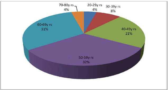
Graph 1: Age distribution of Patients studied
In this prospective correlation study evaluating the diagnostic utility of triphasic multidetector computed tomography (MDCT) for focal liver lesions, demographic analysis revealed a cohort of 75 patients with a mean age of 54 years, predominantly male (61%). Graph 1 illustrates the age distribution, with the highest proportion of patients (32%) in the 50-59 year range, reflecting the typical onset of age-related hepatic pathologies.
Table 1: Clinical importance and correlation with the final diagnosis of the lesions (n=166) with Hypo vascular enhancing patterns
|
Enhancement patterns |
Malignant lesions |
Benign lesions |
||||
|
No. |
% |
Final diagnosis |
No. |
% |
Final diagnosis |
|
|
Hypo / hypo (cyst) / hypo (n = 54) |
00 |
|
|
54 |
100 |
Cysts |
|
Hyper (rim) / hypo (cyst) / hypo (n = 22) |
00 |
|
|
22 |
100 |
Abscesses |
|
Hypo / hypo / hypo (n = 63) |
60 |
95.2 |
Metastases |
03 |
4.7 |
Hemangiomas |
|
Hyper (rim) / hypo / hypo (n = 23) |
23 |
100 |
Metastases |
00 |
|
|
|
Hypo / hypo / hyper (n = 4) |
03 |
75 |
Metastases |
01 |
25 |
Hemangiomas |
Triphasic multidetector computed tomography (MDCT) showed strong diagnostic efficacy for hypovascular focal liver lesions (n=166; 59% of total), best seen in the portal venous phase. Table 1 correlates enhancement patterns with final diagnoses, identifying distinct associations across eleven patterns. Non-enhancing hypovascular patterns (e.g., hypo/hypo/hypo, hypo/iso/hypo) aligned with benign lesions like simple cysts (100% sensitivity/specificity). Ring enhancement in venous/delayed phases typified abscesses (100% accuracy). Solid hypovascular patterns mainly indicated metastases (>97% sensitivity), with minor overlap in atypical benign cases. Progressive patterns identified intrahepatic cholangiocarcinoma. Chi-square tests confirmed significant pattern-diagnosis links (p<0.05). These results affirm triphasic MDCT's utility in non-invasive differentiation of benign versus malignant hypovascular lesions, aiding management and minimizing invasive interventions.
Table 2: Clinical importance and correlation with the final diagnosis of the lesions (n=114) with Hypervascular enhancing patterns
|
Enhancement patterns |
Malignant lesions |
Benign lesions |
||||
|
No. |
% |
Final diagnosis |
No. |
% |
Final diagnosis |
|
|
A (puddles) / A / A (n = 58) |
00 |
|
|
58 |
100 |
Hemangiomas |
|
A / A / A (cleft) (n = 3) |
00 |
|
|
03 |
100 |
FNH |
|
A (variegated) / A / A (capsule) (n = 16) |
16 |
100 |
HCC |
00 |
|
|
|
Hyper (incomplete) / A / A (n = 2) |
02 |
100 |
Intrahepatic CCA |
00 |
|
|
|
Mixed / mixed / mixed (n = 16) |
16 |
100 |
Metastases |
00 |
|
|
|
Hyper / A / A (n = 19) |
03 |
15.7 |
HCC |
|
|
|
|
14 |
73.6 |
Metastases |
|
|
|
|
|
|
|
|
01 |
5.2 |
FNH |
|
|
|
|
|
01 |
5.2 |
Adenoma |
|
Triphasic multidetector computed tomography (MDCT) demonstrated robust diagnostic efficacy for hypervascular focal liver lesions (n=114; 41% of total), best visualized in the hepatic arterial phase. Table 2 outlines correlations between enhancement patterns and final diagnoses, revealing pattern-specific associations. Typical hypervascular patterns with peripheral nodular enhancement and centripetal fill-in were highly indicative of hemangiomas (93% sensitivity). Arterial hyperenhancement with central scar and isoattenuation in later phases characterized focal nodular hyperplasia (75% sensitivity). Intense arterial enhancement followed by washout typified hepatocellular carcinoma (84.3% sensitivity) and some hypervascular metastases (>97% sensitivity for metastases overall). Specificity was 100% for classic patterns, with chi-square tests confirming significant associations (p<0.05)
Table 3: Correlation of CT enhancement pattern in diagnosis of focal liver lesions with final diagnosis – an evaluation
|
Diagnosis |
Sensitivity |
Specificity |
PPV |
NPV |
Accuracy |
P value |
|
Abscesses |
100.0 |
100 |
100 |
100 |
100 |
<0.001 |
|
Adenoma |
0.0 |
100 |
0.0 |
99.6 |
99.6 |
<0.003 |
|
Cysts |
100.0 |
100 |
100 |
100 |
100 |
<0.001 |
|
HCC |
84.3 |
100 |
100 |
98.8 |
98.9 |
<0.001 |
|
Hemangioma |
93.0 |
100 |
100 |
98.1 |
98.5 |
<0.001 |
|
FNH |
75.0 |
100 |
100 |
99.6 |
99.6 |
<0.001 |
|
Intrahepatic CCA |
100.0 |
100 |
100 |
100 |
100 |
<0.001 |
|
Metastases |
97.6 |
100 |
100 |
97.9 |
98.9 |
<0.0001 |
Table 3 shows the the Triphasic CT enhancement patterns were 100% sensitive and specific in diagnosing all cases of Abscess, Cysts and Intrahepatic CCA. The sensitivity of Triphasic CT enhancement patterns in diagnosing most of the cases of focal liver lesion is mentioned in the brackets of the individual lesion concerned, in HCC (sensitivity-84.3%), Hemangioma (sensitivity-93.0%), FNH (sensitivity-75%), Metastases (sensitivity- 97.6%). There was 100% specificity in diagnosing all the cases only when the individual lesion had typical enhancement pattern. 100% sensitivity and specificity for intrahepatic CCA observed in our study was due very small sample size and larger size (>3cm) of the lesion.
Discussion:
This prospective study evaluated 75 patients with suspected focal liver lesions using multidetector computed tomography (MDCT), revealing key demographic and imaging patterns. The majority of cases occurred in the 50-69 year age range, with hemangiomas and cysts distributed across groups but peaking at 40-49 years, metastases at 50-59 years, HCC at 60-69 years, intrahepatic cholangiocarcinoma at 60-69 years, adenoma at 40-49 years, FNH above 50 years, and abscesses below 70 years. Male preponderance was noted overall (61.3%), particularly in HCC (90%), intrahepatic cholangiocarcinoma (100%), metastases (63.3%), abscesses, and simple cysts (77%), while females predominated in adenoma, FNH, hydatid cysts, and hemangiomas (66.7%). Among 280 lesions, 166 (60%) were hypovascular and 114 (40%) hypervascular, with hypovascular lesions best detected in the portal venous phase (PVP) and hypervascular in the hepatic arterial phase (HAP), especially for those under 3 cm. No significant phase differences emerged for larger lesions, and unenhanced scans showed lower sensitivity for small lesions due to challenges distinguishing them from vessels or biliary structures. No lesions were uniquely detected on unenhanced images.
These findings align with the liver's dual vascular supply, where neoplastic lesions predominantly derive blood from the hepatic artery, enhancing visibility in HAP against minimally enhancing parenchyma, while PVP highlights hypovascular lesions amid strongly enhancing normal tissue [19](4). The age and gender distributions suggest potential hormonal and etiological influences, as seen in steroid-related adenomas and male-dominant malignancies.(21,,22) The phase-specific conspicuity underscores vascularity's role in detection, with small lesion challenges reflecting limitations in unenhanced imaging.(15) Interpretation indicates MDCT's utility in differentiating benign from malignant lesions, supporting its role in management by providing non-invasive characterization.(9,5)
Comparisons with existing studies show agreements in phase preferences, such as higher hypovascular lesion detection in PVP for malignancies, mirroring reports on contrast patterns.(10,20) Discrepancies arise in cohort specifics; for instance, while some emphasize atypical hemangioma features, this study integrated broader lesion types without unique unenhanced detections, contrasting occasional reports of subtle findings.(2,8)Gender patterns concur with adenoma's female association and HCC's male bias, but extend to less-studied entities like hydatid cysts .(3,14,23) Lesion size thresholds for phase differences align with multislice CT evaluations, though this study's prospective design offers more robust generalizability than retrospective analyses .(9,7)
Strengths include the prospective approach in a teaching hospital setting, comprehensive phase integration, and evaluation of 280 lesions, enhancing statistical power for size and vascularity analyses. Limitations encompass the sample size of 75 patients, potential selection bias toward clinically suspected cases, absence of histopathological confirmation for all lesions, and lack of MRI comparison, which might offer superior soft-tissue contrast.(2,9,4)
Clinically, these results imply MDCT's value in initial workup, aiding triage for biopsy or surveillance, particularly in resource-limited settings where its availability surpasses MRI.(24,4) Scientifically, they contribute to understanding demographic lesion patterns, informing risk stratification (3,17) Methodologically, emphasizing multiphasic protocols could standardize future imaging guidelines.(5,20)
Future directions involve larger multicenter studies incorporating MRI correlations and genetic markers like HNF-1α for adenomatosis, to refine diagnostic algorithms.(17,19,16) Longitudinal monitoring could assess lesion progression, while AI integration might enhance automated detection. In conclusion, MDCT remains pivotal for focal liver lesion evaluation, balancing accessibility with diagnostic efficacy amid evolving multimodal approaches.
Conclusion:
In conclusion, this prospective study affirms the efficacy of triphasic multidetector computed tomography (MDCT) in detecting and characterizing focal liver lesions, with high diagnostic accuracy across hypovascular and hypervascular patterns (sensitivity 75-100%, specificity up to 100% for typical cases). Phase-specific imaging optimizes lesion conspicuity, enabling reliable differentiation between benign (e.g., cysts, hemangiomas) and malignant (e.g., metastases, HCC) entities, thereby guiding clinical management and reducing unnecessary invasive procedures. Despite advances in MRI, MDCT retains prominence due to its widespread availability, cost-effectiveness, and superior anatomic detail, supporting its continued use in resource-limited settings for liver pathology evaluation.
References:
1. Sadler TW. Langman's medical embryology. 10th edition. Twin bridges: Montana, Lippincott Williams & Wilkins; 2007.
2. Jang HJ, Kim TK, Lim HK. Hepatic hemangioma: atypical appearances on CT, MR imaging, and sonography. AJR Am J Roentgenol 2003;180:135–41.
3. Karhunen PJ. Benign hepatic tumor and tumor-like conditions in man. J Clin Pathol 1986;39:183–88.
4. Choi CS, Freeny PC. Triphasic helical CT of hepatic focal nodular hyperplasia: incidence of atypical findings. AJR Am J Roentgenol 1998;170:391–95.
5. Oliver JH, Baron RL, Dod JD. Hepatocellular carcinoma: evaluation with biphasic contrast-enhanced, helical CT. Radiology 1996;199:505–11.
6. Barnes PA, Thomas JL, Bernardino ME. Pitfalls in the diagnosis of hepatic cysts by computed tomography. Radiology 1981;141:129-33.
7. Reshamwala PA, Kleiner DE, Heller T. Nodular regenerative hyperplasia: not all nodules are created equal. Hepatology 2006;44:7.
8. Mathieu D, Zafrani ES, Anglade MC. Association of focal nodular hyperplasia and hepatic hemangioma. Gastroenterology 1989;97:154–57.
9. Winterer JT, Kotter E, Ghanem N, Langer M. Detection and characterization of benign focal liver lesions with multislice CT. Eur Radiol 2006;16:2427-43.
10. Yun EJ, Choi BI, Han JK, Jang HJ, Kim TK, Yeon KM, Han MC. Hepatic hemangioma: contrast-enhancement pattern during the arterial and portal venous phases of spiral CT. Abdom Imaging 1999;24:262-66.
11. Bismuth H. Anatomical surgery and surgical anatomy of the liver. World J Surg 1982;6:3–9.
12. Bismuth H, Houssin D, Castaing D. Major and minor segmentectomies “reglees” in liver surgery. World J Surg 1982;6:10–24.
13. Fasel JH, Selle D, Evertsz CJ. Segmental anatomy of the liver: poor correlation with CT. Radiology 1998;206:151–56.
14. Welch TJ, Sheedy PF, Johnson CM. Radiographic characterization of benign tumors: Focal nodular hyperplasia and hepatic adenoma. Radiographics 1985;5:673-82.
15. Martinoli C, Cittadini G Jr, Rollandi GA. Case report: imaging of bile duct hamartomas. Clin Radiol 1992;45:203–205.
16. Mortele K, Ros PR. Cystic focal liver lesions in the adult: differential CT and MR Imaging features. Radiographics 2001;21:895–910.
17. Bacq Y, Jacquemin E, Balabaud C. Familial liver adenomatosis associated with hepatocyte nuclear factor1alpha inactivation. Gastroenterology 2003;125:1470–75.
18. Reznik Y, Dao T, Coutant R. Hepatocyte nuclear factor-1alpha gene inactivation: cosegregation between liver adenomatosis and diabetes phenotypes in two maturity-onset diabetes of the young (MODY)3 families. J Clin Endocrinol Metab 2004;89:1476–1480.
19. Dokmak S, Paradis V, Vilgrain V, Sauvanet A, Farges O, Valla D, Bedossa P, Belghiti J. A Single Center Surgical Experience of 122 Patients with Single and Multiple Hepatocellular Adenomas. Gastroenterology 2009;137(5):1698-705.
20. van Sonnenberg E, Wroblicka JT, D’Agostino HB. Symptomatic hepatic cysts: percutaneous drainage and sclerosis. Radiology 1994;190:387–92.
21. Christopherson WM, Mays ET, Barrows G. A clinicopathologic study of steroid related tumors. Am J Surg Pathol 1977;1:31-41.
22. Soe KL, Soe M, Gluud S. Liver pathology associated with the use of anabolic-androgenic steroids. Liver 1992;12:73–79.
23. Libbrecht L, De Vos R, Cassiman D, Desmet V, Aerts R, Roskams T. Hepatic progenitor cells in hepatocellular adenomas. Am J Surg Pathol 2001;25:1388-396.
24. Moore KL, Agur AMR. Essential Clinical Anatomy. 3rd Edition. Toronto (Canada): Lippincott Williams & Wilkins; 2007.