Comparative Ultrasound Evaluation of Intima-Media Thickness of Common Carotid Arteries and Abdominal Aorta in Normal Term Newborns and Small for Gestational Age Term Newborns
- Arjun Kamath H , Assistant Professor, S R Patil Medical College & Research Centre, Badagandi, India
- M L Arkesh , Assistant Professor, S R Patil Medical College & Research Centre, Badagandi, India
- Siddaling B Mindolli , Consultant Radiologist at Aditya Diagnostic Centre, Gokak, India
Article Information:
Abstract:
Background: Small-for-gestational-age (SGA) newborns exhibit prenatal circulatory adaptations to suboptimal intrauterine environments, potentially programming lifelong cardiovascular risks. Non-invasive ultrasound measurement of intima-media thickness (IMT) in the common carotid artery (cIMT) and abdominal aorta (aIMT) serves as a marker for cardiovascular morbidities. Objectives: To compare cIMT and aIMT in SGA term newborns versus appropriate-for-gestational-age (AGA) term newborns using non-invasive high-resolution ultrasonography. Methods: This observational comparative study was conducted from November 2018 to June 2020 at Shri B. M. Patil Medical College Hospital & Research Centre, Vijayapura. A total of 200 term newborns (100 SGA, 100 AGA) were included based on Lubchenco growth charts. Exclusion criteria included preterm/post-term` births and maternal conditions like diabetes or infections. IMT measurements were performed using Philips HD11XE ultrasound with linear (5-12 MHz) and convex (2-6 MHz) transducers. Data were analyzed using unpaired t-tests and chi-square tests, with p<0.05 considered significant. Results: Mean aIMT was significantly higher in SGA (0.48±0.36 mm) than AGA (0.47±0.60 mm) newborns (p=0.002). Mean cIMT was also higher in SGA (0.39±0.38 mm) versus AGA (0.37±0.40 mm) (p<0.001). Birth weight, length, and head circumference were lower in SGA newborns (p<0.001 each). Conclusion: Elevated IMT in SGA newborns indicates early vascular changes predisposing to atherosclerosis. Screening SGA newborns with ultrasound may facilitate early risk factor modification.
Keywords:
Article :
Introduction:
Atherosclerosis remains a leading cause of mortality and morbidity worldwide, primarily contributing to cardiovascular and cerebrovascular disorders1. The measurement of carotid intima-media thickness (cIMT) serves as a validated marker for subclinical atherosclerosis in adults, with increments correlating to increased risks of myocardial infarction and stroke2,3. Arterial intima-media thickness (IMT) reflects structural and morphological alterations in the arterial wall, where initial atherosclerotic signs involve lipid deposits forming fatty streaks1. These lesions progress during childhood and adolescence, accelerated by cardiovascular risk factors4.
The clinical manifestations of atherosclerosis typically emerge in adulthood, but atherogenesis begins early in life5. Autopsy studies indicate that atherosclerotic lesions originate in the abdominal aorta4,6, suggesting that abdominal aorta IMT (aIMT) may provide a superior indicator of preclinical atherosclerosis in high-risk children compared to cIMT 7. Small-for-gestational-age (SGA) newborns display prenatal circulatory adaptations, such as vessel wall thickening and impaired vasodilatory function, in response to suboptimal intrauterine environments8,9 . These adaptations may alter cardiovascular development, programming the fetus for lifelong morbidities including glucose intolerance and dyslipidemia10,11. Fetal programming hypotheses, such as Barker's "fetal origins of adult disease," posit that intrauterine undernutrition induces permanent changes increasing susceptibility to cardiovascular disease 12.
Non-invasive high-resolution ultrasound enables early detection of vascular modifications 13. Given the correlation between ultrasound-measured IMT and histological vessel thickness in atherosclerosis-prone arteries14,15, evaluating IMT in newborns could identify at-risk individuals for preventive interventions.
While studies have reported increased IMT in SGA neonates16,17, limited research compares aIMT and cIMT in term SGA versus appropriate-for-gestational-age (AGA) newborns18. Existing data from autopsy and pediatric cohorts suggest intrauterine factors influence early atherosclerosis19,20, but prospective ultrasound-based evaluations in newborns are scarce.
So, this study is done to compare cIMT and aIMT in SGA term newborns with AGA term newborns using high-resolution ultrasonography. We hypothesized that SGA newborns would exhibit significantly higher IMT values, reflecting early atherosclerotic changes.
Materials and Methods:
This is comparative cross-sectional study which was conducted at the Department of Radio-Diagnosis, Shri B. M. Patil Medical College Hospital & Research Centre, Vijayapura, Karnataka, India, from November 2018 to June 2020. On the basis of a study the anticipated mean±sd of AGA vs SGA was 0.54±0.06 and 0.5±0.04 respectively, with 95% level of significance and 90% power2 & using this
formula 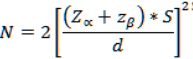 sample size is 100 per group.
Participants included 100 term newborns born between 37 weeks to 42 weeks of gestation, appropriate for gestational age (AGA) normal term newborn with birth weight of 50-90th centile and 100 Small for gestational age (SGA) term newborns with birth weight less than 10th centile as per Lubchenco‟s growth charts. Newborns with maternal diabetes, hypercholesterolemia, shock, major congenital anomalies, or infections were excluded. Informed written consent was taken from all the mothers of the newborns and the non-invasiveness of the scan was clearly explained to the mothers in their own language.
Newborns underwent ultrasound within 120 hours of birth. Maternal data (age, parity, haemoglobin in third trimester) and newborn anthropometrics (weight, length, head circumference) were recorded. Gestational age was assessed via last menstrual period, New Ballard score, and first-trimester ultrasound.
Ultrasound was performed using Philips HD11XE with 2-6 MHz convex and 5-12 MHz linear transducers. aIMT was measured on the dorsal wall of the distal 10 mm abdominal aorta. cIMT was measured bilaterally at 0.5 cm intervals, 1 cm proximal to the carotid bulb. Measurements were taken by a single observer, with images reviewed by a senior radiologist. Reproducibility was assessed via intraclass correlation coefficient.
Statistical Analysis
Data entered in Microsoft Excel and analysed using the CDC’s Epi Infor Software version 7.1. Data were presented as mean ± SD or percentages. Quantitative differences were analyzed using unpaired t-tests; qualitative differences via chi-square or Fisher's exact test. Significance was set at p<0.05. Sample size (100 per group) was calculated based on anticipated mean aIMT differences (0.54±0.06 mm AGA vs. 0.5±0.04 mm SGA), yielding 95% confidence and 90% power.
Ethical Considerations
The study received approval from the Institutional Ethical Committee (dated 13-11-2018). Informed written consent was obtained from all the participants in their language. Procedures adhered to ethical standards, ensuring non-invasiveness and confidentiality.
Results:
In this study among 200 term newborns, Table no 1 show, majority were between 19-24yrs (57.5%) of age, 32.0% were in the 25–29 yrs and 10% were in the age group of 30-34yrs. This predominance of younger maternal age shows pattern of reproductive age in Vijayapura, where early childbearing remains relatively common and may influence fetal growth and development.
Regarding parity, second-order pregnancies formed the largest group (55.5%), indicating that most mothers had previous childbirth experience. First-time mothers accounted for 23.0%. This distribution suggests a largely multiparous population.
In Table no 1, most newborns were delivered at early term, with 42.0% born at 37 weeks and 36.0% at 38 weeks, while deliveries at 39 weeks and later were progressively less frequent. Only a small number of infants were born at 40 or 41 weeks. This emphasis on term births and indicates a predominance of early-term deliveries, which may reflect local obstetric practices
Figure no 1 show, neonatal birth weights were predominantly within the 2.1–3.0 kg range, with the largest proportion in the 2.1–2.5 kg category. While 10% of them had lower birth weights (1.5–2.0 kg), suggestive of possible intrauterine growth restriction.
Table 1: Distribution of Demographic details of mothers
|
Determinants |
Group |
Frequency |
Percentage |
|
Mother's Age (years) |
19-24 |
115 |
57.5 |
|
25-29 |
64 |
32 |
|
|
30-34 |
21 |
10.5 |
|
|
Parity |
1 |
46 |
23 |
|
2 |
111 |
55.5 |
|
|
3 |
38 |
19 |
|
|
4 |
5 |
2.5 |
|
|
Gestational Age (weeks) |
37 |
84 |
42 |
|
38 |
72 |
36 |
|
|
39 |
32 |
16 |
|
|
40 |
9 |
4.5 |
|
|
41 |
3 |
1.5 |
Table 2: DISTRIBUTION OF aIMT/cIMT IN NEONATES
|
aIMT/cIMT |
SGA (n=100) |
AGA (n=100) |
Total |
P Value |
|
Mean aIMT |
||||
|
<0.41 |
0 |
4 |
4 |
<0.001
|
|
0.42-0.48 |
45 |
69 |
114 |
|
|
>0.48 |
55 |
27 |
82 |
|
|
Mean cIMT |
||||
|
<0.35 |
9 |
35 |
44 |
<0.001
|
|
0.35-0.45 |
87 |
62 |
149 |
|
|
>0.45 |
4 |
3 |
7 |
|
Table no 2 show, 45 SGA infants show and 69 AGA infants mean aIMT is 0.42-0.48 thickness & 55 SGA & 27 AGA infants show >0.48 mean aIMT. Meanwhile mean cIMT is more among SGA infants(0.35-0.45) than among AGA infants, which suggest statistical significance. The significantly(p<0.001) greater aortic wall thickness observed in SGA infants indicates that impaired foetal growth is strongly associated with increased aortic intima–media thickness at birth.
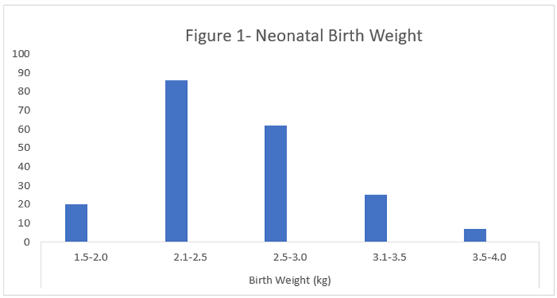
Figure 1 Distribution of Neonatal birth weight
Table 3: Comparison of study variables in SGA and AGA newborns
|
Variables |
SGA group (Mean ± SD) |
AGA group (Mean ± SD) |
P value |
|
Weight of baby at birth (kg) |
2.14 ± 0.42 |
2.87 ± 0.35 |
< 0.001** |
|
Mean aIMT (mm) |
0.48 ± 0.36 |
0.47 ± 0.60 |
< 0.002 |
|
Mean cIMT (mm) |
0.39 ± 0.38 |
0.37 ± 0.40 |
< 0.001** |
|
Length (cm) |
44.71 ± 1.5 |
45.13 ± 1.7 |
< 0.001** |
|
Head circumference (cm) |
31.9 ± 2.5 |
33.4 ± 3.3 |
< 0.001** |
As shown in Table 3, birth weight, abdominal aortic intima media thickness (aIMT), carotid intima media thickness (cIMT), length, and head circumference differed significantly between SGA and AGA newborns (all P < 0.001).
Discussion:
In both developed and developing countries, atherosclerosis is a major cause of premature death and fatality. Barker et al.,21gave a hypothesis, foetal origin of adult disease it states that cardiovascular disorders originate in the early phase in life. Apart from, postnatal events, decreased intrauterine growth affects arterial improvement. The idea of prenatal factors affecting the vascular wall and inflicting atherosclerosis in early in life has already been established 21,22. Studies shows that SGA is aften associated with increased risk in cardiovascular disorders in adult life21,23-25. Newborns with poor size at birth is said to have a correlation with elevated BP, dyslipidemia and alterations in the blood glucose level in adulthood 26-28.
Koklu et al., 29 and Skilton et al.30 proposed some mechanistic pathways that showed that rise in the maternal and postnatal leptin levels, dyslipidemia and elevated sympathetic tone trigger the thickening of the vessel wall in SGA term newborns. This study highlights the need of management of risk factors in order to prevent atherosclerosis in long term and its various complications that begin in childhood. The development and improvement of non-invasive examinations might help in identifying the high risk patients and their treatment. Not many researches have been done to study the aIMT and cIMT in newborns.31
The development of the primary atherosclerotic lesions is believed to be developed in the abdominal aorta first as observed in autopsy studies 23,32. Improved technologies, resolution & increased accuracy and resolution in the imaging technology have helped in noninvasively detecting the early vascular lesions using a USG 32,33 83,84. Measurement of the thickness of intima-media in carotid arteries is a marker in subclinical atherosclerosis in adults, as proven by researchers. USG is a non-invasive, easily accessible and affordable way of measuring the abdominal aortic IMT and carotid IMT 32, 34-36 83,85-87.
Few studies in children with hypercholesterolemia have confirmed an elevated carotid intima- media complex in contrast with the control group32,36 . Carotid arteries are superficially located on the neck and can be accessed with the help of ultrasound and thus have been targeted in most of these researches32 . It has been proven that we are able to reveal altered vascular dynamics in Small for gestation newborns like the elevated the thickness intima-media of the abdominal aorta and carotid artery.
In the study done by Skilton et al. 26 , observed the thickness intima-media of the abdominal aorta was considerably high in the neonates having restricted intrauterine development (0.81 ± 0.11 mm) in comparison with controls (0.74 ± 0.08 mm, P = 0.02). Mean IMT of the abdominal aorta was observed to be similar among these groups. In their research weight adjusted thickness intima-media of the abdominal aorta was correlated; noticeable alterations were seen in growth restricted neonates in comparison to the controls. In our study, IMT of the abdominal aorta was measured a one centimetre longitudinal section of the distal part of the abdominal aorta.
The results of our study show that the mean IMT of the abdominal aorta and carotid artery have been considerably higher in the small-for-gestational-age compared to appropriate for gestation age term newborns. Results of the study show the need for more longitudinal investigations and further research in order to discover if some adverse effect intrauterine factors may lead to potential complications or alter the body metabolism. Only a few drawbacks have been observed in this research. Firstly, cases ought to be followed for long run to understand the development of atherosclerosis. Secondly, additional work might be required to validate our findings in bigger cohort.
Conclusion:
The use of non-invasive, cheap and effortlessly available high resolution ultrasound measurement of the thickness of aortic and carotid artery intima- media is an important modality by which we are able to additionally discover and define the intrauterine elements that are related to increased atherosclerosis and to evaluate the physical effect on the arterial wall. As there has been an increasing emphasis on the early detection of these at an increased threat of developing disease in order to present them with early preventive and treatment choices thereby arresting or delaying the onset of morbidity and mortality. Therefore, screening of all SGA newborns is critical, in order to suggest them concerning the need for life-style modification and correction of various modifiable risk factors which predispose for atherosclerosis and in addition follow up of such patients in adulthood with early commencement of therapy will assist to forestall complication.
References:
1. Libby P. The pathogenesis, prevention, and treatment of atherosclerosis. In: Kasper DL, Fauci AS, Hauser SL, Longo DL, Jameson JL, Loscalzo J, eds. Harrison's Principles of Internal Medicine. 19th ed. New York: McGraw-Hill; 2015:291e-1.
2. Sodhi K, Hondappanavar A, Saxena A, Dutta S, Khandelwal N. Intima-media complex thickness: preliminary workup of comparative evaluation of abdominal aorta and carotid artery of small-for-gestational-age term newborns and normal size term newborns. Acta Cardiol. 2015;70(3):351-357.
3. Lorenz MW, Markus HS, Bots ML, et al. Prediction of clinical cardiovascular events with carotid intima-media thickness: a systematic review and meta-analysis. Circulation. 2007;115(4):459-467.
4. Amato M, Montorsi P, Ravani A, et al. Carotid intima-media thickness by B-mode ultrasound as surrogate of coronary atherosclerosis: correlation with quantitative coronary angiography and coronary intravascular ultrasound findings. Eur Heart J. 2007;28(17):2094-2101.
5. Revkin JH, Shear CL, Pouleur HG, Ryder SW, Orloff DG. Biomarkers in the prevention and treatment of atherosclerosis: need, validation, and future. Pharmacol Rev. 2007;59(1):40-53.
6. Wang JG, Staessen JA, Li Y, et al. Carotid intima-media thickness and antihypertensive treatment: a meta-analysis of randomized controlled trials. Stroke. 2006;37(7):1933-1940.
7. Nathan DM, Lachin J, Cleary P, et al.; Diabetes Control and Complications Trial; Epidemiology of Diabetes Interventions and Complications Research Group. Intensive diabetes therapy and carotid intima-media thickness in type 1 diabetes mellitus. N Engl J Med. 2003;348(23):2294-2303.
8. Yokoyama H, Katakami N, Yamasaki Y. Recent advances of intervention to inhibit progression of carotid intima-media thickness in patients with type 2 diabetes mellitus. Stroke. 2006;37(9):2420-2427.
9. McGill HC Jr, McMahan CA, Herderick EE, et al. Effects of coronary heart disease risk factors on atherosclerosis of selected regions of the aorta and right coronary artery. PDAY Research Group. Arterioscler Thromb Vasc Biol. 2000;20(3):836-845.
10. Espeland MA, O'Leary DH, Terry JG, et al. Carotid intimal-media thickness as a surrogate for cardiovascular disease events in trials of HMG-CoA reductase inhibitors. Curr Control Trials Cardiovasc Med. 2005;6:3.
11. Hurst RT, Ng DW, Kendall C, Khandheria B. Clinical use of carotid intima-media thickness: review of the literature. J Am Soc Echocardiogr. 2007;20(8):907-914.
12. Stary HC. Evolution and progression of atherosclerotic lesions in coronary arteries of children and young adults. Arteriosclerosis. 1989;9(1 suppl):I19-32.
13. Cohen E, Wong FY, Horne RS, Yiallourou SR. Intrauterine growth restriction: impact on cardiovascular development and function throughout infancy. Pediatr Res. 2016;79(6):821-830.
14. Law CM, de Swiet M, Osmond C, et al. Initiation of hypertension in utero and its amplification throughout life. BMJ. 1993;306(6871):24-27.
15. Forsen T, Eriksson JG, Tuomilehto J, Osmond C, Barker DJ. Growth in utero and during childhood among women who develop coronary heart disease: longitudinal study. BMJ. 1999;319(7222):1403-1407.
16. Wong M, Edelstein J, Wollman J, Bond MG. Ultrasonic-pathological comparison of the human arterial wall. Verification of intima-media thickness. Arterioscler Thromb. 1993;13(4):482-486.
17. Gamble G, Beaumont B, Smith H, et al. B-mode ultrasound images of the carotid artery wall: correlation of ultrasound with histological measurements. Atherosclerosis. 1993;102(2):163-173.
18. Raitakari OT. Imaging of subclinical atherosclerosis in children and young adults. Ann Med. 1999;31 Suppl 2:33-40.
19. Kirkby NS, Hadoke PW, Bagnall AJ, Webb DJ. The endothelin system as a therapeutic target in cardiovascular disease: great expectations or bleak house? Br J Pharmacol. 2008;153(6):1105-1119.
20. Libby P, Ridker PM, Maseri A. Inflammation and atherosclerosis. Circulation. 2002;105(9):1135-1143.
21. Barker DJ, Winter PD, Osmond C, Margetts B, Simmonds SJ. Weight in infancy and death from ischaemic heart disease.
22. Gordon T, Kannel WB. Premature mortality from coronary heart disease. The Framingham study. JAMA 1971; 215: 1617-25.
23. Lubchenco LC, Hansman C, Dresseler M, Boyd E. Intrauterine growth as estimated from liveborn birth-weight data 24 to 42 weeks of gestation.
24. 24 . Rich-Edwards JW, Stampfer MJ, Manson JE, Rosner B, Hankinson SE,Colditz GA, Willett WC, Hennekens CH. Birth weight and risk of cardiovascular disease in a cohort of women followed up since 1976.
25. Leon D, Lithell HO, Vagero D, Koupilová I, Mohsen R, Berglund L, Lithell UB, McKeigue PM. Reduced fetal growth rate and increased risk of death from ischaemic heart disease: cohort study of 15000 Swedish men and women born 1915-29. BMJ 1998; 317: 241-5.
26. Skilton MR, Evans N, Griffiths KA, et al. Aortic wall thickness in newborns with intrauterine growth restriction. Lancet. 2005; 365: 1484-86.
27. Pauciullo P, Iannuzzi A, Sartorio R, et al. Increased intima-media thickness of the common carotid artery in hypercholesterolemic children. Arterioscler Thromb Vasc Biol. 1994; 14: 1075–79.
28. Koklu E, Kurtoglu S, Akcakus M, Koklu S, Buyukkayhan D, Gumus H, Yikilmaz A. Increased aortic intima-media thickness is related to lipid profile in newborns with intrauterine growth restriction. Horm Res 2006; 65: 269-75.
29. Bernard Rosner (2000), Fundamentals of Biostatistics, 5th Edition, Duxbury, page 80-240
30. Skilton MR. Intrauterine risk factors for precocious atherosclerosis. Pediatrics 2008; 121: 570-4.
31. Hondappanavar A, Sodhi KS, Dutta S, Saxena AK, Khandelwal N. Quantitative ultrasound measurement of intima-media thickness of abdominal aorta and common carotid arteries in normal term newborns. Pediatr Cardiol 2013; 34: 364-9.
32. Järvisalo MJ, Jartti L, Näntö-Salonen K, Irjala K, Rönnemaa T, Hartiala JJ, Celermajer DS, Raitakari OT. Increased aortic intima- media thickness: a marker of preclinical atherosclerosis in high-risk children. Circulation 2001;104: 2943-7.
33. Sodhi KS, Sidhu R, Gulati M, Saxena A, Suri S, Chawla Y. Role of tissue harmonic imaging in focal hepatic lesions: comparison with conventional sonography. J Gastroenterol Hepatol 2005; 20: 1488-93.
34. Dawson JD, Sonka M, Blecha MB, Lin W, Davis P H. Ris k factors associated with aortic and carotid intima- media thickness in adolescents and young adults : the Muscatine Offspring Study. J Am Coll Cardiol 2009: 53: 2273- 9.
35. Tonstad S, Joakimsen O, Stensland-Bugge E, Leren TP, Ose L, Russell D, Bønaa KH. Risk factors related to carotid intima- media thickness and plaque in children with familial hypercholesterolemia and control subjects. Arterioscler Thromb 1996; 16: 984-91.
36. Cosmi E, Visentin S, Fanelli T, Mautone AJ, Zanardo V. Aortic intimamedia thickness in fetuses and children with intrauterine growth restriction.ObstetGynecol 2009;114: 1109-14.