Clinical and Radiological Correlation of Neurovascular Conflict in Trigeminal Neuralgia: A Prospective MRI-Based Study
- D. Harinath , Professor, Department of Radiology, Government Medical College, Nandyal, Andhra Pradesh, India
- Ramavath Ravi Naik , Assistant Professor, Department of Radiology, Kurnool Medical College, Kurnool, Andhra Pradesh, India
- Bandi Arun Kumar , Assistant Professor, Department of Radiology, Kurnool Medical College, Kurnool, Andhra Pradesh, India
- Gangula Shravani , Senior Resident, Department of Radiology, SVS Medical College, Mahabubnagar, Telangana, India
Article Information:
Abstract:
Background: Trigeminal neuralgia is a debilitating neuropathic pain disorder characterized by sudden, severe, unilateral facial pain, most commonly attributed to NVC (Neurovascular Conflict) at the trigeminal nerve root entry zone. MRI (Magnetic Resonance Imaging) plays a pivotal role in identifying vascular compression and correlating radiological findings with clinical manifestations. Establishing a clear clinicoradiological correlation is essential for accurate diagnosis and appropriate management planning. Methods: This prospective study was conducted over 22 months (March 2023–December 2024) in the Department of Radiodiagnosis, Government General Hospital, Kurnool. Thirty patients with classical clinical features of trigeminal neuralgia were included. MRI of the brain was performed using a 1.5 Tesla superconducting scanner with a dedicated trigeminal nerve protocol, including T1- and T2-weighted sequences, high-resolution balanced fast field echo (B-FFE/FIESTA) sequences, and MR angiography. Clinical parameters such as age, sex, side of involvement, symptomatology, and trigeminal nerve branch involvement were correlated with MRI findings, including site, grade, and vascular source of neurovascular compression. Statistical analysis was performed using SPSS version 20. Results: The majority of patients were aged 21–40 years (56.67%) with a marked male predominance (86.67%). Right-sided facial involvement was more common (73.33%). All patients presented with sudden, severe unilateral facial pain, while tingling or numbness (43.33%), headache (33.33%), and allodynia (23.33%) were additional symptoms. The mandibular division (V3) was the most frequently involved branch (43.33%). MRI revealed neurovascular compression most commonly at lateral–caudal and medial locations (26.67% each). The superior cerebellar artery was the most frequently implicated vessel (46.67%). A statistically significant association was observed between the site of compression and the affected trigeminal nerve branch (p < 0.001), whereas no significant association was found between the involved vessel and nerve branch (p = 0.36). Conclusion: MRI serves as a vital diagnostic tool in trigeminal neuralgia, enabling precise identification of neurovascular conflict and meaningful clinicoradiological correlation. The anatomical site of compression plays a more critical role in determining clinical manifestations than the specific vessel involved, underscoring the importance of high-resolution MRI in diagnosis and treatment planning.
Keywords:
Article :
Introduction:
Trigeminal neuralgia is a chronic neuropathic pain disorder characterized by recurrent, brief episodes of unilateral, electric shock–like pain that begin and end abruptly. These painful paroxysms occur within the sensory distribution of one or more branches of the fifth cranial (trigeminal) nerve and are typically triggered by innocuous stimuli such as speaking, chewing, or light touch.[1]
Clinically, trigeminal neuralgia is defined by sudden attacks of severe, sharp, stabbing pain lasting from a fraction of a second up to two minutes, with pain-free intervals between episodes. The condition predominantly affects the sensory territory of the trigeminal nerve (cranial nerve V). The reported prevalence of classical trigeminal neuralgia ranges from 1 to 2 per 10,000 individuals. Although the exact etiology remains uncertain, several hypotheses have been proposed, with NVC (Neurovascular Conflict) at the cisternal segment of the trigeminal nerve being the most widely accepted cause.[2,3]
The pathophysiology underlying vascular compression–induced facial pain remains a subject of debate. However, there is general consensus that vascular contact results in focal demyelination of the trigeminal nerve, particularly at the root entry zone (REZ). This demyelination facilitates aberrant cross-talk between adjacent nerve fibers, leading to altered pain transmission and neuropathic pain. The REZ represents the proximal cisternal segment of the trigeminal nerve where central myelination transitions to peripheral myelination. Central myelin is derived from oligodendrocytes, whereas peripheral myelin originates from Schwann cells, rendering the REZ particularly susceptible to external compression. This vulnerability is not observed in the more distal segments of the nerve. Studies have also demonstrated that the medial portion of the trigeminal nerve root exhibits shorter central myelination compared to the lateral portion, with minimal variation in REZ length (3–7 mm).[4,5]
MRI (Magnetic Resonance Imaging) of the brain is the primary diagnostic modality for evaluating patients with trigeminal neuralgia. MRI plays a crucial role in identifying neurovascular conflict and correlating radiological findings with clinical symptoms. Establishing this clinicoradiological correlation is essential for selecting the most appropriate therapeutic approach.
Aims And Objectives
The study aimed to evaluate the clinical and radiological correlation of neurovascular conflict in patients with trigeminal neuralgia. The objectives include assessing the role of MRI as an investigative modality in identifying the underlying cause of trigeminal neuralgia, correlating clinical findings with radiological evidence of neurovascular conflict, and evaluating the effectiveness of various treatment modalities employed in the management of trigeminal neuralgia.
Materials and Methods:
Study Design
This was a prospective study conducted over a period of 22 months, from March 2023 to December 2024. The study included a total of 30 patients who were referred to the Department of Radiodiagnosis, Government General Hospital (GGH), Kurnool, with a clinical suspicion of trigeminal neuralgia. Clinical and radiological data were collected and analyzed to assess the presence of neurovascular conflict and to correlate imaging findings with clinical presentation.
Inclusion and Exclusion Criteria
All patients presenting with classical clinical symptoms of trigeminal neuralgia were included in the study. Patients were excluded if they had contraindications to magnetic resonance imaging, including a history of claustrophobia, presence of metallic implants, cardiac pacemakers, or metallic foreign bodies in situ.
Data Collection Procedure
All patients underwent MRI of the brain using a standardized imaging protocol for trigeminal neuralgia. The MRI examination included T1- and T2-weighted sequences of the whole brain, balanced fast field echo (B-FFE/FIESTA) sequences through the brainstem acquired in sagittal, coronal, and axial planes, and magnetic resonance angiography to assess for neurovascular conflict. Imaging parameters for T1-weighted sagittal sequences included a repetition time (TR) of 600 ms with minimum echo time (TE), a slice thickness of 5 mm with a 1 mm interslice gap, a matrix of 256 × 192, a number of excitations (NEX) of 2, and a field of view (FOV) of 22 cm. High-resolution FIESTA sequences were acquired with minimum TR/TE, a flip angle of 65°, slice thickness of 1 mm, matrix size of 384 × 256, and an FOV ranging from 18 to 20 cm. These sequences enabled detailed evaluation of the trigeminal nerve and surrounding vascular structures, and identification of neurovascular conflict.
Statistical Analysis
Data were entered into Microsoft Excel and subsequently analyzed using the SPSS (Statistical Package for the Social Sciences) software, version 20. Descriptive statistical analysis was performed to calculate the mean, standard deviation, and percentages. Inferential statistical tests were applied based on the nature of the variables, with the chi-square test used for qualitative data and the unpaired t-test employed for quantitative data. Additional appropriate statistical tests were conducted as required, depending on the distribution of the data. All statistical analyses were performed using a 1.5 Tesla superconducting Philips MRI system–derived dataset.
Results:
|
Age Group (in years) |
Frequency |
Percentage |
|
< 20 |
3 |
10.00% |
|
21–30 |
9 |
30.00% |
|
31–40 |
8 |
26.67% |
|
41–50 |
6 |
20.00% |
|
51–60 |
4 |
13.33% |
|
Total |
30 |
100% |
|
Table 1: Age Distribution of Patients with Trigeminal Neuralgia (n = 30) |
||
Table 1 illustrates the age-wise distribution of patients with trigeminal neuralgia. The majority of patients (56.67%) were between 21 and 40 years of age, with the highest incidence seen in the 21–30-year age group (30%). This indicates a predominance of trigeminal neuralgia in young to middle-aged adults in the present study.
|
Sex |
Frequency |
Percentage |
|
Male |
26 |
86.67% |
|
Female |
4 |
13.33% |
|
Total |
30 |
100% |
|
Table 2: Sex Distribution of Patients (n = 30) |
||
Table 2 observes the sex distribution among the study population. A marked male predominance was noted, with males constituting 86.67% of cases, while females accounted for only 13.33%.
|
Side Affected |
Frequency |
Percentage |
|
Right |
22 |
73.33% |
|
Left |
8 |
26.67% |
|
Total |
30 |
100% |
|
Table 3: Side of the Face Affected (n = 30) |
||
Table 3 demonstrates the laterality of trigeminal neuralgia. The right side of the face was more commonly affected (73.33%) compared to the left side (26.67%), indicating a clear right-sided predominance.
|
Clinical Feature |
Frequency |
Percentage |
|
Sudden severe unilateral facial pain |
30 |
100.00% |
|
Tingling/numbness |
13 |
43.33% |
|
Headache |
10 |
33.33% |
|
Allodynia |
7 |
23.33% |
|
Table 4: Clinical Presentation of Trigeminal Neuralgia (n = 30) |
||
Table 4 illustrates the clinical presentation of trigeminal neuralgia. All patients (100%) experienced sudden severe unilateral facial pain. Additional symptoms included tingling or numbness (43.33%), headache (33.33%), and allodynia (23.33%).
|
Nerve Branch Involved |
Frequency |
Percentage |
|
V1 + V2 |
2 |
6.67% |
|
V2 |
11 |
36.67% |
|
V2 + V3 |
4 |
13.33% |
|
V3 |
13 |
43.33% |
|
Total |
30 |
100% |
|
Table 5: Trigeminal Nerve Branch Involvement (n = 30) |
||
Table 5 highlights the distribution of trigeminal nerve branch involvement. The mandibular division (V3) was the most commonly affected branch (43.33%), followed by the maxillary division (V2) (36.67%). Ophthalmic division involvement was minimal.
|
Site of Compression |
Frequency |
Percentage |
|
Cranial |
1 |
3.33% |
|
Lateral |
5 |
16.67% |
|
Lateral + Caudal |
8 |
26.67% |
|
Lateral + Cranial |
2 |
6.67% |
|
Medial |
8 |
26.67% |
|
Medial + Caudal |
1 |
3.33% |
|
Medial + Cranial |
5 |
16.67% |
|
Total |
30 |
100% |
|
Table 6: Site of Neurovascular Compression on MRI (n = 30) |
||
Table 6 depicts the MRI-determined sites of neurovascular compression. The most common locations were lateral and caudal (26.67%) and medial (26.67%), indicating frequent involvement around the root entry zone of the trigeminal nerve.
|
Vessel Involved |
Frequency |
Percentage |
|
Superior cerebellar artery (SCA) |
14 |
46.67% |
|
Anterior inferior cerebellar artery (AICA) |
6 |
20.00% |
|
SCA + AICA |
3 |
10.00% |
|
No NVC detected |
7 |
23.33% |
|
Total |
30 |
100% |
|
Table 7: Blood Vessels Involved in Neurovascular Compression on MRI (n = 30) |
||
Table 7 shows the vascular structures involved in neurovascular compression. The superior cerebellar artery was the most frequently implicated vessel (46.67%). In 23.33% of patients, no definite neurovascular compression was detected on MRI.
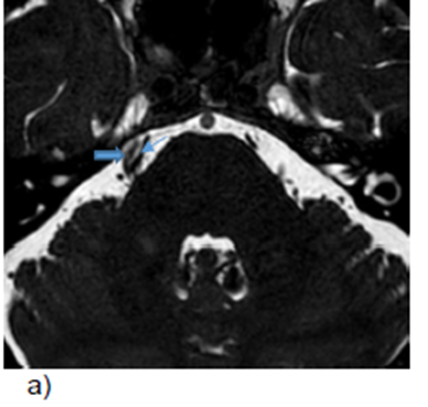
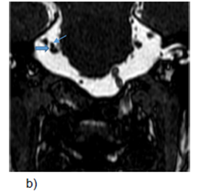
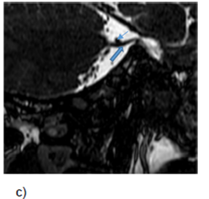 Case 1. 54 yr male patient presented with complaints of headache, tingling sensation and sudden onset pain over right side of forehead since 2 months. MRI BFFE images a) axial b) coronal c) sagittal images showing compression of cisternal segment of right trigeminal nerve (Double arrow) by SCA (Single arrow) causing simple contact with nerve (GRADE 1).
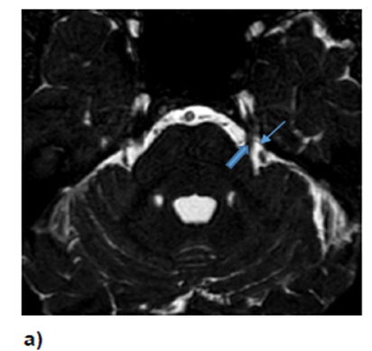
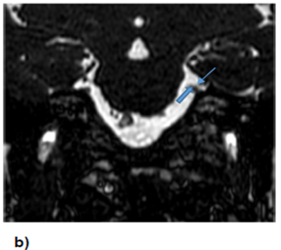
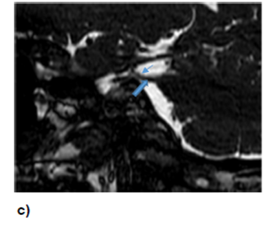
Case 2: 56yrs old female patient complaints of left sided jaw pain increased during eating since 4months. MRI BFFE images a) axial b) coronal c) sagittal images showing aberrant branch of left SCA (Single arrow) abutting left trigeminal nerve (Double arrow). (GRADE-1)
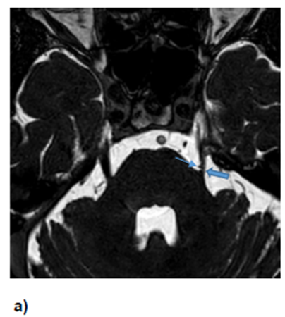
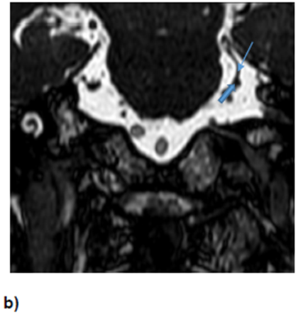
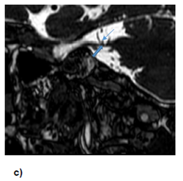 Case 3: 75 yrs male patient presented with sudden paroxysmal electric shock like pain on left cheek and jaw region 6-7 times per day since 5 months. MRI BFFE images a) axial b) coronal c) sagittal images showing compression of cisternal segment of left trigeminal nerve (Double arrow) by vascular loop of AICA (Single arrow) causing slight distortion of nerve. (Grade 2)
Discussion:
Trigeminal neuralgia is a severe neuropathic pain disorder most commonly attributed to NVC at the root entry zone of the trigeminal nerve. The present study aimed to establish a clinicoradiological correlation of NVC in patients with TN using magnetic resonance imaging and to evaluate the relevance of anatomical factors influencing clinical presentation.
Demographic Characteristics
In the present study, TN was most frequently observed in younger adults, with 56.67% of patients belonging to the 21–40-year age group. This finding contrasts with earlier reports suggesting a higher prevalence in individuals above 50 years of age.(6) The relatively younger age of presentation in this cohort may reflect improved access to imaging facilities, early diagnosis, or regional epidemiological variations. A marked male predominance (86.67%) was observed, which differs from several studies reporting female preponderance.[7-11] This discrepancy may be related to sociocultural factors, healthcare-seeking behaviour, or sampling bias due to the limited sample size.
Laterality and Clinical Presentation
Right-sided facial involvement was predominant, observed in 73.33% of cases, consistent with previous studies that have reported a right-side predilection in TN.[12] Although the exact mechanism remains unclear, anatomical asymmetry of vascular structures or posterior fossa configuration may contribute to this lateral dominance.
All patients in the study presented with sudden, severe unilateral facial pain, reaffirming this symptom as the hallmark clinical feature of TN.(1) Associated symptoms such as tingling or numbness (43.33%), headache (33.33%), and allodynia (23.33%) highlight the complexity of sensory involvement and suggest possible central sensitization mechanisms.[13,14]
Trigeminal Nerve Branch Involvement
The mandibular division (V3) was the most commonly affected branch (43.33%), followed by the maxillary division (V2) (36.67%). Minimal involvement of the ophthalmic division (V1) was noted. This pattern aligns with earlier literature reporting increased susceptibility of V3 due to its anatomical course and spatial orientation.[8,15]
MRI Findings and Neurovascular Conflict
MRI demonstrated that compression most frequently occurred at lateral-caudal and medial locations (26.67% each), emphasizing the vulnerability of the root entry zone.(4,5) The SCA (Superior Cerebellar Artery) was the most commonly implicated vessel (46.67%), followed by the anterior inferior cerebellar artery (AICA) (20%), consistent with previous reports.[16,17]
Notably, 23.33% of patients showed no definite NVC on imaging, despite classical clinical features. Similar observations have been reported in earlier studies, underscoring the limitations of conventional MRI and the possibility of alternative mechanisms such as microstructural nerve changes or transient vascular contact.[18-20]
Clinicoradiological Correlations
No statistically significant association was observed between the specific vessel involved and the affected trigeminal nerve branch (p = 0.36), indicating that vascular identity alone does not determine symptom distribution. However, a highly significant association was found between the site of compression and the clinical manifestation (p < 0.001). Medial compression was predominantly associated with V2 involvement, whereas lateral and caudal compression correlated strongly with V3 involvement. These findings emphasize that the anatomical location of compression plays a more critical role than the type of vessel involved.[16,21]
Clinical Implications
The results highlight the importance of high-resolution MRI in accurately identifying NVC and predicting clinical symptomatology, which is crucial for treatment planning, particularly in candidates for microvascular decompression.[18,22,23] Limitations of the study include the small sample size and lack of long-term follow-up. Incorporation of advanced imaging techniques such as diffusion tensor imaging may further enhance diagnostic accuracy.[24,25]
Limitations
The relatively small sample size of 30 patients limits the generalisability of the findings of this study. Conducting larger, multicentric studies would strengthen the validation of the observed associations. In addition, the absence of follow-up data restricts the ability to correlate radiological findings with treatment outcomes. Furthermore, the radiological evaluation was limited to conventional MRI techniques; the inclusion of advanced imaging modalities may have improved the sensitivity for detecting neurovascular compression.
Conclusion:
This study underscores the importance of comprehensive clinical evaluation combined with high-resolution magnetic resonance imaging in the diagnosis and management of trigeminal neuralgia. The predominance of mandibular nerve (V3) involvement, right-sided facial pain, and male preponderance highlights a distinct clinical and demographic pattern. While neurovascular compression involving the superior cerebellar artery is frequently observed, its absence in a subset of patients emphasizes the limitations of current imaging techniques and the need for meticulous interpretation. Importantly, the anatomical location of nerve compression was found to be a more decisive factor in symptom distribution than the specific vessel involved. These findings reinforce the pivotal role of MRI in preoperative assessment and treatment planning and contribute to a deeper understanding of the clinicoradiological spectrum of trigeminal neuralgia. Further studies with larger cohorts and standardized imaging protocols are warranted to validate these observations and improve clinical outcomes.
References:
1. Bukholm G, Tannaes T, Kjelsberg AB, Smith-Erichsen N. An outbreak of multidrug-resistant Pseudomonas aeruginosa associated with increased risk of patient death in an intensive care unit. Infect Control Hosp Epidemiol. 2002;23:441-6.
2. Colvin KM, Gordon VD, Murakami K, Borlee BR, Wozniak DJ, Wong GC, et al. The pel polysaccharide can serve a structural and protective role in the biofilm matrix of Pseudomonas aeruginosa. PLoS Pathog. 2011;7:e1001264.
3. Douraghi M, Ghasemi F, Dallal MMS, Rahbar M, Rahimiforoushani A. Molecular identification of Pseudomonas aeruginosa recovered from cystic fibrosis patients. J Prev Med Hyg. 2014;55:1-3.
4. Ghadaksaz A, Fooladi AAI, Hosseini HM, Amin M. The prevalence of some Pseudomonas virulence genes related to biofilm formation and alginate production among clinical isolates. J Appl Biomed. 2014;13:61-8.
5. Davies JC. Paediatric Respiratory Reviews: Pseudomonas aeruginosa in Cystic Fibrosis: Pathogenesis and Persistence. Paediatr Respir Rev. 2002; 3(2):128-34.
6. Boisvert AA, Cheng MP, Sheppard DC and Nguyen D. Microbial Biofilms in Pulmonary and Critical Care Diseases. Ann Am Thorac Soc. 2016; 13(9):1615– 1623.
7. Strohl WA, Rouse H, Fisher DB. Lippincott, s illustrated reviews: Microbiology. 3rd Ed. Baltimore, Lippincott Williams and Wilkins; 2013: 137-139.
8. Fraise AP, Lambert PA, Maillard JY. Principles and Practice of Disinfection, Preservation and Sterilization. USA, Blackwell; 2004: 556-619.
9. Aloush V, Navon-Venezia S, Seigman-Igra Y, Cabili S, Carmeli Y. Multidrugresistant Pseudomonas aeruginosa: risk factors and clinical impact. Antimicrob. Agents Chemother. 2006; 50: 43–48.
10. Dash M, Padhi S, Narasimham MV, Pattnaik S. Antimicrobial resistance pattern of Pseudomonas aeruginosa isolated from various clinical samples in a tertiary care hospital, South Odisha, India. Saudi J Health Sci. 2014; 3:15-9.
11. Sambrook J, Russel DW. Molecular Cloning: A Laboratory Manual. 3rd Ed. New York, Cold Spring Harbor; 2001: 1- 4.
12. Da Costa Lima JL, Alves LR, Pereira da Paz JN, et al. Analysis of biofilm production by clinical isolates of Pseudomonas aeruginosa from patients with ventilator-associated pneumonia. Rev Bras Ter Intensiva. 2017; 29(3): 310–316.
13. Faraji F, Mahzounieh M, Ebrahimi AO, et al. Molecular detection of virulence genes in Pseudomonas aeruginosa isolated from children with Cystic Fibrosis and burn wounds in Iran. Microbial Pathogenesis. 2016; 99:1-4.
14. Saeed M, Rasheed F, Afzal RK et al. Pseudomonas aeruginosa: Evaluation of Pathogen Burden and Drug-Resistance Trends in a Tertiary Care Hospital. Journal of the College of Physicians and Surgeons Pakistan. 2018; 28(4): 279-283.
15. Unan D, Gnseren F. The resistance of P. aeruginosa strains isolated from nosocomial infections against various antibiotics. Mikrobiyol Bult. 2000; 34: 255–60.
16. Corehtash ZG, Khorshidi A, Firoozeh F, Akbari H, Aznaveh AM. Biofilm Formation and Virulence Factors Among Pseudomonas aeruginosa Isolated From Burn Patients. Jundishapur J Microbiol. 2015; 8(10): e22345.
17. Hoque MM, Ahmad M, Khisa S, Uddin MN, Jesmine R . Antibiotic Resistance Pattern in Pseudomonas Aeruginosa Isolated from Different Clinical Specimens. JAFMC Bangladesh. 2015; 11(1):45-49.
18. Direkel Ş, Uzunoglu E, Uzalp Ç, Findik E, Tontak S, and Ahmadli C. Determination of Piperacillin/Tazobactam and Ticarcillin/Clavulanate Susceptibilities in Pseudomonas aeruginosa Isolates in Hospitalised Patients by E-test Gradient Method and Comparison of Results with Disk Diffusion Tests. Clin Microbiol. 2017; 6:1.
19. Abidi SH, Sherwani SK, Siddiqui TR, Bashir A, and Kazmi SU. Drug resistance profile and biofilm forming potential of Pseudomonas aeruginosa isolated from contact lenses in Karachi-Pakistan. BMC Ophthalmol. 2013; 13: 57.
20. Yekani M, Memar MY, Alizadeh N, Safaei N, Ghotaslou R. Antibiotic Resistance Patterns of Biofilm-Forming Pseudomonas aeruginosa Isolates from Mechanically Ventilated Patients. International Journal of Scientific Study. 2017; 5(5): 1-5.
21. Costa Limaa JLD, Alvesb LR, Araújo Jacoméb PRL et al. Biofilm production by clinical isolates of Pseudomonas aeruginosa and structural changes in LasR protein of isolates non biofilm-producing. braz j infect dis. 2018;2 2(2):129-136.
22. Pournajaf A, Razavi S, Irajian G, et al. Integron types, antimicrobial resistance genes, virulence gene profile, alginate production and biofilm formation in Iranian cystic fibrosis Pseudomonas aeruginosa isolates. Le Infezioni in Medicina. 2018; 3: 226- 236