What Lies Beneath the Scar? A Clinicopathological Study of Incisional Hernia Sac
- D. K. Uma Shankar , Assistant Professor, Department of General Surgery, TRR Institute of Medical Sciences, Sangareddy District, Telangana, India
- Kumar Manish Gopal Prasad2 , Assistant Professor, Department of General Surgery, TRR Institute of Medical Sciences, Sangareddy District, Telangana, India
Article Information:
Abstract:
Background: Incisional hernia is a common long-term complication following abdominal surgery. Although surgical repair is routinely performed, the pathological changes within incisional hernia sacs are often overlooked. Objectives: To study the histopathological spectrum of incisional hernia sacs and assess their clinical relevance. Materials and Methods: This prospective clinicopathological study included 51 patients undergoing elective incisional hernia repair at TRR Institute of Medical Sciences, Sangareddy district, Telangana, over a one-year period (January 2024–January 2025). All excised hernia sacs were subjected to histopathological examination, and findings were correlated with clinical data. Results: The mean age was 46.8 years with female predominance (60.8%). Chronic inflammation was the most common histopathological finding (58.8%), followed by fibrosis with hyalinization (21.6%), foreign-body granuloma (9.8%), mesothelial hyperplasia (7.8%), and incidental pathology (2.0%). Conclusion: Incisional hernia sacs frequently demonstrate active pathological changes. Routine histopathological examination provides valuable insights into hernia biology and may influence surgical decision-making and postoperative outcomes.
Keywords:
Article :
Introduction:
Incisional hernia is a frequent complication of abdominal surgery, with an incidence reported between 10% and 20% depending on patient-related and surgical factors1. Known risk factors include wound infection, obesity, advanced age, poor nutritional status, and increased intra-abdominal pressure2.
Conventionally, the hernia sac is discarded without further evaluation. However, recent studies suggest that incisional hernia sacs may harbor significant pathological changes such as chronic inflammation, fibrosis, foreign body reactions, or even incidental lesions3,4. These changes may play a role in impaired wound healing and recurrence5.
Histopathological examination of hernia sacs can therefore serve as a useful tool for understanding the underlying pathophysiology of hernia formation6. This study was undertaken to analyze the histopathological features of incisional hernia sacs and correlate them with clinical parameters in a small teaching hospital.
Materials and Methods:
Study Design and Setting - This prospective clinicopathological study was conducted in the Department of General Surgery in collaboration with the Department of Pathology at TRR Institute of Medical Sciences, Sangareddy district, Telangana, India.
Study Period - January 2024 to January 2025
Sample Size - A total of 51 patients undergoing elective incisional hernia repair.
Inclusion Criteria
· Patients aged ≥18 years
· Elective incisional hernia repair
· Informed consent obtained
Exclusion Criteria
· Congenital hernias
· Recurrent incisional hernias
· Emergency surgeries
The study was conducted after obtaining approval from the Institutional Ethics Committee of TRR Institute of Medical Sciences, Sangareddy district, Telangana, and informed consent was obtained from all participants.
Histopathological Examination - Excised hernia sacs were fixed in 10% buffered formalin, processed routinely, and stained with hematoxylin and eosin. Slides were examined for inflammatory changes, fibrosis, granulomatous reaction, mesothelial alterations, and incidental pathology.
Results:
The study population comprised 51 patients undergoing elective incisional hernia repair. The majority of patients belonged to the 41–50 year age group (29.4%), followed by those aged 31–40 years (23.5%) and 51–60 years (21.6%). Patients above 60 years constituted 13.7% of cases, while younger adults aged 21–30 years formed the smallest group (11.8%). This distribution indicates that incisional hernia is most frequently encountered in middle-aged individuals, likely reflecting cumulative surgical exposure and impaired wound healing with advancing age.(Table 1)
Table 1: Age distribution of patients with incisional hernia (n = 51)
|
Age Group (Years) |
Number (n) |
Percentage (%) |
|
21–30 |
6 |
11.8 |
|
31–40 |
12 |
23.5 |
|
41–50 |
15 |
29.4 |
|
51–60 |
11 |
21.6 |
|
>60 |
7 |
13.7 |
A clear female predominance was observed in the present study, with females accounting for 60.8% (n = 31) of cases, compared to 39.2% (n = 20) males. This gender difference may be attributed to a higher frequency of obstetric and gynecological surgeries among women, particularly caesarean sections and hysterectomies, which are well-recognized risk factors for incisional hernia formation.( Table 2)
Table 2: Gender distribution of patients undergoing incisional hernia repair (n = 51)
|
Gender |
Number (n) |
Percentage (%) |
|
Male |
20 |
39.2 |
|
Female |
31 |
60.8 |
Previous Surgical Procedures- Analysis of prior surgical history revealed that lower segment caesarean section was the most common antecedent surgery, seen in 35.3% of patients, followed by exploratory laparotomy (27.5%) and hysterectomy (21.6%). Appendectomy accounted for 15.6% of cases. These findings underscore the association between lower abdominal incisions and subsequent development of incisional hernia, especially following gynecological procedures. (Table 3) (Figure 1)
Table 3: Distribution of previous surgical procedures among patients with incisional hernia (n = 51)
|
Previous Surgery |
Number (n) |
Percentage (%) |
|
Lower segment caesarean section |
18 |
35.3 |
|
Hysterectomy |
11 |
21.6 |
|
Exploratory laparotomy |
14 |
27.5 |
|
Appendectomy |
8 |
15.6 |
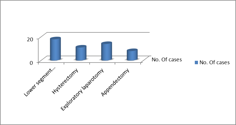
Figure 1 : Bar chart showing distribution of previous surgical procedures among patients with incisional hernia (n = 51)
Histopathological examination of excised incisional hernia sacs demonstrated a wide spectrum of pathological alterations. Chronic inflammatory changes were the predominant finding, observed in 30 cases (58.8%). Fibrosis with hyalinization was noted in 11 cases (21.6%), reflecting long-standing tissue injury and abnormal healing. Foreign-body granulomatous reaction was identified in 5 cases (9.8%), likely related to retained suture material from previous surgeries. Mesothelial hyperplasia was observed in 4 cases (7.8%). Incidental pathology was detected in 1 case (2.0%).(Figure 2)
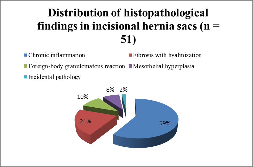
Figure 2: Pie chart showing distribution of histopathological findings in incisional hernia sacs (n = 51)
Discussion:
Incisional hernia continues to represent a significant postoperative complication, with multifactorial etiopathogenesis involving mechanical failure, patient-related risk factors, and biological alterations in wound healing. The present study provides valuable insight into the often-neglected pathological changes occurring within incisional hernia sacs.
The female predominance observed aligns with earlier studies by Mudge and Hughes and subsequent authors, who have consistently reported a higher incidence of incisional hernia following obstetric and gynecological procedures7,13. Lower segment caesarean section and hysterectomy were the most common antecedent surgeries in this study, reinforcing the vulnerability of lower abdominal incisions to herniation due to increased intra-abdominal pressure, repeated stretching, and wound infection.
Chronic inflammation emerged as the most frequent histopathological finding, affecting nearly three-fifths of cases. Persistent inflammatory infiltrates within the hernia sac may interfere with normal collagen synthesis and remodeling, resulting in weakened fascial strength and predisposition to hernia formation and recurrence. Similar findings have been documented by Agarwal et al., Singh et al., and Rosch et al., emphasizing inflammation as a central biological driver rather than a secondary phenomenon8,9,5.
Fibrosis with hyalinization, the second most common finding, reflects repeated cycles of tissue injury and repair. Altered collagen metabolism, particularly an imbalance between type I and type III collagen, has been demonstrated in patients with incisional hernia, lending support to the concept of hernia as a disorder of connective tissue biology10,14. These microscopic changes may have implications for mesh integration and long-term surgical outcomes.
Foreign-body granulomatous reactions were identified in nearly 10% of cases, likely secondary to non-absorbable suture material used during previous operations. Klinge et al. highlighted the role of foreign-body response in perpetuating inflammation and fibrosis, which may compromise tissue planes and complicate subsequent repairs11,15.
Mesothelial hyperplasia, though less frequent, represents a reactive change to chronic irritation and mechanical stress. While usually benign, it may pose diagnostic challenges and underscores the importance of histopathological evaluation to differentiate reactive changes from neoplastic processes16.
The detection of incidental pathology, although rare in the present study, is clinically significant. Previous reports have documented unexpected findings such as endometriosis, tuberculosis, and even malignancy within hernia sacs12,17. Such findings may alter postoperative management and warrant further investigation, reinforcing the value of routine histopathological examination.
Conclusion:
The incisional hernia sac is biologically active rather than inert.Routine histopathological examination reveals frequent and clinically relevant changes such as chronic inflammation, fibrosis, and foreign-body reactions that reflect impaired wound healing and altered tissue biology. From a surgeon’s perspective, submitting hernia sacs for histopathological analysis is a simple, low-cost practice that enhances surgical audit, deepens understanding of hernia pathogenesis, and may ultimately contribute to improved repair strategies and reduced recurrence rates.
References:
1. Luijendijk RW, et al. Incisional hernia recurrence following repair. N Engl J Med. 2000;343:392–398.
2. Burger JW, et al. Long-term follow-up of incisional hernia repair. Ann Surg. 2004;240:578–585.
3. Read RC. Herniology: past, present, and future. Hernia. 2003;7:1–4.
4. Basak S, et al. Histopathological evaluation of hernia sacs. Indian J Pathol Microbiol. 2016;59:39–42.
5. Rosch R, et al. Hernia disease and wound healing. Hernia. 2002;6:171–175.
6. Franz MG. The biology of hernia formation. Surg Clin North Am. 2008;88:1–15.
7. Mudge M, Hughes LE. Incisional hernia: a 10-year prospective study. Br J Surg. 1985;72:70–71.
8. Agarwal BB, et al. Pathological changes in incisional hernia sacs. J Clin Diagn Res. 2017;11:EC20–EC23.
9. Singh A, et al. Role of chronic inflammation in incisional hernia. Int Surg J. 2019;6:1456–1460.
10. Klinge U, et al. Altered collagen metabolism in incisional hernia. World J Surg. 1998;22:808–815.
11. Klinge U, et al. Foreign body reaction and mesh repair. World J Surg. 1998;22:808–815.
12. Leibl BJ, et al. Unexpected findings in hernia sacs. Surg Endosc. 2000;14:1068–1071.
13. Ellis H, Gajraj H, George CD. Incisional hernias: when do they occur? Br J Surg. 1983;70(5):290–291.
14. Bellón JM, Buján J, Honduvilla NG, Jurado F, Gimeno MJ, Turnay J. Altered collagen metabolism in incisional hernia. Hernia. 2001;5(2):82–87.
15. Junge K, Rosch R, Klinge U, Schwab R, Peiper C, Schumpelick V. Risk factors related to recurrence in incisional hernia repair. Hernia. 2002;6(3):113–118.
16. Ordóñez NG. Mesothelial hyperplasia and its differential diagnosis. Adv Anat Pathol. 2006;13(4):173–186.
17. Al-Hawary MM, Kaza RK, Wasnik AP, Platt JF. Unexpected pathology encountered in hernia sacs: clinical significance. Hernia. 2005;9(4):388–391.