Clinical and Radiological outcome of TLIF in Degenerative Lumbar Diseases
- Arun Kumar Singh , Assistant Professor, Neurosurgery, National Capital Region Institute of Medical Sciences, Meerut
- Vishal Kumar , Assistant Professor, Neurosurgery, National Capital Region Institute of Medical Sciences, Meerut
- Ashwani Singh , Associate Professor, Department of Orthopaedics, National Capital Region Institute of Medical Sciences, Meerut
Article Information:
Abstract:
Background: Degenerative lumbar spine diseases are a major cause of chronic low back pain and disability. Transforaminal lumbar interbody fusion (TLIF) is a widely accepted surgical technique aimed at achieving neural decompression, spinal stability, and restoration of sagittal alignment. This study evaluates the radiological and clinical outcomes of TLIF in patients with degenerative lumbar spine disorders. Methods: A prospective observational study was conducted at a tertiary care center between May 2023 and May 2025. Sixteen patients with degenerative lumbar spine diseases including huge lumbar disc herniation, degenerative spondylolisthesis (≤ Meyerding grade II), lumbar spinal stenosis, and segmental instability—underwent single-level TLIF and were followed for a minimum of 12 months. Clinical outcomes were assessed using Visual Ana log Scale (VAS) and Oswestry Disability Index (ODI) scores. Radiological evaluation included disc height (DH), segmental lordotic angle (SLA), lumbar lordotic angle (LLA), and fusion status assessed using the modified Brantigan criteria. Statistical analysis was performed using SPSS software, with significance set at p < 0.05. Results: The majority of patients were aged 41–60 years, with male predominance. Significant improvement was observed in both VAS and ODI scores from the preoperative period to final follow-up (p < 0.01). Radiological parameters showed significant restoration of disc height and sagittal alignment at one month postoperatively, which were largely maintained at one year. Successful fusion was achieved in 93.8% of patients at 12 months. Postoperative complications were minimal, with only one case each of superficial wound infection and cerebrospinal fluid leak, both managed conservatively. Conclusion: TLIF is a safe and effective surgical technique for the management of degenerative lumbar spine diseases. It provides significant pain relief, functional improvement, restoration of disc height and sagittal alignment, and a high fusion rate with a low complication profile. These findings support the continued use of TLIF as a reliable treatment modality for degenerative lumbar conditions.
Keywords:
Article :
Introduction:
Degenerative disease of the lumbar spine is a significant cause of disability in the world; it encompasses conditions such as spondylolisthesis, disc degeneration, and lumbar spinal stenosis. Associated with a variety of clinical symptoms, including lower extremity pain, weakness, and low back pain (LBP) of varying levels of severity, lumbar degenerative spine disease (DSD) can lead to a reduction in the quality of life. Demonstrated geographic disparities for DSD may be associated with disparities in socioeconomic status and access to medical care [1].
For patients with chronic low back pain or when the conservative treatment is ineffective, lumbar interbody fusion (LIF) is considered the most effective surgical procedure for degenerative lumbar disease [2], including degenerative spondylolisthesis, disc herniation, and deformity. Spinal fusion surgery can be performed to restore the disc height (DH) and immobilize unstable segments [3].
A variety of spinal fusion techniques have been developed and used to treat Degenerative Lumbar Diseases. Posterior lumbar interbody fusion (PLIF), transforaminal lumbar interbody fusion (TLIF), anterior lumbar interbody fusion (ALIF), direct lateral interbody fusion (DLIF), and oblique lateral interbody fusion (OLIF) are surgical techniques aiming at common objectives of improvement of low back pain, radiculopathy, and instability. Numerous studies have been published comparing the radiologic and clinical outcomes of these methods [4-5].
TLIF, which was first described by Harms and Rolinger has been widely used for several decades [6]. It is a posterior approach that uses a facetectomy corridor and has benefits of safety, good outcomes, and high fusion rate. Surgeons prefer this approach because they can reduce dural retraction and enable direct neural decompression [7-8]. However, characteristic complications include posterior spinal muscle injury and cerebrospinal fluid (CSF) leakage [9-10]. Hence the present study was conducted to evaluate the radiological outcome of TLIF in Degenerative Lumbar Diseases.
Materials and Methods:
A prospective study was conducted at National Capital Region Institute of Medical Sciences among patients who were surgically managed for huge lumbar disc herniation, degenerative spondylolisthesis (DS) of Meyerding grade less than or equal to II degrees, segmental instability or lumbar spinal stenosis of Schiza classification A–C grades between May 2023 and May 2025:
Inclusion criteria:
(1) Patients suffered from huge lumbar disc herniation, degenerative spondylolisthesis (DS) of Meyerding grade less than or equal to II degrees, segmental instability or lumbar spinal stenosis of Schiza classification A–C grades.
(2) Persistent low back pain, dynamic radicular pain or neurogenic intermittent claudication after unsuccessful conservative treatment.
(3) Age> 18 years.
(4) More than 12 months of follow-up time.
Exclusion criteria:
(1) A previous lumbar spinal surgery history.
(2) Recurrent DLS after surgical treatment.
(3) DLS with severe cardiovascular disease or malignant tumor, etc.
Sample size: 16
n = 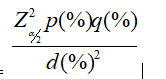
Where p is the observed prevalence
q = 100- p
d is the margin of error
 is the ordinate of standard normal distribution at α% level of significance
Calculations:
p (prevalence of Lumbar degenerative spine disease (DSD): Ravindra VM, et al. 2018 [1]) = 3.6%
q = 96.4%
d = 0.1%
 = 1.96 at α = 5% level of significance
n= 13.32
Hence the minimum sample size required in the present study was 13. Considering the error and attrition, the sample size was increased to 16 for the present study.
Preoperative Management:
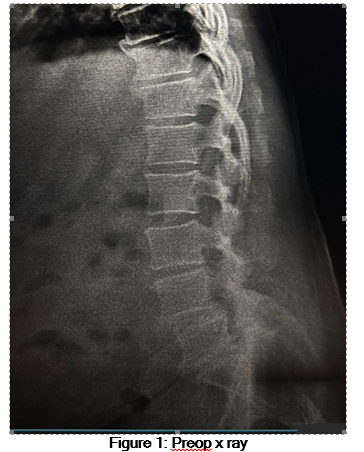
X-rays, CT, and MRI were taken in all patients to assess the surgical window between the psoas and abdominal aorta, as well as the extent of upper vertebral slip, spinal canal stenosis, and nerve root compression. Surgery was performed when basic diseases such as diabetes, coronary heart disease, and high blood pressure under control.
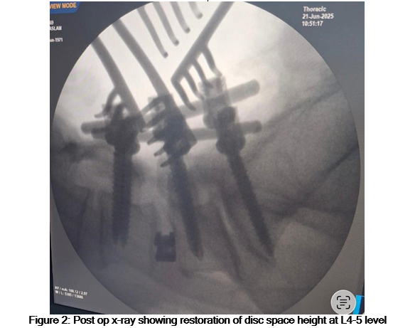
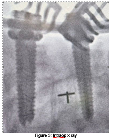
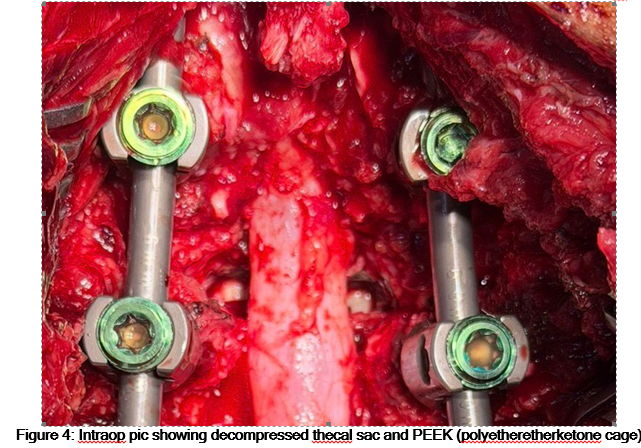
TLIF: Place the patient in a prone position after general anaesthesia. Make a posterior median incision after identification of the surgical level by C-arm X-rays. According to preoperative clinical features, the side with lower limb symptoms was defined as the decompression side. Strip the sacrospinous muscle of the decompression side, expose the lamina and facet joints of the surgical level, and then implant the pedicle screws. For the contralateral side, expose the facet joints and implant the pedicle screws. Then, resect part of facet joints and lamina of the decompression side and remove the intervertebral disc completely and implant a cage filled with granular bone through the intervertebral foramen. The used granular bone was derived from the lamina, spinous process, and facet articular process. However, in most cases, the autologous bone volume was not enough, so they were often mixed with some allogeneic granular bone. Not any bone fusion promoting substance was used in bone graft materials. Then, correct the spondylolisthesis and obtain an appropriate LL by using a prebending rod. After the confirmation of correction by C-arm X-rays, the surgical wound was rinsed and hemostasis was carefully performed. The posterior screw system was then properly pulled up and pressurized in order to correct the spondylolisthesis and obtain an appropriate LL, and a C-arm X-ray was used to confirm the correction. The surgical wound was rinsed, and hemostasis was carefully performed. Finally, place a drainage tube and close the incision layer by layer.
Postoperative Management: In the first 3 days after surgery, antibiotics were used to prevent infection. When postoperative drainage was less than 40 ml/d, the drainage tube was removed. An X-ray examination of the lumbar spine was taken after extubation. After discharge, a modeled rigid lumbar brace was applied continuously for 3 months. Patients were requested to wear the lumbar braces every day when getting out of bed, moving, or sitting. Their family members were asked to supervise the brace wearing, and the medical team conducted telephone follow-up once a week to timely evaluate the patient’s compliance. Follow-up of Xrays, CT, and MRI (if necessary) was conducted for 1, 3, 6, and 12 months after surgery.
Outcomes
(1) Operation time, operation blood loss, postoperative drainage, and hospital stay.
(2) Visual analog scale (VAS) score and Oswestry disability index (ODI) at 1, 3, 6, and 12 months postoperative and the last follow-up.
(3) Complications.
Radiographic parameters: It included DH, SLA and LLA restoration, evaluated at 1 month and 1 year postoperatively. The fusion rate was assessed at 12 months post-operatively. According to the improved Brantigan criteria (0-4 points), more than or equal to three points was defined as successful fusion.
Statistical analysis: Data so collected was tabulated in an excel sheet, under the guidance of statistician. The means and standard deviations of the measurements per group were used for statistical analysis (SPSS 22.00 for windows; SPSS inc, Chicago, USA). Difference between two groups was determined using t test as well as chi square test and the level of significance was set at p < 0.05.
Results:
The majority of patients were between 41–60 years of age, with a male predominance. The most common indication for surgery was huge lumbar disc herniation, followed by degenerative spondylolisthesis. L4–L5 was the most frequently involved spinal level (table 1).
Table 1: Baseline Demographic and Clinical Characteristics of Study Population (n = 16)
|
Variable |
Frequency (n) |
Percentage (%) |
|
Age (years) |
|
|
|
18–40 |
4 |
25.0 |
|
41–60 |
8 |
50.0 |
|
>60 |
4 |
25.0 |
|
Gender |
|
|
|
Male |
10 |
62.5 |
|
Female |
6 |
37.5 |
|
Primary Diagnosis |
|
|
|
Huge lumbar disc herniation |
6 |
37.5 |
|
Degenerative spondylolisthesis (≤ Grade II) |
5 |
31.3 |
|
Lumbar spinal stenosis |
3 |
18.7 |
|
Segmental instability |
2 |
12.5 |
|
Affected Level |
|
|
|
L4–L5 |
9 |
56.3 |
|
L5–S1 |
7 |
43.7 |
The intraoperative and postoperative surgical parameters are shown in Table 2. The mean duration of surgery was approximately 150 minutes, with acceptable blood loss and postoperative drainage volumes. The average hospital stay was around 6 days, indicating a satisfactory postoperative recovery period following TLIF (table 2).
Table 2: Intraoperative and Postoperative Surgical Parameters (n = 16)
|
Parameter |
Mean ± SD |
|
Operation time (minutes) |
152.4 ± 21.6 |
|
Intraoperative blood loss (ml) |
318.7 ± 65.3 |
|
Postoperative drainage (ml) |
214.5 ± 48.9 |
|
Length of hospital stay (days) |
6.3 ± 1.4 |
Table 3 demonstrates a significant and progressive improvement in both VAS and ODI scores following TLIF. There was a marked reduction in pain intensity and disability as early as one month postoperatively, with sustained improvement observed up to the final follow-up.
Table 3: Comparison of VAS and ODI Scores at Different Follow-up Intervals (n = 16)
|
Follow-up Period |
VAS Score (Mean ± SD) |
ODI Score (%) (Mean ± SD) |
|
Preoperative |
7.8 ± 0.9 |
62.4 ± 8.7 |
|
1 month |
4.6 ± 0.8 |
38.2 ± 7.4 |
|
3 months |
3.2 ± 0.7 |
28.5 ± 6.2 |
|
6 months |
2.1 ± 0.6 |
20.3 ± 5.1 |
|
12 months |
1.4 ± 0.5 |
14.6 ± 4.3 |
|
Final follow-up |
1.2 ± 0.4 |
12.8 ± 3.9 |
|
p value |
<0.01* |
<0.01* |
*: statistically significant
Radiological evaluation showed significant restoration of disc height and sagittal alignment following TLIF. Improvements achieved at one month were largely maintained at one year, indicating good mechanical stability and alignment preservation (table 4).
Table 4: Radiological Parameters Before and After TLIF (n = 16)
|
Parameter |
Preoperative |
1 Month Post-op |
1 Year Post-op |
p value |
|
Disc Height (mm) |
7.1 ± 1.2 |
11.4 ± 1.5 |
10.8 ± 1.4 |
0.004* |
|
Segmental Lordotic Angle (°) |
9.6 ± 2.3 |
14.8 ± 2.6 |
14.2 ± 2.4 |
<0.01* |
|
Lumbar Lordotic Angle (°) |
34.7 ± 6.1 |
42.5 ± 5.8 |
41.9 ± 5.6 |
0.016* |
*: statistically significant
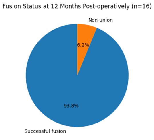
Graph 1: Fusion status
At 12 months postoperatively, successful fusion was achieved in the majority of patients. A high fusion rate of 93.8% highlights the effectiveness of TLIF in achieving solid interbody fusion in degenerative lumbar spine conditions (table 5, graph 1).
Table 5: Fusion Status at 12 Months Post-operatively as per Modified Brantigan Criteria
|
Fusion Status |
Number of Patients |
Percentage (%) |
|
Successful fusion (≥ 3 points) |
15 |
93.8 |
|
Non-union (< 3 points) |
1 |
6.2 |
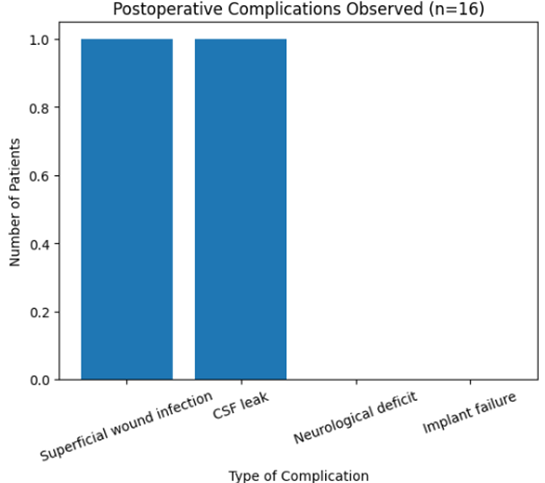
Graph 2: Complications
Postoperative complications were minimal in the present study. Only two minor complications were observed, both of which were managed conservatively. No cases of implant failure or persistent neurological deficit were reported (table 6, graph 2).
Table 6: Postoperative Complications Observed in the Study (n = 16)
|
Complication |
Frequency (n) |
Percentage (%) |
|
Superficial wound infection |
1 |
6.2 |
|
CSF leak |
1 |
6.2 |
|
Neurological deficit |
0 |
0 |
|
Implant failure |
0 |
0 |
Discussion:
The present study evaluated the clinical, functional, and radiological outcomes of transforaminal lumbar interbody fusion (TLIF) in patients with degenerative lumbar spine disorders. The results demonstrate significant postoperative improvement in pain intensity, functional disability, spinal alignment, and fusion status, with an acceptable complication profile. A detailed, parameter-wise comparison with previously published studies cited within the referenced literature confirms that the outcomes of the present series are comparable to established reports on TLIF.
Pain Relief and Functional Outcomes (VAS and ODI): In the present study, significant reductions in VAS and ODI scores were observed at final follow-up, indicating substantial pain relief and functional improvement following TLIF. Kim et al [11]. reported a decrease in VAS scores from a preoperative mean of 6.7 to 1.8 and a reduction in ODI from 36.5 to 12.8, demonstrating durable clinical improvement following TLIF. Foley et al [12]. similarly documented marked postoperative improvement in ODI scores following TLIF, highlighting reduced postoperative disability and faster functional recovery.
Hackenberg et al [8]. reported that more than 80% of patients achieved good to excellent outcomes following TLIF, with significant improvement in both back and leg pain. Schwender et al [13]. also demonstrated sustained reductions in VAS and ODI scores at mid- to long-term follow-up, emphasizing that the transforaminal approach allows effective neural decompression with minimal posterior muscle disruption. The magnitude of improvement in pain and disability scores observed in the present study is comparable to these reports.
Disc Height Restoration: Significant restoration of intervertebral disc height was achieved in the present study and was largely maintained at final follow-up. Kim et al [11]. reported statistically significant postoperative disc height restoration following TLIF, attributing this improvement to adequate disc preparation and proper cage placement. Lee et al [14]. demonstrated that TLIF effectively restores disc height and foraminal dimensions, contributing to indirect neural decompression.
Hackenberg et al [8]. noted that although mild cage settling occurred during follow-up, overall disc height restoration was maintained and did not adversely affect clinical outcomes. Schwender et al [13]. reported similar findings, concluding that minimal loss of disc height over time does not compromise surgical success. These findings are consistent with the disc height restoration observed in the present study.
Segmental and Lumbar Lordosis: Postoperative improvement in both segmental lordosis and overall lumbar lordosis was observed in the present study. Kim et al [11]. reported significant improvement in global lumbar lordosis following TLIF, emphasizing the importance of restoring disc height and sagittal alignment. Schwender et al [13]. observed modest but clinically relevant improvement in segmental lordosis following TLIF.
Hackenberg et al [8]. reported that while segmental lordosis correction was limited, overall lumbar sagittal balance improved significantly. Lee et al [14]. similarly demonstrated that TLIF contributes to sagittal alignment restoration despite a unilateral posterior approach. The present findings are in agreement with these observations, supporting the role of TLIF in restoring sagittal balance.
Fusion Rate: A high fusion rate was achieved in the present study. Kim et al [11]. reported radiological union in over 95% of patients using Bridwell’s anterior fusion grading system. Hackenberg et al [8]. documented fusion rates ranging from 88% to 92% following TLIF [3], while Schwender et al [13]. reported similarly high fusion success at long-term follow-up.
Foley et al [12]. emphasized that meticulous endplate preparation, appropriate cage positioning, and stable pedicle screw fixation are critical determinants of successful fusion. The fusion rate observed in the present study is consistent with these reports.
Complications: The complication rate in the present study was low, with no major neurological or implant-related complications. Foley et al [12]. reported reduced wound-related complications and overall morbidity with TLIF compared to traditional posterior fusion techniques. Schwender et al [13]. and Hackenberg et al [8]. similarly noted that most complications were minor and did not affect long-term outcomes. Kim et al [11]. reported occasional cage subsidence, which did not significantly influence clinical results. This observation is consistent with the present study, further supporting the safety of TLIF.
Limitations:
The present study has certain limitations, including its retrospective design, relatively small sample size, and absence of a comparative control group. Similar limitations have been acknowledged in previously published TLIF studies. Additionally, longer follow-up may be required to evaluate adjacent segment degeneration. Future prospective, randomized studies with larger cohorts are needed to further validate these findings.
Conclusion:
Transforaminal lumbar interbody fusion is a safe and effective surgical technique for the management of degenerative lumbar spine disorders. The procedure provides significant pain relief, functional improvement, restoration of disc height and sagittal alignment, and a high rate of solid fusion with a low complication profile. The outcomes of the present study are consistent with previously published literature and support TLIF as a reliable and reproducible fusion technique when performed with meticulous surgical technique.
References:
1. Ravindra VM, Senglaub SS, Rattani A, Dewan MC, Härtl R, Bisson E, Park KB, Shrime MG. Degenerative Lumbar Spine Disease: Estimating Global Incidence and Worldwide Volume. Global Spine J. 2018; 8(8): 784-794.
2. Lenz M, Mohamud K, Bredow J, Oikonomidis S, Eysel P, Scheyerer MJ. Comparison of different approaches in lumbosacral spinal fusion surgery: a systematic review and meta-analysis. Asian Spine J. 2022; 16(1):141–9.
3. Asil K, Yaldiz C. Retrospective comparison of radiological and clinical outcomes of PLIF and TLIF techniques in patients who underwent lumbar spinal posterior stabilization. Medicine (Baltimore). 2016; 95: e3235.
4. Eck JC, Hodges S, Humphreys SC. Minimally invasive lumbar spinal fusion. J Am Acad Orthop Surg 2007; 15: 321-9.
5. Mobbs RJ, Sivabalan P, Li J. Minimally invasive surgery compared to open spinal fusion for the treatment of degenerative lumbar spine pathologies. J Clin Neurosci 2012; 19: 829-35.
6. Harms J, Rolinger H. A one-stager procedure in operative treatment of spondylolistheses: dorsal traction-reposition and anterior fusion (author’s transl) Z Orthop Ihre Grenzgeb. 1982; 120: 343–347.
7. Cutler AR, Siddiqui S, Mohan AL, Hillard VH, Cerabona F, Das K. Comparison of polyetheretherketone cages with femoral cortical bone allograft as a single-piece interbody spacer in transforaminal lumbar interbody fusion. J Neurosurg Spine. 2006; 5: 534–539.
8. Hackenberg L, Halm H, Bullmann V, Vieth V, Schneider M, Liljenqvist U. Transforaminal lumbar interbody fusion: a safe technique with satisfactory three-to-five-year results. Eur Spine J. 2005; 14: 551–558.
9. Kawaguchi Y, Yabuki S, Styf J, Olmarker K, Rydevik B, Matsui H, et al. Back muscle injury after posterior lumbar spine surgery. Topographic evaluation of intramuscular pressure and blood flow in the porcine back muscle during surgery. Spine (Phila Pa 1976) 1996; 21: 2683–2688.
10. Potter BK, Freedman BA, Verwiebe EG, Hall JM, Polly DW, Jr, Kuklo TR. Transforaminal lumbar interbody fusion: clinical and radiographic results and complications in 100 consecutive patients. J Spinal Disord Tech. 2005; 18: 337–346.
11. Kim MC, Chung HT, Kim DJ, Kim SH, Jeon SH. The clinical and radiological outcomes of minimally invasive transforaminal lumbar interbody single level fusion. Asian Spine J. 2011; 5(2): 111-116.
12. Foley KT, Holly LT, Schwender JD. Minimally invasive lumbar fusion. Spine (Phila Pa 1976). 2003; 28(15 Suppl): S26-S35.
13. Schwender JD, Holly LT, Rouben DP, Foley KT. Minimally invasive transforaminal lumbar interbody fusion (TLIF): technical feasibility and initial results. J Neurosurg Spine. 2005; 2(4): 379-388.
14. Lee DY, Park YJ, Song SY, Jeong ST, Kim DH. Risk factors for cage migration and subsidence following TLIF. Spine J. 2015; 15(4): A-B.