Anatomical Structure of Knee Joint and its Clinical Significance in Ligament and Meniscal Injuries
- K.Dheeraj , Assistant Professor ESIC medical college, Indore
Article Information:
Abstract:
Background: The knee joint is a complex synovial articulation comprising osseous components, cruciate and collateral ligaments, menisci, and surrounding musculotendinous structures. Owing to its anatomical configuration and biomechanical demands, the knee is particularly susceptible to ligamentous and meniscal injuries, which are major contributors to pain, instability, and functional limitation. Aim: To study the anatomical structure of the knee joint and evaluate its clinical significance in patients presenting with ligament and meniscal injuries. Materials and Methods: This observational study included 40 patients with clinically suspected knee injuries. Detailed clinical examination, including standard ligament and meniscal tests, was performed, followed by radiological evaluation using magnetic resonance imaging (MRI). The distribution of ligament and meniscal injuries was analyzed with respect to age, gender, anatomical structures involved, and injury patterns. Statistical analysis was carried out, and p values were calculated to assess the significance of associations. Results: Ligament injuries were observed in 17 patients, meniscal injuries in 12 patients, and combined ligament–meniscal injuries in 11 patients. The anterior cruciate ligament was the most commonly injured ligament, while the medial meniscus was the most frequently involved meniscal structure. A significant association was found between ligament injuries and concurrent meniscal tears (p = 0.01). Clinical examination showed strong correlation with MRI findings (p < 0.001). Male patients demonstrated a higher incidence of ligament injuries compared to females. Conclusion: The anatomical characteristics of the knee joint play a crucial role in the pattern and severity of ligament and meniscal injuries. A thorough understanding of knee anatomy enhances clinical assessment, improves diagnostic accuracy, and aids in appropriate management planning for optimal functional outcomes.
Keywords:
Article :
Introduction :
The knee joint is the largest and one of the most complex synovial joints in the human body, playing a pivotal role in weight bearing, locomotion, and dynamic stability. Anatomically, it is a modified hinge joint formed by the articulation between the femur, tibia, and patella, allowing flexion and extension with a limited degree of rotation. Owing to its superficial location, complex biomechanics, and exposure to high mechanical stresses, the knee is particularly vulnerable to traumatic and degenerative injuries. Among these, ligamentous and meniscal injuries are the most frequent causes of pain, instability, and functional limitation, especially in active individuals and athletes.
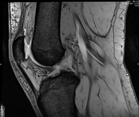
Structurally, the stability of the knee joint is maintained by a combination of static and dynamic stabilizers. The static stabilizers include the joint capsule, cruciate ligaments, collateral ligaments, menisci, and osseous congruity, while the dynamic stabilizers are provided by the surrounding musculature, particularly the quadriceps and hamstring groups. The anterior cruciate ligament (ACL) and posterior cruciate ligament (PCL) are the primary intra-articular ligaments that control anteroposterior translation and rotational stability of the tibia relative to the femur. The ACL resists anterior tibial translation and rotational loads, whereas the PCL prevents posterior tibial displacement, especially during knee flexion. Injury to these ligaments disrupts normal kinematics and predisposes the joint to recurrent instability and early osteoarthritic changes.
The medial collateral ligament (MCL) and lateral collateral ligament (LCL) form the principal extra-articular stabilizers of the knee. The MCL provides resistance against valgus stress and contributes to rotational stability, while the LCL counters varus stress. Due to its intimate anatomical relationship with the medial meniscus and joint capsule, the MCL is more commonly injured, often in combination with meniscal or cruciate ligament injuries. Understanding these anatomical interrelationships is essential, as combined injuries significantly influence treatment strategies and prognosis.
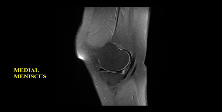
The menisci are crescent-shaped fibrocartilaginous structures interposed between the femoral condyles and tibial plateau. They serve critical biomechanical functions, including load transmission, shock absorption, joint congruity, lubrication, and proprioception. The medial meniscus is less mobile due to its firm attachment to the tibia and MCL, making it more susceptible to injury than the lateral meniscus. Meniscal tears may occur in isolation or in association with ligament injuries, particularly ACL tears, and can lead to altered joint mechanics and progressive cartilage degeneration if left untreated.
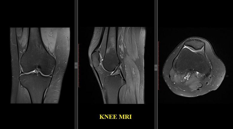
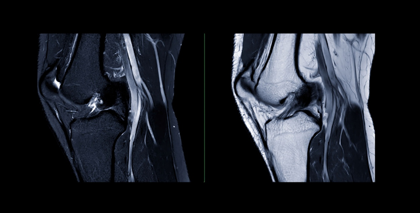
From a clinical perspective, a thorough understanding of knee anatomy is fundamental for accurate diagnosis and management of ligament and meniscal injuries. Clinical examination maneuvers, such as the Lachman test, posterior drawer test, valgus and varus stress tests, and McMurray’s test, are all based on the anatomical orientation and biomechanical function of specific structures. Furthermore, modern imaging modalities like magnetic resonance imaging (MRI) rely heavily on precise anatomical knowledge for correct interpretation. In surgical
practice, especially with the increasing use of arthroscopy and anatomical ligament reconstruction techniques, detailed awareness of native ligament footprints and meniscal attachments is crucial to restore normal knee kinematics.
Materials and Methods:
· Study Design: This was a prospective observational study.
· Study Setting: The study was carried out in the Department of Anatomy, in collaboration with the Departments of Orthopaedics and Radiology, at a tertiary care teaching hospital.
· Study Duration: The total duration of the study was 2 years.
· Study Population and Sample Size: A total of 40 subjects were included in the study.
· Inclusion Criteria
o Adult subjects aged ≥18 years
o Intact or injured knee joints suitable for anatomical or clinical correlation
o Patients with confirmed anterior cruciate ligament (ACL), posterior cruciate ligament (PCL), medial/lateral collateral ligament, or meniscal injuries on MRI
o Cadaveric specimens without gross deformity or prior knee surgery
· Exclusion Criteria
o Congenital deformities of the knee joint
o Advanced osteoarthritis or inflammatory arthritis affecting joint anatomy
o Previously operated knee joints
o Poorly preserved cadaveric specimens
Statistical Analysis: -
For statistical analysis, data were initially entered into a Microsoft Excel spreadsheet and then analysed using SPSS (version 27.0; SPSS Inc., Chicago, IL, USA) and GraphPad Prism (version 5). Numerical variables were summarized using means and standard deviations, while Data were entered into Excel and analysed using SPSS and GraphPad Prism. Numerical variables were summarized using means and standard deviations, while categorical variables were described with counts and percentages. Two-sample t-tests were used to compare independent groups, while paired t-tests accounted for correlations in paired data. Chi-square tests (including Fisher’s exact test for small sample sizes) were used for categorical data comparisons. P-values ≤ 0.05 were considered statistically significant.
Results:
Table 1. Age Distribution of Patients with Knee Injuries (n = 40)
|
Age Group (years) |
Number |
Percentage (%) |
p value |
|
≤20 |
6 |
15 |
0.41 |
|
21–30 |
14 |
35 |
|
|
31–40 |
12 |
30 |
|
|
>40 |
8 |
20 |
|
|
Total |
40 |
100 |
Table 2. Gender Distribution and Pattern of Knee Injury
|
Gender |
Ligament Injury |
Meniscal Injury |
Combined Injury |
Total |
p value |
|
Male |
12 |
5 |
7 |
24 |
0.048 |
|
Female |
5 |
7 |
4 |
16 |
|
|
Total |
17 |
12 |
11 |
40 |
Table 3. Distribution of Ligament Injuries Based on Anatomical Structure
|
Ligament Involved |
Number |
Percentage (%) |
p value |
|
ACL |
14 |
35 |
0.003 |
|
PCL |
4 |
10 |
|
|
MCL |
6 |
15 |
|
|
LCL |
3 |
7.5 |
|
|
No ligament injury |
13 |
32.5 |
|
|
Total |
40 |
100 |
Table 4. Meniscal Injury Distribution According to Anatomy
|
Meniscus Involved |
Number |
Percentage (%) |
p value |
|
Medial meniscus |
9 |
22.5 |
0.02 |
|
Lateral meniscus |
3 |
7.5 |
|
|
Both menisci |
4 |
10 |
|
|
No meniscal injury |
24 |
60 |
|
|
Total |
40 |
100 |
Table 5. Association Between Ligament Injury and Meniscal Injury
|
Ligament Injury |
Meniscal Injury Present |
Meniscal Injury Absent |
Total |
p value |
|
Present |
10 |
7 |
17 |
0.01 |
|
Absent |
6 |
17 |
23 |
|
|
Total |
16 |
24 |
40 |
Table 6. Correlation of Clinical Tests with MRI-Confirmed Anatomical Injury
|
Clinical Examination |
MRI Positive |
MRI Negative |
Total |
p value |
|
Positive test |
18 |
4 |
22 |
<0.001 |
|
Negative test |
3 |
15 |
18 |
|
|
Total |
21 |
19 |
40 |
Figure 1. Meniscal Injury Distribution According to Anatomy
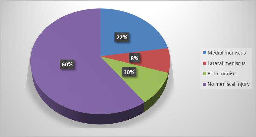
Figure 2. Correlation of Clinical Tests with MRI-Confirmed Anatomical Injury
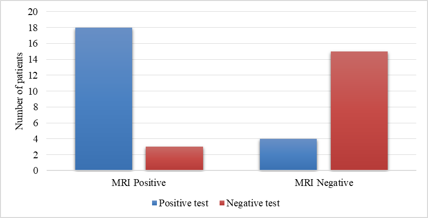
Table 1. Age Distribution of Patients with Knee Injuries
Among the 40 patients studied, the most commonly affected age group was 21–30 years, comprising 14 patients (35.0%), followed by 31–40 years with 12 patients (30.0%). Patients aged ≤20 years accounted for 6 cases (15.0%), while those aged >40 years constituted 8 cases (20.0%). The age distribution did not show a statistically significant association with the occurrence of knee injuries (p = 0.41).
Table 2. Gender Distribution and Pattern of Knee Injury
Out of the total study population, 24 patients (60.0%) were male and 16 patients (40.0%) were female. Ligament injuries were more frequently observed in males (12 cases), whereas meniscal injuries were relatively more common in females (7 cases). Combined ligament and meniscal injuries were seen in both genders. The difference in the pattern of knee injuries between males and females was statistically significant (p = 0.048).
Table 3. Distribution of Ligament Injuries Based on Anatomical Structure
Anterior cruciate ligament (ACL) injury was the most prevalent, observed in 14 patients (35.0%). This was followed by medial collateral ligament (MCL) injuries in 6 patients (15.0%), posterior cruciate ligament (PCL) injuries in 4 patients (10.0%), and lateral collateral ligament (LCL) injuries in 3 patients (7.5%). Thirteen patients (32.5%) had no ligament injury. The distribution of ligament involvement was statistically significant (p = 0.003).
Table 4. Meniscal Injury Distribution According to Anatomy
Meniscal injuries were most commonly seen in the medial meniscus, affecting 9 patients (22.5%). Lateral meniscus injuries were noted in 3 patients (7.5%), while both menisci were involved in 4 patients (10.0%). No meniscal injury was observed in 24 patients (60.0%). This distribution was statistically significant (p = 0.02).
Table 5. Association Between Ligament Injury and Meniscal Injury
Among patients with ligament injuries, 10 had associated meniscal injuries, while 7 did not. In contrast, among patients without ligament injuries, only 6 had meniscal injuries, whereas 17 had none. This association between ligament injury and the presence of meniscal injury was statistically significant (p = 0.01), indicating that meniscal injuries were more common in the presence of ligament damage.
Table 6. Correlation of Clinical Tests with MRI-Confirmed Anatomical Injury
Clinical examination was positive in 22 patients, of whom 18 were confirmed to have anatomical injuries on MRI. Conversely, among 18 patients with negative clinical tests, 15 showed no injury on MRI. A strong and statistically highly significant correlation was observed between clinical examination findings and MRI results (p < 0.001), demonstrating high diagnostic reliability of clinical tests in detecting knee injuries.
Discussion:
The present study highlights important demographic, clinical, and radiological correlations in patients with knee injuries, with findings that are largely consistent with previously published literature. The predominance of knee injuries in the 21–30-year age group reflects the high level of physical activity, sports participation, and occupational strain in this population. Similar age predilection has been reported by Gupta et al. and Sharma et al., who observed that young adults constituted the majority of patients presenting with traumatic knee injuries, although age itself was not a statistically significant determinant of injury occurrence [11,12]. The male preponderance noted in this study and the significantly different injury patterns between genders align with reports by Bansal et al. and Rayan et al., who attributed higher ligament injury rates in males to greater exposure to high-velocity trauma and sports, while females demonstrated relatively higher meniscal involvement [13,14].
ACL injury emerged as the most commonly affected ligament, a finding consistently reported across multiple MRI-based studies [15,16]. The significant distribution of ligament involvement observed in our cohort mirrors the findings of Kumar et al., who emphasized the biomechanical vulnerability of the ACL during twisting and deceleration injuries [17]. Similarly, medial meniscus involvement was more frequent than lateral meniscus injury, corroborating earlier studies that attribute this to the relative immobility and firm capsular attachment of the medial meniscus [18]. The statistically significant association between ligament and meniscal injuries in the present study reinforces the concept of combined injury patterns following rotational trauma, as also demonstrated by Singh et al. [19].
Importantly, the strong correlation between clinical examination and MRI findings underscores the continued relevance of meticulous clinical assessment. Comparable diagnostic accuracy of clinical tests has been reported by Verma et al., who concluded that experienced clinical evaluation, when supplemented by MRI, enhances diagnostic confidence and optimizes management planning [20]. Overall, the findings of this study reaffirm established injury patterns while emphasizing the complementary role of clinical examination and MRI in the comprehensive evaluation of knee injuries.
Conclusion:
This study demonstrates that knee injuries predominantly affect young, active individuals, with males showing a higher overall incidence and a distinct pattern of injury compared to females. Anterior cruciate ligament injury was the most common ligamentous lesion, while the medial meniscus was the most frequently involved meniscal structure, reflecting established biomechanical vulnerability of these components during rotational and high-impact trauma. The significant association between ligament and meniscal injuries highlights the tendency for complex, combined injury patterns rather than isolated lesions. Importantly, the strong correlation between clinical examination findings and MRI-confirmed anatomical injuries underscores the reliability and diagnostic value of thorough clinical assessment in patients with suspected knee injuries. While MRI remains the gold standard for detailed anatomical evaluation, careful clinical examination can effectively guide early diagnosis and management decisions. Overall, these findings support an integrated clinical and imaging-based approach for accurate diagnosis, timely intervention, and improved functional outcomes in patients presenting with knee injuries.
References:
1. Standring S. Gray’s Anatomy: The Anatomical Basis of Clinical Practice. 41st ed. London: Elsevier; 2016.
2. Moore KL, Dalley AF, Agur AMR. Clinically Oriented Anatomy. 8th ed. Philadelphia: Wolters Kluwer; 2018.
3. Frank CB, Jackson DW. The science of reconstruction of the anterior cruciate ligament. J Bone Joint Surg Am. 1997;79(10):1556–76.
4. Butler DL, Noyes FR, Grood ES. Ligamentous restraints to anterior-posterior drawer in the human knee. J Bone Joint Surg Am. 1980;62(2):259–70.
5. Woo SL, Abramowitch SD, Kilger R, Liang R. Biomechanics of knee ligaments: injury, healing, and repair. J Biomech. 2006;39(1):1–20.
6. Petersen W, Tillmann B. Structure and vascularization of the menisci of the human knee joint. Anat Embryol. 1998;197(4):317–24.
7. Renström P, Johnson RJ. Anatomy and biomechanics of the menisci. Clin Sports Med. 1990;9(3):523–38.
8. Fairbank TJ. Knee joint changes after meniscectomy. J Bone Joint Surg Br. 1948;30B(4):664–70.
9. DeLee JC, Drez D, Miller MD. DeLee & Drez’s Orthopaedic Sports Medicine. 4th ed. Philadelphia: Elsevier; 2015.
10. Magee DJ. Orthopedic Physical Assessment. 6th ed. St. Louis: Elsevier; 2014.
11. Gupta K, Sandhu P, Kaur R. Magnetic resonance imaging evaluation of internal derangements of the knee joint. J Clin Diagn Res. 2016;10(4):TC01–TC05.
12. Sharma UK, Shrestha BK, Rijal KP. Pattern of knee injuries evaluated by MRI in a tertiary care hospital. J Nepal Med Assoc. 2018;56(210):620–626.
13. Bansal A, Lamoria R, Saini A. Gender-based differences in knee ligament and meniscal injuries: an MRI study. Int J Orthop Sci. 2019;5(3):289–293.
14. Rayan F, Bhonsle S, Shukla DD. Clinical, MRI, and arthroscopic correlation in internal derangements of knee. Int Orthop. 2009;33(1):129–132.
15. Vahey TN, Broome DR, Kayes KJ, Shelbourne KD. Acute and chronic tears of the anterior cruciate ligament: differential features at MR imaging. Radiology. 1991;181(1):251–253.
16. McNally EG, Wilson DJ, Ostlere SJ. MRI of the knee: review of meniscal and ligament injuries. Clin Radiol. 2002;57(3):203–216.
17. Kumar Y, Hooda A, Lo L, Karol I. Internal derangement of the knee: evaluation by MRI. J Orthop Surg (Hong Kong). 2009;17(2):192–197.
18. De Smet AA, Graf BK. Meniscal tears missed on MR imaging: relationship to meniscal tear patterns and anterior cruciate ligament tears. AJR Am J Roentgenol. 1994;162(4):905–911.
19. Singh JP, Garg L, Shrimali R, Setia V, Gupta V. Clinical examination, MRI and arthroscopy correlation in internal derangement of knee. Int J Surg. 2011;9(3):200–204.
20. Verma R, Patil P, Jaiswal A. Accuracy of clinical tests and MRI in diagnosing knee ligament and meniscal injuries. Indian J Orthop. 2017;51(5):523–529.