Diagnostic value of Forced Vital Capacity%/Diffusing Capacity of the Lungs for Carbon Monoxide% (FVC%/DLCO%) in predicting Pulmonary Hypertension in patients with Connective Tissue Disease associated Interstitial Lung Disease
- Alimath Nafeena , PG Resident, Subbaiah Institute of Medical Sciences & Research Centre, Shivamogga, Karnataka, India.
- Sujith H2 , Associate Professor, Subbaiah Institute of Medical Sciences & Research Centre, Shivamogga, Karnataka, India.
- Madhusudhan Y , Associate Professor, Subbaiah Institute of Medical Sciences & Research Centre, Shivamogga, Karnataka, India.
- Sudheer K , Lecturer, Dept. of Biostatistics, Subbaiah Institute of Medical Sciences, Shivamogga, Karnataka, India.
Article Information:
Abstract:
Introduction: Interstitial lung disease (ILD) is one of the most frequent manifestations in connective tissue diseases (CTDs). CTD associated Pulmonary Hypertension (CTD-PH) is the second most common cause of group 1 Pulmonary Arterial Hypertension (PAH) after idiopathic PAH. Among patients with CTDs, those with concurrent PH have a significantly worse prognosis and lower survival rates compared to those without PH. Therefore, the early detection of PH in CTDs and proactive intervention are of utmost importance. Pulmonary function tests (PFTs) especially the FVC%/ DLCO% ratio reflects disproportionate gas-exchange impairment relative to volume restriction with studies suggesting that an elevated ratio >1.35 correlates with PH-ILD. Methods And Materials: This was a prospective, cross sectional study conducted at Medical College Hospital from June 2024 to May 2025 including a total of 35 patients. The objectives of the study were to evaluate the predictive value of FVC/DLCO ratio for PH in ILD and examine the correlation between FVC/DLCO and RVSP. Patients aged >18 years with CTD-ILD were included while those hemodynamically unstable or contraindications to perform PFT were excluded from the study. Eligible patients were subjected to Pulmonary Function Test (Spirometry & DLCO) and Transthoracic Echocardiography. Data was collected in excel sheet and analysed using SPSS 20 version. Results: With a total of 35 patients, mean age was 47.8 ± 11.3 years and majority were females (n=28, 80%). Mixed CTD (37%) was predominant subtype followed by Rheumatoid arthritis(25.7%). PH was detected in 12 patients (34.3%). The mean FVC/DLCO ratio was 1.11. Using a cut-off >1.11, sensitivity and specificity for PH were 62.5% and 31.6% respectively. With >1.35, sensitivity remained 62.5% but specificity decreased to 27.3%. Pearson’s correlation coefficient between FVC/DLCO% ratio and PH was 1.03, showing negative correlation. Conclusion: The FVC%/DLCO% ratio has a screening tool failed to predict PH in patients with CTD-ILD in our study.
Keywords:
Article :
Introduction:
Connective Tissue Disease (CTD) refers to a large and heterogeneous group of immunologically mediated disorders characterised by inflammation, tissue damage and abnormal repair, often leading to degeneration of the target organ, replacement by fibrotic tissue and loss of function.1 Interstitial lung disease is a significant manifestation of connective tissue diseases (CTD-ILD) with an overall incidence of 15% and estimated prevalence of 30-40% in autoimmune myopathies, SSc, Sjögren’s syndrome and 10-12% in Rheumatoid arthritis, systemic lupus erythematosus (SLE) respectively.2-4
CTD associated Pulmonary Hypertension (CTD-PH) is the second most common cause of group 1 Pulmonary Arterial Hypertension (PAH) after idiopathic PAH.5 The ‘2022 ESC/ERS Guidelines for the diagnosis and treatment of pulmonary hypertension’ define a hemodynamic criterion for diagnosing PH as a mean pulmonary artery pressure (mPAP) >20 mmHg, measured during resting right heart catheterization.6 In CTD-ILD, PH may develop due to combination of Pulmonary vascular remodelling, hypoxic vasoconstriction, destruction of pulmonary capillary bed and left heart involvement.7 It occurs in systemic sclerosis, SLE, and mixed connective tissue disease, and less commonly in RA, inflammatory myopathies, and Sjögren’s syndrome.8 PH in CTD has a significantly worse prognosis and lower survival rates compared to those without PH.9 Hence early detection of PH in CTD and proactive intervention are of utmost importance.
Right Heart Catheterisation (RHC) is the diagnostic gold standard for diagnosis of PH, but its invasive nature, cost, and limited accessibility restrict widespread use. Transthoracic Echocardiography is recommended as the primary non-invasive screening and evaluation method for PH using Right Ventricular Systolic Pressure (RVSP).6
Pulmonary function tests (PFTs) especially the FVC/ DLCO ratio reflects disproportionate gas-exchange impairment relative to volume restriction.10 Several studies suggests that an elevated ratio (>1.35) correlates with PH-ILD as effective early predictor of PH with some studies questioning its specificity and validity.
Objectives:
1. To measure FVC /DLCO ratio with spirometry.
2. To assess Right Ventricular Systolic Pressure (RVSP) by Transthoracic Echocardiography.
3. To evaluate the FVC/DLCO ratio in predicting Pulmonary Hypertension in ILD patients.
4. To correlate with FVC/DLCO ratio and Right Ventricular Systolic Pressure (RVSP) in PH-ILD patient
Materials and Methods:
This was a prospective, cross sectional study conducted in a Tertiary Care Centre over a period of one year from June 2024 – May 2025. A total of 35 patients were included in study after obtaining informed written consent. Eligible patients were subjected to Spirometry, DLCO and Transthoracic Echocardiography for RVSP measurement. Patients unable to or with any contradications to perform PFT and those with hemodynamic unstability were excluded from the study.
All data was entered into Microsoft Excel and analysed using SPSS (Statistical Package for the Social Sciences) version 20 for Windows. Descriptive statistics—including percentages, diagrams and mean ± standard deviations were used to summarize the data. Receiver Operating Characteristic (ROC) curves was applied to evaluate the performance of the FVC%/DLCO% ratio in detecting PH. The Sensitivity, Specificity, Positive Predictive Value (PPV), and Negative Predictive Value (NPV) were calculated for the FVC%/DLCO% ratio and RVSP. Pearson’s correlation applied to assess the relationship between FVC/DLCO ratio and Pearson’s correlation applied to assess the relationship between FVC/DLCO ratio and RVSP.
Results:
A total of 35 participants were analysed with Mean age was 47.8 ± 11.3 years and majority were females (n=28, 80%). Mixed CTD (37%) was predominant subtype followed by Rheumatoid arthritis(25.7%). UIP was most common ILD pattern. PH was detected in 12 patients (34.3%). The mean FVC/DLCO ratio was 1.11. Using a cut-off >1.11, sensitivity and specificity for PH were 62.5% and 31.6% respectively. With >1.35, sensitivity remained 62.5% but specificity decreased to 27.3%. Pearson’s correlation coefficient between FVC/DLCO% ratio and PH was 1.03, showing negative correlation.
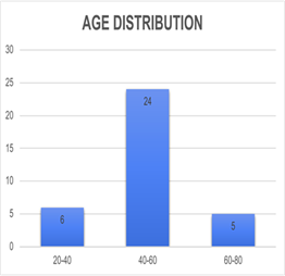
Fig 1: Age Group Distribution
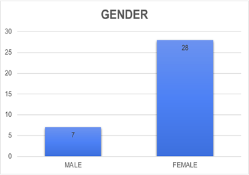
Fig 2: Gender Distribution
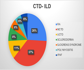
Fig 3: CTD Distribution
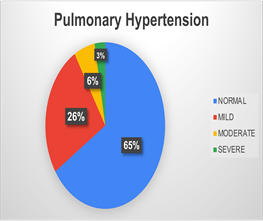
Fig 4: Pulmonary HTN Severity
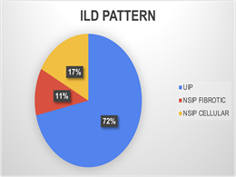
Fig 4: Pulmonary HTN Severity
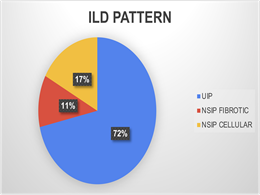
Fig 5: ILD Imaging Patterns
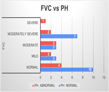
Fig 6: PH Distribution with FVC severity
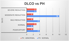
Fig 7: PH Distribution with DLCO severity
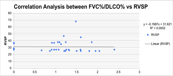
Fig 8: Correlation Analysis between FVC%/DLCO% vs RVSP
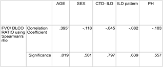
Table 1: FVC%/DLCO% Ratio using Spearman’s rho
Discussion:
CTD- ILD associated PH is often underdiagnosed significantly affecting morbidity, mortality and quality of life. The FVC%/DLCO% ratio reflects disproportionate gas exchange impairment, a hallmark of PH in ILD. Our study showed negative correlation, when combined use of FVC%/DLCO% ratio and echocardiography for suspecting PH.
Study by Xiong et al. also showed that the FVC/DLCO of more than 50% patients with CTD-PH was below 1.4, and only 10% of the patients had a ratio above 1.9, suggesting that the value of FVC%/DLCO% ratio as a predictor of PH had a limited value. 11 Similar study by Abdelwahab et al. demonstrated FVC/DLCO ratio with PH had no statistically significant association but concluded that the 6-MWT showed high validity in the prediction of PH.12
Nadia Sivova et al demonstrated that in Systemic sclerosis patients with PH-ILD, though elevated FVC/DLCO ratio is sensitive for PH, its specificity is limited and it should be interpreted in conjunction with other clinical parameters.13
However in a narrative review by Motschwiller et al described disproportionate reduction in DLCO relative to preserved FVC (elevated FVC/DLCO ratio) is emphasized as a key screening marker, while right heart catheterization remains essential for definitive diagnosis and for guiding multidisciplinary, phenotype-based management.14
Another study, Pulmonary Hypertension Assessment and Recognition of Outcomes in Scleroderma (PHAROS) cohort showed disproportionate reduction in DLCO with relatively preserved FVC and elevated FVC/DLCO ratio were early feature preceding PH diagnosis.15
LIMITATIONS:
This being a single centre study with small sample size and limited number of PH cases may not be generalizable to all CTD-ILD populations. The population of study was heterogeneous comprising different CTD and ILD patterns, which can influence the pulmonary function parameters. PH was assessed using Transthoracic Echocardiography rather than RHC, which may have led to misclassification of PH.
Conclusion:
The FVC%/DLCO% ratio has a screening tool failed to predict PH in patients with CTD-ILD in our study. More multicentre studies with larger sample size may be required to confirm its diagnostic utility as we had less patients with PH.pathogens like P.aeruginosa. Prudent antibiotic usage, detection of biofilm formation and high standards of hospital infection control practices aid in combating the Pseudomonas aeruginosa infection as well as preventing the development of resistant strains.
References:
1. Ersnet.org. [cited 2026 Jan 13]. Available from: https://publications.ersnet.org/content/erj/47/5/1535
2. Gutsche M, Rosen GD, Swigris JJ. Connective tissue disease-associated interstitial lung disease: A review. Curr Respir Care Rep [Internet]. 2012;1(4):224–32. Available from: http://dx.doi.org/10.1007/s13665-012-0028-7
3. Storrer KM, Müller C de S, Pessoa MC de A, Pereira CA de C. Connective tissue disease-associated interstitial lung disease. J Bras Pneumol [Internet]. 2024;50(1):e20230132. Available from: http://dx.doi.org/10.36416/1806-3756/e20230132
4. Fischer A, Strek ME, Cottin V, Dellaripa PF, Bernstein EJ, Brown KK, et al. Proceedings of the American college of rheumatology/association of physicians of great Britain and Ireland connective tissue disease-associated interstitial lung disease summit: A multidisciplinary approach to address challenges and opportunities. Arthritis Rheumatol [Internet]. 2019;71(2):182–95. Available from: http://dx.doi.org/10.1002/art.40769
5. Cansu DÜ, Korkmaz C. Pulmonary hypertension in connective tissue diseases: epidemiology, pathogenesis, and treatment. Clin Rheumatol [Internet]. 2023;42(10):2601–10. Available from: http://dx.doi.org/10.1007/s10067-022-06446-y
6. Humbert M, Kovacs G, Hoeper MM, Badagliacca R, Berger RMF, Brida M, et al. 2022 ESC/ERS Guidelines for the diagnosis and treatment of pulmonary hypertension. Eur Respir J [Internet]. 2023;61(1):2200879. Available from: http://dx.doi.org/10.1183/13993003.00879-2022
7. Patel D, Modugula SR, Rosa B, Niranjan S, Gutierrez L, Ramanujam D, et al. Pulmonary hypertension in connective tissue disease-interstitial lung disease (ctd-ild) combined clinic: A review. Chest [Internet]. 2024;166(4):A5605–6. Available from: http://dx.doi.org/10.1016/j.chest.2024.06.3325
8. Rhee RL, Gabler NB, Sangani S, Praestgaard A, Merkel PA, Kawut SM. Comparison of treatment response in idiopathic and connective tissue disease-associated pulmonary arterial hypertension. Am J Respir Crit Care Med [Internet]. 2015;192(9):1111–7. Available from: http://dx.doi.org/10.1164/rccm.201507-1456OC
9. Clements PJ, Tan M, McLaughlin VV, Oudiz RJ, Tapson VF, Channick RN, et al. The pulmonary arterial hypertension quality enhancement research initiative: comparison of patients with idiopathic PAH to patients with systemic sclerosis-associated PAH. Ann Rheum Dis [Internet]. 2012;71(2):249–52. Available from: http://dx.doi.org/10.1136/annrheumdis-2011-200265
10. Shi H, Gao P, Liu H, Su J, He X. Diagnostic value of combined FVC%/DLCO% and echocardiography in connective tissue disorder associated pulmonary hypertension. Med Int (Lond) . 2024;4(1):8.
11. Xiong J, Li J, Huang Y, Yang F, Wu R. The role of pulmonary function test for pulmonary arterial hypertension in patients with connective tissue disease. Dis Markers [Internet]. 2022;2022:6066291.
12. Abdelwahab HW, Hamoda HO, Shalabi NM, Arafa S, Morsy NE. Prediction of pulmonary hypertension in patients with interstitial lung disease using functional parameters. Egypt J Chest Dis Tuberc. 2025;74(3):620–628.
13. Guillén-Del-Castillo A, Meseguer ML, Fonollosa-Pla V, Giménez BS, Colunga-Argüelles D, Revilla-López E, et al. Impact of interstitial lung disease on the survival of systemic sclerosis with pulmonary arterial hypertension. Sci Rep [Internet]. 2022;12(1). Available from: http://dx.doi.org/10.1038/s41598-022-09353-z
14. Sivova N, Launay D, Wémeau-Stervinou L, De Groote P, Remy-Jardin M, Denis G, et al. Relevance of partitioning DLCO to detect pulmonary hypertension in systemic sclerosis. PLoS One [Internet]. 2013;8(10):e78001. Available from: http://dx.doi.org/10.1371/journal.pone.0078001
15. Hsu VM, Chung L, Hummers LK, Wigley F, Simms R, Bolster M, et al. Development of pulmonary hypertension in a high-risk population with systemic sclerosis in the Pulmonary Hypertension Assessment and Recognition of Outcomes in Scleroderma (PHAROS) cohort study. Semin Arthritis Rheum [Internet]. 2014;44(1):55–62. Available from: http://dx.doi.org/10.1016/j.semarthrit.2014.03.002