Does Mucin Matter? A Histopathological Study Comparing Mucinous and Non-Mucinous Colon Adenocarcinoma in a Tertiary Care Centre.
- Dr. Deepika , Assistant Professor Department of Pathology Karnataka Medical College and Research Institute (KMC&RI), Hubballi, 580021, Karnataka
- Dr. Sudeep , Post Graduate Department of Pathology Karnataka Medical College and Research Institute (KMC&RI), Hubballi, 580021, Karnataka
- Dr. Hito Jakhalu , Post Graduate, Department of Pathology Karnataka Medical College and Research Institute (KMC&RI), Hubballi, 580021, Karnataka
- Dr. Sunita Vernekar , Professor and Head of the Department Department of Pathology Karnataka Medical College and Research Institute (KMC&RI), Hubballi, 580021, Karnataka
Article Information:
Abstract:
Background: Colorectal carcinoma is a heterogeneous disease with distinct histopathological subtypes, including mucinous and non-mucinous adenocarcinoma. Mucinous adenocarcinoma (MAC) accounts for approximately 10–15% of colorectal cancers and has been associated with distinct clinicopathological characteristics compared with non-mucinous adenocarcinoma (NMAC). However, data from Indian populations are limited. Objective: To compare clinicopathological features of mucinous versus non-mucinous colon adenocarcinoma diagnosed in the Department of Pathology at KMC&RI, Hubballi between June 2023 and June 2025. Methods: A retrospective cross-sectional study was conducted. All histopathologically confirmed colon adenocarcinomas were classified into MAC (≥50% extracellular mucin) and NMAC (<50% mucin). Clinicopathological parameters including age, sex, tumor grade, tumor stage, lymphovascular invasion, and perineural invasion were compared. Data were entered into Microsoft Excel and analyzed using SPSS software. Results: Among 180 patients, 36 (20%) had MAC and 144 (80%) had NMAC. MAC patients were younger (mean age: 52.4 vs 59.1 years; p = 0.02). MAC showed higher frequency of advanced T stage (T3/T4: 83.3% vs 65.3%; p = 0.04) and lymphovascular invasion (58.3% vs 36.8%; p = 0.03). Higher proportions of advanced stage disease and poor differentiation were also observed in MAC, though not all differences were statistically significant. Conclusion: Mucinous adenocarcinoma exhibits distinct clinicopathological characteristics compared with non-mucinous tumors, including younger age at presentation and higher frequency of aggressive pathological features. Recognition of mucinous histology is important for pathological assessment and may have implications for clinical management.
Keywords:
Article :
Introduction :
Colorectal carcinoma (CRC) is a leading cause of cancer morbidity and mortality worldwide. Histologically, CRC is heterogeneous, with adenocarcinoma being the predominant type. Within this, mucinous adenocarcinoma (MAC) represents a distinct subtype characterized by abundant extracellular mucin comprising ≥50% of the tumor volume5. MAC accounts for approximately 10–15% of CRCs in Western populations 1,6.
Previous studies suggest that MAC has distinct clinicopathological features, including younger age of onset and advanced stage at diagnosis compared to non-mucinous adenocarcinoma (NMAC)3,7. However, data from Indian tertiary care centers are limited, and the clinical significance of mucin production remains debated8.
We conducted a histopathological comparison of MAC and NMAC in the Department of Pathology at Karnataka Medical College and Research Institute (KMC&RI), Hubballi over a period of two years (June 2023–June 2025) to evaluate whether mucinous histology correlates with clinically relevant differences.
Materials and Methods:
Study Design & Setting
This retrospective observational study was conducted at the Department of Pathology, Karnataka Medical College and Research Institute (KMC&RI), Hubballi.
Study Population
Inclusion criteria:
Patients with histologically confirmed primary colon adenocarcinoma diagnosed between June 2023 and June 2025 (2 years).
Exclusion criteria:
Rectal cancer, metastatic carcinoma to the colon, and inadequate histological material.
Histopathological Evaluation
Formalin-fixed paraffin-embedded (FFPE) sections stained with H&E were reviewed.
Tumors were classified as:
Mucinous adenocarcinoma (MAC): ≥50% mucin pools containing malignant cells5
Non-mucinous adenocarcinoma (NMAC): <50% mucin
Tumor grading (well, moderate, poor), staging (AJCC 8th edition), lymphovascular invasion (LVSI), and perineural invasion (PNI) were recorded.
Statistical Analysis
The data were analyzed using chi-square (χ²) test or Student’s t-test where appropriate. A p-value <0.05 was considered statistically significant.
Results:
MAC patients were significantly younger than NMAC patients (p = 0.02). Advanced tumor stage (T3/T4) and lymphovascular invasion were significantly more frequent in MAC (p = 0.04 and 0.03 respectively), suggesting a more aggressive pathological profile. No statistically significant difference was observed in lymph node metastasis, perineural invasion, sex distribution, or tumor differentiation, although trends toward higher poor differentiation and nodal positivity were noted in MAC [Table 1].
Table 1. Demographic and Clinicopathological Features of MAC and NMAC
|
Feature |
MAC (n=36) |
NMAC (n=144) |
p-value |
|
Age (mean ± SD), years |
52.4 ± 10.3 |
59.1 ± 11.8 |
0.02 |
|
Male sex, n (%) |
22 (61.1) |
89 (61.8) |
0.93 |
|
T3/T4 stage, n (%) |
30 (83.3) |
94 (65.3) |
0.04 |
|
Lymph Node Positive, n (%) |
19 (52.8) |
62 (43.1) |
0.29 |
|
LVSI present, n (%) |
21 (58.3) |
53 (36.8) |
0.03 |
|
PNI present, n (%) |
12 (33.3) |
38 (26.4) |
0.41 |
|
Poor differentiation, n (%) |
16 (44.4) |
42 (29.2) |
0.10 |
*MAC = mucinous adenocarcinoma; NMAC = non-mucinous adenocarcinoma; LVSI= lymphovascular invasion; PNI = perineural invasion.
Histopathological examination demonstrated characteristic morphological features of mucinous adenocarcinoma, including abundant extracellular mucin pools containing floating malignant epithelial cells. [ Figure 1]
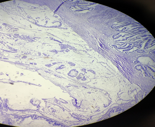
Figure 1: Photomicrograph showing mucinous adenocarcinoma of colon with abundant extracellular mucin pools containing floating malignant epithelial cells (Hematoxylin and Eosin stain, ×40).
Conventional adenocarcinoma cases showed well-formed glandular architecture with tumor infiltration into the muscularis propria in advanced lesions. [Figure 2]
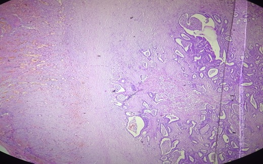
Figure 2:Photomicrograph showing well differentiated adenocarcinoma of colon forming glandular structures infiltrating into muscularis propria (Hematoxylin and Eosin stain, ×40).
MAC demonstrated a higher proportion of advanced stage disease (Stage III and IV combined: 61.1%) compared with NMAC (50.7%). Early-stage disease (Stage I and II) was relatively less common in MAC. This distribution supports the observation that mucinous tumors tend to present at a more advanced stage at diagnosis [Table 2].
Table 2. Stage Distribution Between MAC and NMAC
|
Stage |
MAC (n=36) |
NMAC (n=144) |
|
Stage I |
4 (11.1) |
22 (15.3) |
|
Stage II |
10 (27.8) |
49 (34.0) |
|
Stage III |
14 (38.9) |
53 (36.8) |
|
Stage IV |
8 (22.2) |
20 (13.9) |
Discussion:
This study demonstrates significant clinicopathological differences between mucinous and non-mucinous colon adenocarcinoma in a tertiary care setting. The proportion of MAC in our cohort (20%) is slightly higher than the commonly reported 10–15% in Western literature, which may reflect geographic or referral bias, or population-specific tumor biology1,6.
A key finding in our study was the younger age at presentation in MAC compared to NMAC. Similar observations have been reported in earlier studies suggesting mucinous tumors may develop through distinct molecular pathways, often associated with microsatellite instability and specific genetic alterations3,8.
We also observed significantly higher rates of advanced T stage and lymphovascular invasion in MAC. These findings align with Lagoudianakis et al., who reported that mucinous tumors often show deeper bowel wall invasion and aggressive local behavior 3. Mucin pools may facilitate tumor spread by acting as a physical medium for tumor cell migration and by modifying tumor microenvironment interactions, potentially aiding immune evasion 7.
Although lymph node positivity and perineural invasion were higher in MAC, these differences did not reach statistical significance in our study. Similar mixed findings have been reported in literature, with some studies demonstrating higher nodal metastasis in MAC while others show no significant difference after stage adjustment 9.
Stage distribution analysis showed a greater proportion of Stage III and IV disease in MAC. This supports prior observations that mucinous tumors tend to present late, possibly due to their infiltrative growth pattern and subtle early clinical presentation 6.
Regarding tumor differentiation, although not statistically significant, a higher proportion of poorly differentiated tumors was observed in MAC. Previous studies have suggested mucinous tumors may be associated with poor differentiation and signet ring morphology, both of which are linked to aggressive tumor biology 5.
From a clinical standpoint, recognition of mucinous histology is important because it may:
· Indicate potentially aggressive tumor biology
· Influence decisions regarding surveillance intensity
· Suggest the need for molecular testing such as MSI status and RAS mutation profiling
Our findings support the concept that mucin is not merely a histological feature but may reflect underlying biological differences influencing tumor behavior.
Conclusion:
In this study, mucinous colon adenocarcinoma demonstrated distinct clinicopathological features compared to non-mucinous tumors. Mucin appears to matter in terms of tumor behavior and pathological aggressiveness
References:
1. Bosman FT, Carneiro F, Hruban RH, Theise ND. WHO Classification of Tumours of the Digestive System. 4th ed. Lyon: IARC; 2010.
2. Siegel RL, Miller KD, Jemal A. Cancer statistics, 2023. CA Cancer J Clin. 2023;73(1):17–48.
3. Lagoudianakis EE, Kataki A, Papadima A, et al. Mucinous histology in colorectal cancer: epidemiology and prognostic value. World J Gastrointest Oncol. 2014;6(12):462–469.
4. Mei Z, Shi L, Wang B, et al. Prognostic Role of Mucinous Histology in Colorectal Cancer: A Systematic Review and Meta-Analysis. J Natl Cancer Inst. 2018;110(8):831–840.
5. Compton C, Fenoglio-Preiser CM, Pettigrew N, Field MP. Colorectal carcinoma: pathologic aspects. Arch Pathol Lab Med. 2000;124(7):979–994.
6. Hugen N, van de Velde CJ, de Wilt JH, Nagtegaal ID. Metastatic pattern in colorectal cancer is strongly influenced by histological subtype. Ann Oncol. 2014;25(3):651–657.
7. Ueno H, Mochizuki H, Hashiguchi Y, et al. Prognostic factors for colorectal cancer: use of factor combinations and histologic type. Dis Colon Rectum. 2004;47(7): 1006–1014.
8. Zaanan A, Shi Q, Taieb J, et al. Prognostic effect of mucinous histology in colon cancer: a population-based study. Ann Oncol. 2018;29(2):343–350.
9. Verhulst J, Ferdinande L, Demetter P, et al. Mucinous subtype as prognostic factor in colorectal cancer: a systematic review and meta-analysis. J Clin Pathol. 2012;65(5):381–388.