Histomorphological Spectrum of Peripheral Nerve Sheath Tumours Diagnosed in a Tertiary Care Hospital of Northeast India
- Shreyoshi Dhar , MD Pathology, Senior Resident, Dept. Of Pathology, Agartala Government Medical College & Gobind Ballabh Pant Hospital, Agartala (Tripura), 799006, India
- Tushar Ghosh , MD Pathology, Senior Resident, Dept. Of Pathology, Agartala Government Medical College & Gobind Ballabh Pant Hospital, Agartala (Tripura), 799006, India
- Prasenjit Roy , Post Graduate Trainee, Dept. Of Pathology, Agartala Government Medical College & Gobind Ballabh Pant Hospital, Agartala (Tripura), 799006, India
- Kulashekhar Bhattacharjee , MD Pathology, Associate Professor, Dept. Of Pathology, Agartala Government Medical College & Gobind Ballabh Pant Hospital, Agartala (Tripura), 799006
- Alpana Banerjee , MD Pathology, Associate Professor, Dept. Of Pathology, Agartala Government Medical College & Gobind Ballabh Pant Hospital, Agartala (Tripura), 799006,
Article Information:
Abstract:
Background: Peripheral nerve sheath tumours (PNSTs) comprise a heterogeneous group of neoplasms arising from the supporting cells of peripheral nerves. These tumours range from benign entities such as Schwannoma and Neurofibroma to malignant peripheral nerve sheath tumours (MPNSTs). Due to their diverse histomorphological patterns and variable clinical behavior, accurate diagnosis is essential for appropriate management. Data regarding the histomorphological spectrum of PNSTs from Northeast India remains limited. Objectives: To study the histomorphological spectrum of peripheral nerve sheath tumours and analyse their distribution with respect to age, sex, and anatomical site in a tertiary care hospital. Materials and Methods: This retrospective descriptive study included 65 histopathologically confirmed cases of peripheral nerve sheath tumours diagnosed over a defined study period in a tertiary care hospital of Northeast India. Clinical details, including age, sex, and tumour location, were retrieved from records. Hematoxylin and Eosin–stained sections were reviewed, and tumours were classified according to World Health Organization criteria. Descriptive statistical analysis was performed. Results: Of the 65 cases, 57 (87.7%) were benign, and 8 (12.3%) were malignant. Neurofibroma was the most common benign tumour, followed by Schwannoma. All malignant cases were diagnosed as MPNST. The head and neck region was the most frequently involved site. Schwannomas showed male predominance, whereas Neurofibromas and malignant peripheral nerve sheath tumours were more common in females. Benign tumours predominantly occurred in young to middle-aged adults. Conclusion: Benign peripheral nerve sheath tumours constitute the majority of PNSTs, with Neurofibroma being the most common subtype. Histomorphological evaluation remains the cornerstone for accurate diagnosis. Regional studies such as this provide valuable epidemiological data and aid in better understanding the clinicopathological profile of PNSTs.
Keywords:
Article :
Introduction:
Peripheral nerve sheath tumours (PNSTs) comprise a diverse group of neoplasms arising from the cellular components of peripheral nerves, including Schwann cells, perineurial cells, and endoneurial fibroblasts. These tumours account for approximately 10–12% of all soft tissue neoplasms and exhibit a wide spectrum of biological behavior ranging from indolent benign lesions to highly aggressive malignant tumours.1,2 Accurate classification of PNSTs is essential because prognosis and therapeutic strategies differ significantly among tumour subtypes.
According to the World Health Organization (WHO) classification of soft tissue and bone tumours, PNSTs are broadly categorized into benign and malignant forms.3 The most common benign PNSTs include Schwannoma and Neurofibroma, while malignant peripheral nerve sheath tumour (MPNST) represents the malignant counterpart.4 Schwannomas are typically well-circumscribed, encapsulated tumours composed entirely of Schwann cells and demonstrate characteristic Antoni A and Antoni B areas with Verocay bodies.5 In contrast, Neurofibromas are unencapsulated lesions composed of a mixture of Schwann cells, fibroblasts, perineurial-like cells, and axons, often showing an infiltrative growth pattern.6
MPNSTs are rare but clinically significant due to their aggressive nature, high recurrence rates, and poor overall survival.7 These tumours may arise de novo or in association with pre-existing Neurofibromas, particularly in patients with neurofibromatosis type 1 (NF1).8 Histologically, MPNSTs exhibit increased cellularity, nuclear atypia, brisk mitotic activity, necrosis, and loss of normal nerve architecture, often posing diagnostic challenges and requiring careful morphological evaluation supplemented by immunohistochemistry.9,10
Clinically, PNSTs may occur at any anatomical site, with a predilection for the head and neck region, trunk, and extremities.11 Patients typically present with slow-growing, painless masses; however, pain, neurological deficits, or rapid enlargement may suggest malignancy.12 Radiological imaging aids in localization, but definitive diagnosis relies on histopathological examination.13
Despite extensive literature on PNSTs from Western populations, there is limited data from Northeast India, a region with distinct demographic and genetic characteristics.14 Regional studies are crucial to understand variations in tumour distribution, site predilection, and histomorphological patterns. Moreover, most available Indian studies focus on either benign or malignant PNSTs, with few providing a comprehensive spectrum analysis.15,16
Furthermore, advances in immunohistochemistry, including the use of markers such as S100 protein, SOX10, and Ki-67, have significantly enhanced diagnostic accuracy and prognostic assessment.17,18
The present study aims to analyse the histomorphological spectrum of peripheral nerve sheath tumours in a tertiary care hospital in Northeast India, emphasizing their distribution according to age, sex, and anatomical site. Such data will contribute to existing literature and assist pathologists and clinicians in improving diagnostic accuracy and patient management.
OBJECTIVES
1. To study the histomorphological spectrum of diagnosed Peripheral nerve sheath tumours over a span of 5 years.
2. To analyse age, sex, and site distributions of Peripheral nerve sheath tumours.
Results:
A total of 65 cases of peripheral nerve sheath tumours (PNSTs) were analysed during the study period. Of these, 57 cases (87.7%) were benign, while 8 cases (12.3%) were malignant. Among the benign tumours, Schwannoma constituted the majority, followed by Neurofibroma. All malignant tumours were diagnosed as Malignant peripheral nerve sheath tumours (MPNSTs).
Distribution Based on Histological Type
Schwannoma was the most frequently encountered tumour, accounting for the largest proportion of benign PNSTs. Neurofibroma represented the second most common benign lesion. Malignant peripheral nerve sheath tumours formed a smaller subset of cases but constituted the entire malignant category in the present study is depicted in Table 2 and shown in pie chart 1.
Gender-wise distribution of Tumour Types
Schwannoma showed a male predominance with 17 cases in males and 10 cases in females. In contrast, Neurofibroma was more frequently observed in females, accounting for 25 cases, while 5 cases were noted in males. Malignant peripheral nerve sheath tumour (MPNST) demonstrated a slight female predominance, with 5 cases in females compared to 3 cases in males is depicted in Table 3 & shown in bar diagram 1.
Age and Sex distribution
Peripheral nerve sheath tumours were observed across a wide age range, spanning from the second to the seventh decade of life. The highest number of cases was recorded in the 41–50 year age group for both males and females. Female patients outnumbered males across most age groups, while no male cases were observed in the 51–60 year age group, which is depicted in Table 4 and shown in bar diagram 2.
Anatomical Site Distribution
The head and neck region was the most frequently involved anatomical site, accounting for 57% of cases. This was followed by tumours arising from the back (24%). Lesions involving the upper and lower extremities constituted 10% of cases, while the remaining 9% were distributed across other less common anatomical locations. The distribution of PNSTs according to anatomical site is depicted in Pie chart 2.
Table 2: Histopathlogically diagnosed peripheral nerve sheath tumours (PNSTs)
|
Diagnosis |
Number of cases |
Percentage (%) |
|
Schwannoma (benign) |
25 |
38.5 |
|
Neurofibroma (benign) |
32 |
49.2 |
|
Malignant peripheral nerve sheath tumour (MPNST) |
8 |
12.3 |
|
Total |
65 |
100 |
Pie Chart 1: Peripheral Nerve Sheath Tumours
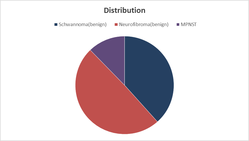
A total of 65 histopathologically confirmed PNST cases were analysed, of which Neurofibroma was the most common subtype (49.2%), followed by Schwannoma (38.5%). Malignant peripheral nerve sheath tumours accounted for 12.3% of cases (Table 2)
Table 3: Gender wise distribution of tumour types:
|
Tumour type |
Males (n) |
Females (n) |
Total |
|
|
Schwannoma |
17 |
10 |
27 |
|
|
Neurofibroma |
5 |
25 |
30 |
|
|
Malignant peripheral nerve sheath tumour (MPNST) |
3 |
5 |
8 |
|
|
Total |
25 |
40 |
65 |
|
Bar diagram 1: Gender wise tumour distribution:
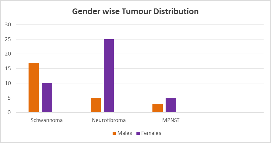
Gender-wise analysis revealed male predominance in Schwannoma, whereas Neurofibroma and MPNST were more common among females (Table 3).
Table 4: Age and Sex distribution
|
Age group (years) |
Males (n) |
Females (n) |
Total |
|
11–20 |
2 |
3 |
5 |
|
21–30 |
5 |
7 |
12 |
|
31–40 |
7 |
10 |
17 |
|
41–50 |
12 |
14 |
26 |
|
51–60 |
0 |
1 |
1 |
|
61–70 |
1 |
3 |
4 |
|
Total |
27 |
38 |
65 |
Most cases were seen in the fourth and fifth decades, with a higher number of female patients across age groups (Table 4).
Table 5: Anatomical site distribution:
|
Anatomical site |
Number of cases (n = 65) |
Percentage (%) |
|
Head and neck |
37 |
57 |
|
Back |
16 |
24 |
|
Upper and lower extremities |
7 |
10 |
|
Other sites |
5 |
9 |
|
Total |
65 |
100 |
Pie chart 2: Anatomical site distribution:
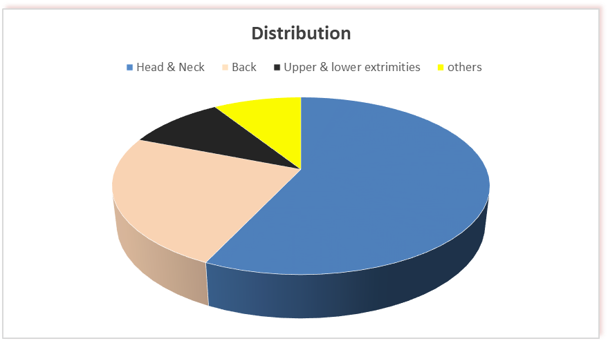
The head and neck region was the most common site of involvement, followed by the back and extremities (Table 5).
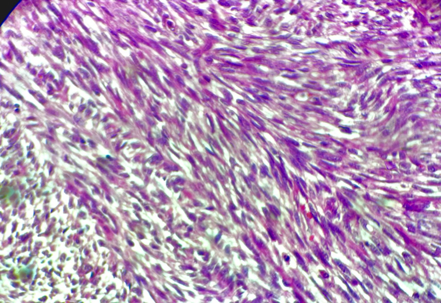
Figure 1:100X view of H&E slide of MPNST showing an interlacing fascicles pattern.
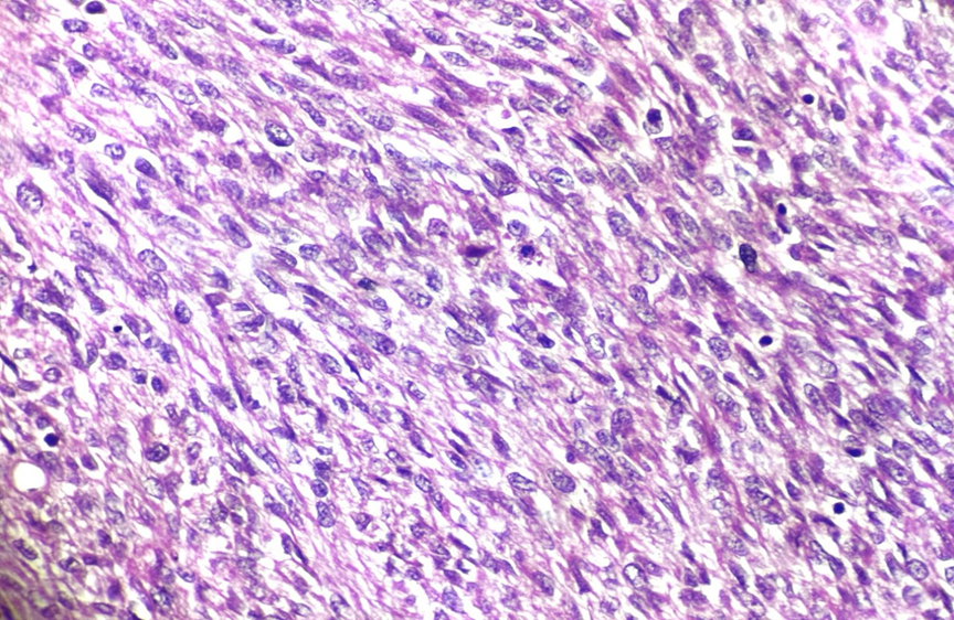
Figure 2: 100X view of H&E slide of MPNST with cellular areas showing elongated hyperchromatic spindle cells, irregular nuclear contours and with moderate to marked nuclear plomorphism. Frequent mitotic figures are also noted.
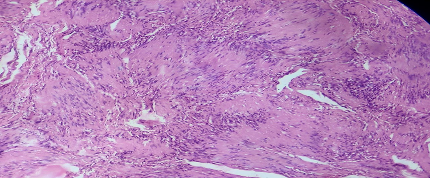
Figure 3: 100X view of H&E slide of schwannoma showing a dimorphic pattern of cellular (Antoni A) and loose myxoid areas (Antoni B)
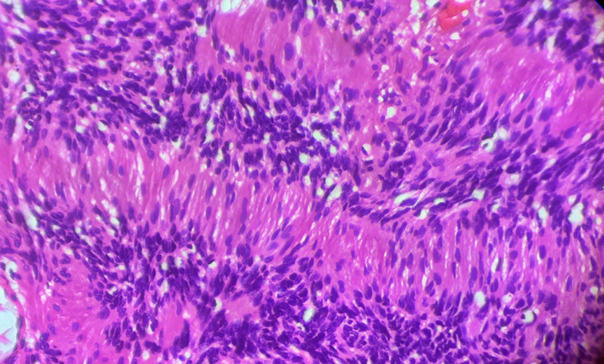
Figure 4: 100X view of H&E slide of schwannoma showing verocay bodies in Antoni A areas (hypercellular areas).
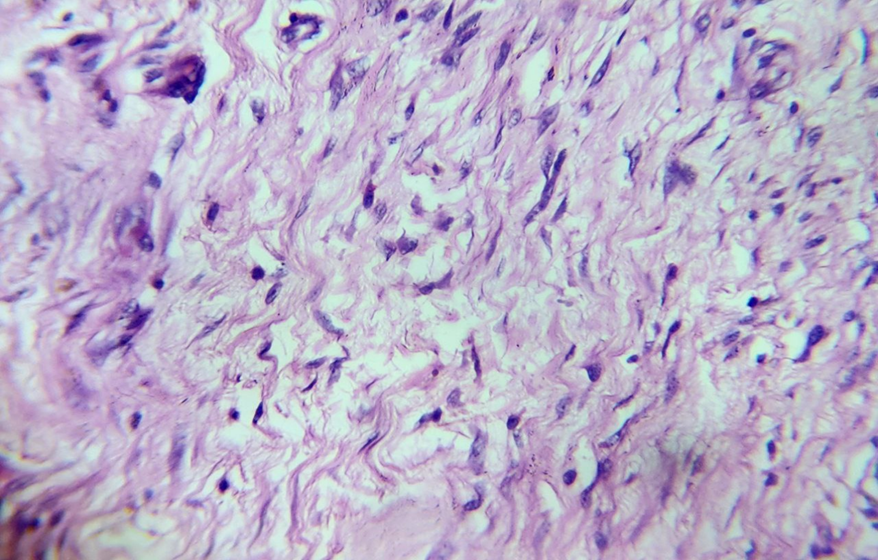
Figure 5: 100X view of H&E slide of Neurofibroma showing spindle cells with wavy nucleus in a background of myxoid matrix.
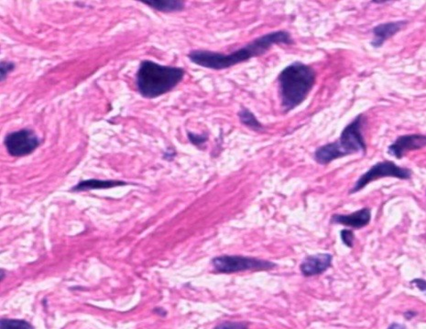
Figure 6: 100x view of H&E slide of neurofibroma showing spindle cells with thin, wavy nuclei and stromal collagen showing shredded carrot appearance.
Discussion:
In the present series, benign peripheral nerve sheath tumours constituted the majority of cases, while malignant peripheral nerve sheath tumours (MPNSTs) formed a smaller but clinically significant subset. Neurofibroma emerged as the most common benign tumour in this study, followed by Schwannoma, reinforcing previously documented trends.
The histomorphological features of Schwannomas in this study were characteristic, including encapsulation. Neurofibromas were predominantly unencapsulated, and Recognition of these classical features is essential for accurate diagnosis, particularly in limited biopsy specimens.
MPNSTs accounted for all malignant cases in this series. Histologically, these tumours exhibited features of malignancy such as increased cellularity, nuclear pleomorphism, mitotic activity, and necrosis. Although MPNSTs are rare, their aggressive nature, potential for local recurrence, and poor prognosis make their identification critical. In the present study, histomorphology remained the primary diagnostic modality, highlighting its importance in routine pathology practice.
Gender-wise analysis revealed a male predominance in Schwannomas, while Neurofibromas and MPNSTs were more common among females.
The head and neck region was the most common anatomical site involved, followed by the back and extremities. This distribution pattern is comparable to several published studies and may be attributed to the rich peripheral nerve supply in the head and neck region. Tumours of the trunk and extremities were less frequent but formed a significant proportion of cases.
Age distribution analysis demonstrated that benign PNSTs predominantly affected young to middle-aged adults, whereas malignant tumours tended to present at a relatively older age. Although syndromic association was not assessed in detail in the present study, its potential contribution cannot be excluded.
Regional studies such as this provide valuable epidemiological data and enhance understanding of disease patterns in specific populations.
Many of these nerve sheath tumours were clinically diagnosed as lipoma, granuloma, etc. Which, after histopathological examination came out to be peripheral nerve sheath tumours.
Discussion comparison to other studies:
|
NAME OF AUTHOR |
CURRENT STUDY |
MARACHAPU.J et al19
|
CHIKKANNAIAH.P et al20
|
SUNITA B PATIL AND CHATURA KR et al21 |
|
Age group |
41-50 |
21-30 |
21-30
|
21-30 |
|
Site of tumour |
Head & Neck |
Head & Neck |
Head & Neck |
Head & Neck |
|
Predominant type |
Neurofibroma |
Neurofibroma |
Neurofibroma |
Neurofibroma |
Limitations
The number of cases was fewer. Moreover, IHC and molecular diagnostics have not been implemented in this study.
Conclusion:
Incidence of benign tumour was higher in our studies, with Neurofibroma being the most common type, followed by Schwannoma. Malignant peripheral nerve sheath tumour, although less frequent show distinct histomorphological features indicative of aggressive behaviour. Female preponderance was noted. 41-50 years is the most common age group. Therefore, histopathological examination is a must to rule out malignancy and also to rule out clinically diagnosed cases like lipoma, granuloma, etc.
References:
1. Enzinger FM, Weiss SW. Soft Tissue Tumors. 6th ed. Philadelphia: Elsevier; 2014.
2. Fletcher CDM, Bridge JA, Hogendoorn PCW, Mertens F. WHO Classification of Tumours of Soft Tissue and Bone. Lyon: IARC; 2020.
3. Goldblum JR, Folpe AL, Weiss SW. Enzinger and Weiss’s Soft Tissue Tumors. 7th ed. Philadelphia: Elsevier; 2019.
4. Ducatman BS, Scheithauer BW, Piepgras DG, Reiman HM, Ilstrup DM. Malignant peripheral nerve sheath tumors. Cancer. 1986;57(10):2006–21.
5. Woodruff JM. Peripheral nerve tumors: histologic classification and clinical significance. Am J Surg Pathol. 1983;7(7):637–52.
6. Rodriguez FJ, Folpe AL, Giannini C, Perry A. Pathology of peripheral nerve sheath tumors. Brain Pathol. 2012;22(1):51–65.
7. Stasik CJ, Tawfik O. Malignant peripheral nerve sheath tumor. Arch Pathol Lab Med. 2006;130(4):586–93.
8. Ferner RE. Neurofibromatosis 1 and neurofibromatosis 2: a twenty-first century perspective. Lancet Neurol. 2007;6(4):340–51.
9. Miettinen M, Antonescu CR, Fletcher CDM, Kim A, Lazar AJ. Diagnostic criteria for malignant peripheral nerve sheath tumors. Mod Pathol. 2016;29(Suppl 1):S91–104.
10. Thway K, Fisher C. Diagnostic pathology of peripheral nerve sheath tumors. Histopathology. 2014;64(1):25–40.
11. Pilavaki M, Chourmouzi D, Kiziridou A, et al. Imaging of peripheral nerve sheath tumors. Eur Radiol. 2004;14(4):748–59.
12. Baehring JM, Betensky RA, Batchelor TT. Malignant peripheral nerve sheath tumor: clinical features and prognosis. J Neurooncol. 2003;61(3):297–306.
13. Kim DH, Murovic JA, Tiel RL, Moes G, Kline DG. A series of peripheral nerve sheath tumors. J Neurosurg. 2005;102(2):246–55.
14. Rekhi B, Gorad BD, Kakade AC, Chinoy RF. Clinicopathological study of peripheral nerve sheath tumors. Indian J Pathol Microbiol. 2010;53(4):630–36.
15. Jain D, Sharma MC, Sarkar C, Suri V, Garg A. Malignant peripheral nerve sheath tumors: clinicopathologic study. J Cancer Res Ther. 2009;5(2):97–101.
16. Gupta G, Maniker A. Malignant peripheral nerve sheath tumors. Neurosurg Focus. 2007;22(6):E12.
17. Nonaka D, Chiriboga L, Rubin BP. Differential diagnosis of peripheral nerve sheath tumors using SOX10 immunohistochemistry. Am J Surg Pathol. 2008;32(9):1293–98.
18. Miettinen M, Shekitka KM, Sobin LH. Schwannomas in soft tissue: a clinicopathologic study of 165 cases. Am J Surg Pathol. 1994;18(9):882–94.
19. Marachapu J. Histomorphological spectrum of peripheral nerve sheath tumors from a secondary care hospital. Indian J Appl Res. 2023;13(3).
20. Chikkannaiah P, Boovalli MM, Nathiyal V, Venkataramappa S. Morphological spectrum of peripheral nerve sheath tumors: An insight into World Health Organization 2013 classification. J Neurosci Rural Pract. 2016;7(3):346-54.
21. Patil SB, Chatura KR. A histopathological study of neurofibroma and malignant peripheral nerve sheath tumors in a medical institute of Central Karnataka. Int J Clin Diagn Pathol. 2021;4(1):81–5.