Histopathological spectrum of large gut lesions on colonoscopic biopsies and their correlated immunohistochemistry
- Dr. Komal Yadav , Post Graduate Resident, Department of Pathology, Govt. Medical College, Jammu, Jammu and Kashmir India
- Dr Akhtar Un Nisa Salaria , Assistant Professor, Department of Pathology, Govt. Medical College, Jammu, Jammu and Kashmir India
- Dr. Pratibha Shalini , Senior Resident, Department of Pathology, Govt. Medical College, Jammu, Jammu and Kashmir India
- Dr. Anu Radha , Assistant Professor, Department of Radiodiagnosis, Rama Medical College Hospital & Research Centre, Hapur. (Corresponding Author)
- Dr. Subash Bhardwaj , Professor. & Ex. Head, Department of Pathology, Govt. Medical College Jammu
Article Information:
Abstract:
Background: The large intestine is affected by a wide range of disorders, including inflammatory, infectious, benign, and malignant conditions. Colonoscopic biopsy offers a minimally invasive diagnostic approach, with histopathology as the gold standard. Immunohistochemistry (IHC) further refines diagnosis, especially in morphologically ambiguous or poorly differentiated cases. Materials and Methods: A retrospective, hospital-based descriptive study was conducted in the Department of Pathology, Government Medical College, Jammu. Archived colonoscopic biopsy specimens of large gut lesions were reviewed. Hematoxylin and eosin (H&E) stained slides and formalin-fixed paraffin-embedded (FFPE) blocks were examined. Lesions were classified as benign, malignant, inflammatory/infective, or developmental. Relevant IHC markers were applied and correlated with histological features. Data were analyzed using descriptive statistics. Results: Among 200 biopsies, 119 were males (59.5%) and 81 females (40.5%), with a peak incidence in the 31–40-year age group. Inflammatory lesions were most frequent (42%), followed by benign (28%), malignant (21.5%), infectious (5%), and developmental (3.5%) lesions. Representative entities included adenomas, adenocarcinomas, carcinoid tumors, GISTs, and melanomas. Conclusion: Inflammatory and benign lesions predominated, though malignant cases were significant. IHC enhanced diagnostic precision and subclassification, underscoring colonoscopic biopsy as a vital tool for early and accurate evaluation of large gut pathologies.
Keywords:
Article :
Introduction:
Diseases of the gastrointestinal (GI) tract are among the most frequently encountered conditions in routine clinical practice [1]. Biopsies obtained from different segments of the GI tract form a significant proportion of the specimens received in the histopathology department of any tertiary care hospital [2].
The large bowel and anal canal are affected by a wide spectrum of pathological conditions, both neoplastic and non-neoplastic, which are frequently encountered in clinical settings [3][4]. The variety of conditions that can arise across different portions of the tract is therefore extensive. These may present as either acute or chronic disorders and include infectious lesions, vascular pathologies, ulcerative processes, chronic inflammatory bowel diseases, as well as benign and malignant tumors. In most cases, colonoscopic biopsy followed by histopathological examination serves as the cornerstone for establishing a definitive diagnosis [5][6].
Among the available diagnostic tools, histopathological examination remains the gold standard, offering the highest sensitivity and specificity, particularly for the detection of malignant lesions. It plays an essential role not only in confirming the nature of the disease but also in guiding timely clinical management [7][8]
The present study is designed to analyze the prevalence of different types of lesions encountered in large gut colonoscopic biopsies and to characterize the broad spectrum of their histopathological features. In addition, it aims to evaluate the diagnostic utility of relevant immunohistochemical markers in differentiating and confirming various large gut lesions. By integrating clinical data with both histological patterns and immunohistochemical findings, the study seeks to achieve a comprehensive correlation that enhances diagnostic accuracy and contributes to better clinical management of patients with large gut pathology.
OBJECTIVES OF THE STUDY
Primary Objective
· To study the histopathological spectrum of large gut lesions in colonoscopic biopsies.
Secondary Objectives
· To classify large gut lesions into inflammatory/infectious, benign, malignant, and developmental categories.
· To evaluate the role of immunohistochemistry in confirming and subtyping large gut lesions, especially in diagnostically challenging cases.
Materials and Methods:
Study Design
This retrospective, observational, hospital-based descriptive observational study was conducted in the Department of Pathology, Government Medical College (GMC), Jammu, using archival histopathology material and reports.
Sample size and sampling technique:
No formal sample size calculation was performed, and convenience sampling was used for 1 year, including all patients diagnosed with large gut lesions on histopathological examination during the study period.
Inclusion criteria:
All histopathologically diagnosed large gut lesions with available H&E-stained slides and FFPE tissue blocks.
Exclusion criteria:
Autolyzed specimens and inadequate tissue samples.
Sampling Procedure and Sample Size
Non-probability convenience sampling was used, including all eligible archived cases. The sample size was 200, determined by the total number of cases available; no statistical formula was applied.
Ethical Clearance
As the study involved archival material with no patient interaction, informed consent was waived. Confidentiality was maintained, and approval was obtained from the Institutional Ethics Committee, GMC Jammu. (IEC/GMCJ/2025/075, dated: 18-01-2025)
Tissue Processing and H&E Staining
Biopsy specimens were fixed in 10% neutral buffered formalin, processed routinely, embedded in paraffin, sectioned at ~3 μm, and stained with H&E using standard protocols.
Histopathological Review and IHC Correlation
Slides were independently reviewed by experienced pathologists. Selected cases underwent immunohistochemistry using appropriate panels (CK7/CK20, CDX2, p53, Ki-67, CD117, DOG1, Chromogranin A, Synaptophysin) to confirm diagnosis.
The lesions were further characterized into the following categories based on histopathological features:
1. Benign lesions
2. Malignant lesions
3. Inflammatory/infectious lesions
4. Developmental lesions
Data Analysis Procedure
All collected data were compiled and entered into Microsoft Excel spreadsheets. Descriptive statistical methods were applied, and data were analyzed using Statistical Package for Social Sciences (SPSS) software.
Results:
Table 1: Distribution of Study Participants based Gender and Age:
|
Variable |
Category |
Frequency (n) |
Percentage (%) |
|
Gender |
Male |
119 |
59.5 |
|
Female |
81 |
40.5 |
|
|
Age group (years) |
0–10 |
11 |
5.5 |
|
11–20 |
8 |
4.0 |
|
|
21–30 |
20 |
10.0 |
|
|
31–40 |
48 |
24.0 |
|
|
41–50 |
43 |
21.5 |
|
|
51–60 |
34 |
17.0 |
|
|
61–70 |
22 |
11.0 |
|
|
71–80 |
14 |
7.0 |
|
|
Total |
200 |
100 |
A total of 200 participants were included in the study, with a male predominance (59.5%) over females (40.5%). The most common age group was 31–40 years (24%), followed by 41–50 years (21.5%) and 51–60 years (17%). Fewer cases were observed at the extremes of age, with 9.5% below 20 years and 18% above 60 years.
Table 2: Distribution of Study Participants based on histopathological features:
|
Histopathology Category |
Frequency (n) |
Percentage (%) |
|
INFLAMATORY |
84 |
42 |
|
BENIGN |
56 |
28 |
|
MALIGNANT |
43 |
21.5 |
|
DEVELOPMENTAL |
7 |
3.5 |
|
INFECTIOUS |
10 |
5 |
The most frequent category was inflammatory lesions, accounting for 84 cases (42%), followed by benign lesions with 56 cases (28%). Malignant lesions were observed in 43 cases (21.5%), while developmental anomalies contributed to 7 cases (3.5%).
In order to distinguish infection-related pathology from non-infectious inflammatory conditions, a separate category of infectious lesions was created, comprising 10 cases (5%). This adjustment highlights that while the majority of specimens were inflammatory in nature.
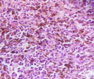
Figure 1: Malignant melanoma: Tumor cells are predominantly polygonal with abundant eosinophilic cytoplasm containing melanin pigment, and nuclei showing prominent eosinophilic nucleoli.
Figure 2 a: Basaloid type of squamous cell carcinoma at the anorectal junction, presenting clinically as a rectal polyp.
2 b: IHC of basaloid variant of squamous cell carcinoma showing strong nuclear/cytoplasmic positivity for p63 (brown) in tumor nests against a hematoxylin counterstain (blue).
Figure 3: a. 400× H&E-stained photomicrograph showing moderately differentiated adenocarcinoma of the colon, demonstrating malignant glands with pleomorphic epithelial cells and increased mitotic activity.
b. IHC photomicrograph of colonic adenocarcinoma showing irregular malignant glands with strong cytoplasmic immunopositivity for Cytokeratin 20 (CK20), confirming the epithelial origin of the neoplasm.
Figure 4: Inflammatory lesions- Photomicrograph of colonic mucosa from a case of ulcerative colitis, stained with H&E at 100× magnification, showing crypt shortening with architectural distortion.
Figure 5: H&E stain photomicrograph showing GIST, characterized by spindle-shaped tumor cells arranged in fascicles with elongated nuclei and eosinophilic cytoplasm.
2a 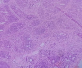 2b
2b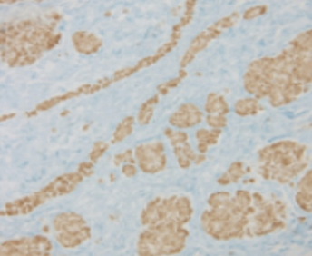
3a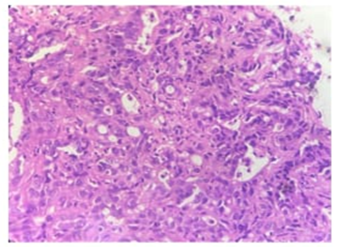 3b
3b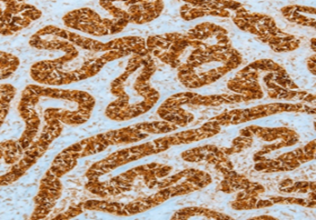
4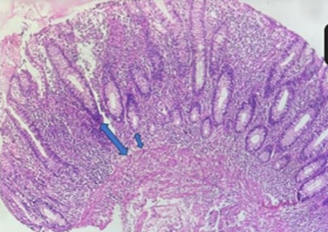 5
5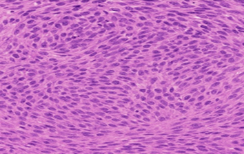
Discussion:
A wide spectrum of inflammatory disorders affects the colon and rectum, presenting as acute or chronic conditions. Non-neoplastic lesions include colitis of various etiologies, inflammatory bowel disease, irritable bowel syndrome, and diverticular disease, while neoplastic lesions range from benign polyps to colorectal carcinoma (CRC) [9]. CRC is among the most common malignancies worldwide and a leading cause of cancer-related mortality [10]. Globally, CRC ranks third in incidence, with an annual increase of approximately 3% [11]. In India, CRC incidence is lower in rural populations, likely due to dietary factors such as higher fiber intake [12].
In the present study of 200 colonoscopic biopsies, males predominated (59.5%), similar to other studies [13,14]. Most patients were middle-aged, supporting the trend of higher prevalence in this age group. Inflammatory lesions were the most frequent (42%), followed by benign (28%) and malignant lesions (21.5%). Infectious lesions were analyzed separately (5%), emphasizing the dominance of inflammatory pathology. Compared with previous studies [15-19], our findings show a higher inflammatory burden and a carcinoma incidence comparable to population-based studies, highlighting the influence of geographic and methodological factors on colorectal pathology patterns.
Conclusion:
Colonoscopic biopsy is an effective tool for evaluating the wide histopathological spectrum of large gut lesions, with inflammatory lesions being the most common, followed by benign and malignant entities. Immunohistochemistry significantly aids in accurate diagnosis and subclassification, particularly in poorly differentiated and ambiguous lesions. The combined use of histopathology and IHC enhances diagnostic precision and supports timely clinical management, aligning well with the study objectives.
Relevance of the study: This study highlights the diagnostic value of combining histopathology with immunohistochemistry for accurate classification and early detection of large gut lesions on colonoscopic biopsies.
Limitation of the study: Being a retrospective, hospital-based study with limited IHC and no follow-up or molecular analysis, the findings may not reflect true population prevalence.
|
Lesion |
IHC Markers |
Diagnostic Role |
|
Adenocarcinoma |
CK20, CDX2 |
Confirms colorectal epithelial origin |
|
Neuroendocrine tumor |
Chromogranin A, Synaptophysin |
Confirms neuroendocrine differentiation |
|
GIST |
CD117, DOG1 |
Distinguishes from other spindle cell tumors |
|
Squamous cell carcinoma |
p63 |
Confirms squamous differentiation |
|
Malignant melanoma |
S-100, HMB-45 |
Confirms melanocytic origin |
|
Inflammatory lesions |
Ki-67 |
Highlights increased proliferative activity |
References:
1. Bamanikar S, Sonkawade D, Bhandari P, Bamanikar A, Chandanwale S, Buch A. Histomorphology Spectrum of Gastrointestinal Lesions in a Tertiary Care Centre. Acta Sci Cancer Biol. 2019;3(12):4-11.
2. Fenoglio-Preiser CM, Noffsinger AE, Stemmerman GN, Lantz PE, Isaacson PG. Gastrointestinal pathology: an atlas and text. 3rd ed. Philadelphia: Lippincott Williams & Wilkins; 2008. Chapter 14, Epithelial neoplasms of the colon; p. 99–1036.
3. Gill MK, Jain K, Manjari M, Kaur T. Expression of Her2/neu in Colon Carcinoma and Its Correlation with the Histological Grades and the Lymph nodes status. Journal of Clinical and Diagnostic Research. 2011; 5(8): 1564-68.
4. Scheull B, Gruenberger T, Scheithauer W, Zielinski Ch, Wrba F. Her 2/neu protein expression in colorectal cancer. BMC Cancer. 2006,6:123-27.
5. Liu C, Crawford JM. The Gastrointestinal tract. In: Kumar, Abbas, Fausto, editors. [1] Robbins and Cotran Pathologic Basis of Disease. 7th ed. Philadelphia. Saunders, 2004; 97: 876.
6. Qayyum A, Sawan AS. Profile of colonic biopsies in King Abdul Aziz University Hospital, Jeddah. J Park Med Assoc. 2009; 59(9):608-11.
7. Dajani YF, Kamal MF. Colorectal juvenile polyps. An epidemiological and histopathological study of 144 cases in Jordanians. Histopathology. 1984;8(5):765–79.
8. Nwafor CC, Nwafor NN, Etuk EB, Kanu O. Histopathological spectrum of gastrointestinal lesions seen in university of uyo teaching hospital, South–South Nigeria. Annals of Tropical Pathology. 2019;10(1):27-33.
9. Sulegaon R, Kulkarni D, Yelikar B. Histological spectrum of large intestinal lesions with clinicopathological correlation. J Clin Diagn Res. 2015;9(11):EC30-4.
10. Heppner BI, Behrens HM, Balschun K, Haag J, Krüger S, Becker T, et al. HER2/neu testing in primary colorectal carcinoma. Br J Cancer. 2014;111(10):1977-84.
11. Bernard L. Nutrition and colorectal cancer. Cancer. 1992;70(6):1723-6.
12. Mohandas KM, Desai DC. Epidemiology of digestive tract cancers in India. V. Large and small bowel. Indian J Gastroenterol. 1999;18(3):118-21.
13. Biswal R, Behera S, Kar A, Sahoo B. Histopathological spectrum of lower gastrointestinal colonoscopic biopsy lesions with special reference to HER2/neu expression in carcinoma colon. Int J Res Pharm Sci. 2020;11(SPL4):1233-40.
14. Deo SVS, Shukla NK, Srinivas G, Mohanti BK, Raina V, Sharma A, et al. Colorectal cancers: a study of 50 cases at a regional cancer centre. Indian J Cancer. 2001;38(1):29-34.
15. Yanik S, Altintas E, Kekilli M, Dagli U, Uygun A, Demir K, et al. Evaluation of results of lower gastrointestinal endoscopic biopsy. Int J Clin Exp Med. 2014;7(12):5820-5.
16. Rajbhandari M, Karmacharya A, Khanal K, Dhakal P, Shrestha R. Histomorphological profile of colonoscopic biopsies and pattern of colorectal carcinoma in Kavre District. Kathmandu Univ Med J. 2015;11(3):196-200.
17. Florea MA, Becheanu G, Niculae A, Dobre M, Costache M. Immunohistochemical insights into the pathogenesis of colonic sessile serrated lesions. Arch Clin Cases. 2025 Mar;12(1):22-28.
18. Paragannavar V, Nidagundi V, Paragannavar L. Clinicopathological spectrum of intestinal lesions: a 2-year study in a tertiary care center. MGM J Med Sci. 2025;12(3):469–75.
19. Hanumanthu S, Reddy VS, Kumar A. Histopathological spectrum of inflammatory and neoplastic colorectal lesions in a tertiary care centre. Int J Acad Med Pharm. 2024;6(4):660