Role of Optic Nerve Sheath Diameter in Anasethesia for Modified Electroconvulsive Therapy
- Dr. Pavithra J , Final Year Postgraduate in Anaesthesia, Department of Anaesthesia, Dhanalakshmi Srinivasan Medical College and Hospital, Siruvachur, Perambalur, Tamil Nadu, India
- Dr. Maivizhi S. , Assistant Professor, Department of Anaesthesia, Dhanalakshmi Srinivasan Medical College and Hospital, Siruvachur, Perambalur, Tamil Nadu, India
- Dr. Geetha J , Professor, Department of Anaesthesia, Dhanalakshmi Srinivasan Medical College and Hospital, Siruvachur, Perambalur, Tamil Nadu, India.
Article Information:
Abstract:
Introduction: The thought and mood disorders contribute to a major class of mental illnesses, around 13.7% of people have affected mental health disorders in India as per 2025 statistics. Modified electroconvulsive therapy has occupied a major role in treating schizophrenia and bipolar disorders. Changes in intracranial pressure noted in these patients and anticipated during electrical stimulation, seizure response or due to the disease process itself causing structural changes in the brain. Anaesthetics and antipsychotics may reduce ICP. Hence monitoring intracranial pressure may give us a lead towards progress in the line of management. Optic nerve sheath diameter has been documented as a sensitive indicator of intra cranial pressure. It is a advantageous being non-invasive, real time and simple. In this study, patients recommended for MECT had three measurements of ONSD before during and after seizure response, the strength of the electrical stimulus and the duration of seizure were also part of the study. Aims And Objectives: To evaluate the role of optic nerve sheath diameter (ONSD) in anesthesia for MECT.To correlate the effects of anesthetic drugs and convulsions on ICP via ONSD. To examine the effects of repetitive MECT on ONSD. To observe correlation between electric charge and ONSD. To evaluate feasibility of ONSD as an indicator of progress or recovery of the disorder. Materials And Methods: Study Population: All patients with bipolar disorder in depression and patients with schizophrenia as diagnosed by psychiatrists and recommended for MECT of both genders were included for the study. Study Place: Dhanalakshmi Srinivasan Medical College and Hospital, Siruvachur, Perambalur. Study Period: January 2025 to December 2025. Study: Prospective Observational Clinical study. Study Sample: 84 observations. Equipments: Ultrasound by LOGIQ e Ref: 5419804 window XP embedded SN 24113WXO June 2012 20v/5A/. ECT Machine: NiviQure INT-5 Integrated ECT system 30 mC= 4.8 J. Methods: All patients included in the study underwent thorough preanesthetic work up for comorbidities, systemic illnesses, and liver functions. As patients were emotionally challenged all underwent measurement of optic nerve sheath diameter on the left eye when they were sedated. After warming up and setting the ultrasound machine for superficial image study. Results: Pre procedure, ONSD showed a p of 0.003 with age, ONSD, immediately after seizure response showed p of 0.029 and post procedure ONSD depicted p of 0.008, higher ONSD with the mean of 6.8 MM was noted in schizophrenics. 84 observations from 12 patients demonstrated excellent inter-measurement agreement with Pearson coefficient of 0.84 to 0.92. Conclusion: Optic nerve sheath diameter appears to be a promising non-invasive real time simple parameter to monitor anticipated changes during anesthesia for modified electroconvulsive therapy. While MECT is a supplement to pharmacotherapy it may also reduce duration of medications and their requirement. Thus ONSD is of great advantage to document Neuro protection in repeated MECT with our findings of normalization of raised ONSD Especially with clinical improvement in schizophrenics, ONSD may play a role as prognostic indicator in future.
Keywords:
Article :
Introduction:
burden and calculation disability adjusted life years (DALY) and years lost to disability. Mental and substance use disorders are the third leading cause of DALY just next to cardiovascular and respiratory diseases.[1] Lifetime prevalence of mental disorders in India is 13.7% as per 2025 statistics. Urban areas have high prevalence and 70-92% people do not receive propre treatment due to lack of awareness, stigma and shortage of professionals.
Modified Electroconvulsive therapy plays a major role in speeding up recovery and reduce pharmacotherapy as an economic burden to the affected families. Mechanism of electroconvulsive therapy include memory disruption and abandoned amnesia hypothesis, neuroendocrine, neuro-plasticity, receptor hypothesis and cytokine hypothesis.[2]
Modulation of neurotransmitter systems and neuropeptides following electroconvulsive therapy have been well documented.
Anesthetic needs for MECT include tolerance of the electrical stimuli, sedation, amnesia without affecting seizure expression, neuroprotection, muscle relaxation, maintenance of airway, protection from aspiration and early recovery with reduced post ECT cognitive impairment.
Variations in intracranial pressure due to ECT and drugs are anticipated while organic changes that do occur with chronic mental illnesses such as schizophrenia also contribute to rise in intracranial pressure due to ventricular dilatation which tends to normalize with clinical improvement.
Ultrasound measurement of optic nerve sheath diameter proves promising monitor of intracranial pressure (ICP). The trend of ONSD seems to correlate with clinical recovery from anesthesia and the mental illness. Thiopentone sodium and succinyl choline have opposing actions on ICP and the effects of convulsions itself are mixed.
In this study, we have attempted to measure ONSD in the peri MECT period and tried to analyze the role of ONSD in correlating response to MECT and course of the mental disorders.
Aims and Objectives
1. To evaluate the role of optic nerve sheath diameter (ONSD) in anesthesia for MECT.
2. To corelate the effects of anesthetic drugs and convulsions on ICP via ONSD.
3. To examine the effects of repetitive MECT on ONSD.
4. To observe correlation between electric charge and ONSD.
5. To evaluate feasibility of ONSD as an indicator of progress or recovery of the disorder.
Materials and Methods:
Study Population
All patients with bipolar disorder in depression and patients with schizophrenia as diagnosed by psychiatrists and recommended for MECT of both genders were included for the study.
Study Place
Dhanalakshmi Srinivasan Medical College and Hospital, Siruvachur, Perambalur.
Study Period
January 2025 to December 2025.
Study
Prospective Observational Clinical study.
Study Sample
84 observations
Equipment
Ultrasound by LOGIQ e Ref: 5419804 window XP embedded SN 24113WXO June 2012 20v/5A/
ECT Machine
NiviQure INT-5 Integrated ECT system 30 mC= 4.8 J.
Methods
All patients included in the study underwent thorough preanesthetic work up for comorbidities, systemic illnesses, and liver functions. As patients were emotionally challenged all underwent measurement of optic nerve sheath diameter on the left eye when they were sedated. After warming up and setting the ultrasound machine for superficial image study.
After lubricating with lignocaine gel the linear probe was placed lightly on left upper eyelid for few seconds, clear image of vitreous humor, optic nerve head and sheath below were obtained and image frozen. The probe was immediately taken off the patient.
Measurements were taken on the frozen image with same operator and an assistant who froze the image.
Operator errors were attempted to be minimized by participation of the same team. Once the measuring cursor was accurately placed the values in CM were given by the equipment on the screen in real time which was noted and converted to millimeters for correlation purposes.
The optic nerve sheath diameter was taken after a sleep dose of thiopentone noted as base value (ONSD 1), after an electric energy administration (EL millicoulmts) by bitemporal electrodes by the psychiatrist immediately after the convulsive response settled (ONSD 2) and following recovery of spontaneous breathing by the patient while he/she was still sedated (ONSD 3). All patients received thiopentone sodium 3mg per kg body weight as sleep dose, injection Glycopyrrolate 0.2 mg, followed by Injection Succinyl choline 50mg. Airway was supported by oxygenation with face mask via adult Bain’s circuit from anesthesia work station. Soft bite block to prevent tongue bite and jaw support with extension of neck by operator’s hand were offered during this convulsion as protective measures.
Hyperventilation by bag mask was done after scoline administration during apnea to threshold. Care of airway with gentle suction during post ictal phase was given. Pulse, blood pressure and ECG monitoring was done throughout the procedure.
The electrical energy applied (millicoulombs) and duration of seizure response in seconds were noted as displayed in the ECT machine. Along with patient demographics, ECT repetition number, electrical energy, duration of seizures, optic nerve sheath diameter at various phases of MECT were all noted, tabulated and analyzed.
Observations & Results:
An ONSD of 5mm corresponds to an ICP of around 20 mmhg.
Study Population and Measurements
A total of 84 observations from 12 patients were analyzed, with a mean age of 36.1 ± 8.9 years. Each patient underwent repeated testing (median 4, range 1–13). ONSD was measured three times per test, and the mean ONSD was used for downstream analysis.
Measurement Reliability
ONSD measurements demonstrated excellent inter-measurement agreement, with Pearson correlations ranging from 0.84 to 0.92 across the three readings. The mean coefficient of variation was 4.7%, indicating low measurement noise and supporting the use of averaged ONSD as a reliable outcome variable.
Distributional Properties
The distribution of mean ONSD was non-normal (Shapiro–Wilk p = 0.0017), justifying the use of non-parametric statistical methods for group comparisons and correlations.
Gender Differences
A statistically significant difference in ONSD was observed between genders (Mann–Whitney U p = 0.0049). The effect size was moderate (Cliff’s delta = −0.36), suggesting a clinically relevant gender-associated difference in ONSD values.
Age Association
ONSD demonstrated a significant positive association with age (Pearson r = 0.33, p = 0.0019; Spearman ρ = 0.52, p < 10⁻⁶), indicating that ONSD increases monotonically with age.
Treatment-Related Factors
● MECT number showed no significant association with ONSD (p > 0.15), suggesting no cumulative test-order effect.
● Energy dose (mC) demonstrated a strong positive association with ONSD (Spearman ρ = 0.52, p < 10⁻⁶), indicating a clear dose–response relationship.
● Duration (seconds) was not significantly associated with ONSD.
Within-Patient Longitudinal Trends
Individual patients showed heterogeneous ONSD trajectories, with some exhibiting significant increasing or decreasing trends. However, the overall mean slope across patients was not significantly different from zero (p = 0.80), indicating that population-level stability masks substantial individual variability.
Additionally, ONSD variability decreased modestly with increasing test number (ρ = −0.23, p = 0.039), suggesting possible physiological adaptation or measurement stabilization over time.
Mixed-Effects Modeling
A mixed-effects model accounting for repeated measures revealed:
● Significant between-patient variability (Group variance = 0.69)
● No independent fixed effects of test number, age, or gender after accounting for patient-level clustering
This indicates that inter-individual differences dominate ONSD variation, exceeding the contribution of measured covariates in the adjusted model.
Overall Interpretation
ONSD is a highly reliable measurement with strong associations to age and energy dose, moderate gender differences, and pronounced patient-specific effects. Population-level averages obscure meaningful individual trajectories, underscoring the importance of longitudinal and mixed-effects approaches in repeated-measure clinical data.
Gender Distribution
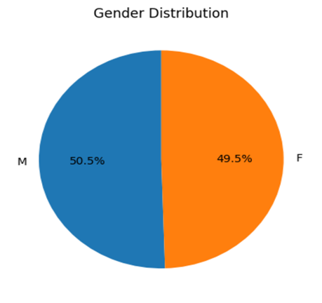
Figure 1: Gender Distribution
In our study group the gender distribution was nearly equal 50.5% males and 49.5% females.
Comparison of the 3 ONSD Values
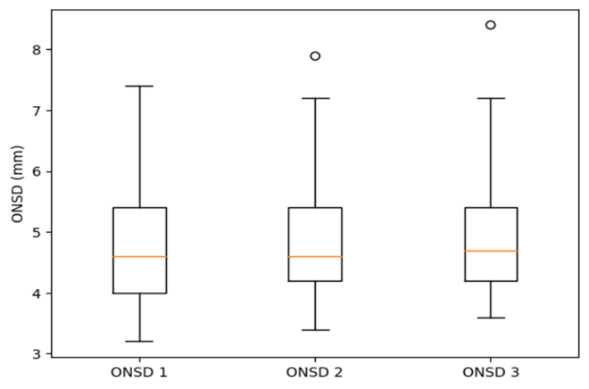
Figure 1: Comparison of the 3 ONSD Values
Though noninvasive ultrasound is the modality the measurement was done when the patients were still under mild sedation as they were emotionally challenged to understand and accept a probe on the left eyelid. Hence the change in ONSD may be more significant than the actual value itself.
The ONSD measurements 1, 2, 3 were pre-ECT, immediate post ECT, post ictal near recovery values.
ONSD 1 was between four and 5.5 MM, with mean at 4.7 5MM, ONSD 2- 4.5 and 5.6 and ONSD 3-4.5 and 5.75 as upper limits.
Mean ONSD versus Age
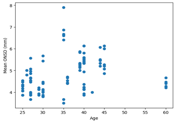
Figure 3: Mean ONSD Versus Age
Scatter plot between age in years and mean ONSD shows maximum values with age 35 years (8) and the most values between 35 to 40 years of age.
Mean ONSD versus Psychiatric Diagnosis
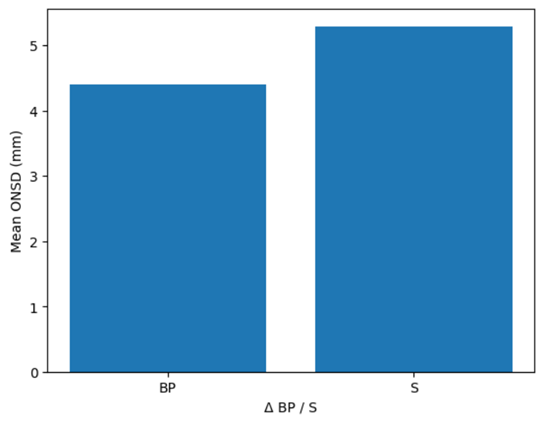
Figure 4: Mean ONSD versus Psychiatric Diagnosis
Namely, bipolar disorder and schizophrenia. The bar diagram between the diagnosis and mean ONSD shows higher values of more than 5.5 MM in schizophrenic patients.
Gender Wise ONSD Differences

Figure 5: Gender Wise ONSD Differences
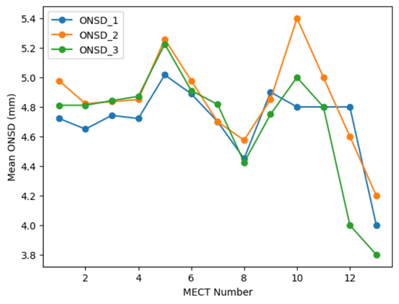
Figure 6: Repetitive MECT and Mean ONSD
Most of our patients needed multiple sittings of MECT. The mean ONSD 1, 2 and 3 values were plotted against the number of sittings. The ONSD 2 which is immediate post ECT reached more the 5.4 peak with 10th sitting of ECT. All the rises reached near normalcy with clinical improvement.
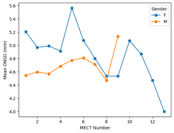
Figure 7: Mean ONSD and Repetitive MECT Gender Wise
The highest mean ONSD was 5.6 MM recorded in female and dipped to 4MM in females showing increased variability in females and lesser peak and nil drop below 4.6 MM in mails.
Correlation Matrix Depicting ‘R’ Values with all Parameter
(a) Poor correlation of ONSD with strength of electric stimuli (E in mc)
(b) Poor correlation of ONSD with duration of seizure response to MECT
(c) Good correlation of ONSD pre-and post MECTs (r=0.87)
(d) Poor correlation between age and ONSD (r=0.3)
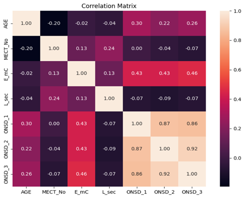
Figure 8: Correlation Matrix Depicting ‘R’ Values with all Parameter
Pearson R and P Values-Correlation Matrix
(a) ONSD 2- ONSD immediately after ECT response showed p of 0.02 with age
(b) p of 0.016 with MECT repetition, numbers and duration of seizure response
(c) ONSD 1 showed a p of 0.003 with age
(d) ONSD 2 showed 0.029 and ONSD 3 0.008
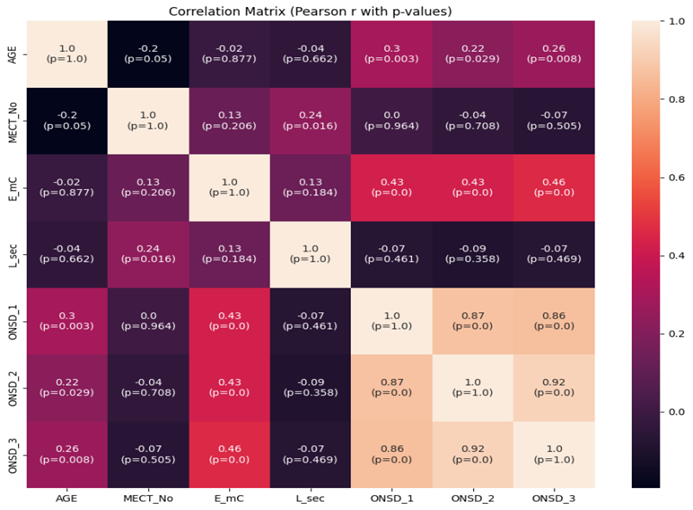
Figure 9: Pearson R and P Values-Correlation Matrix
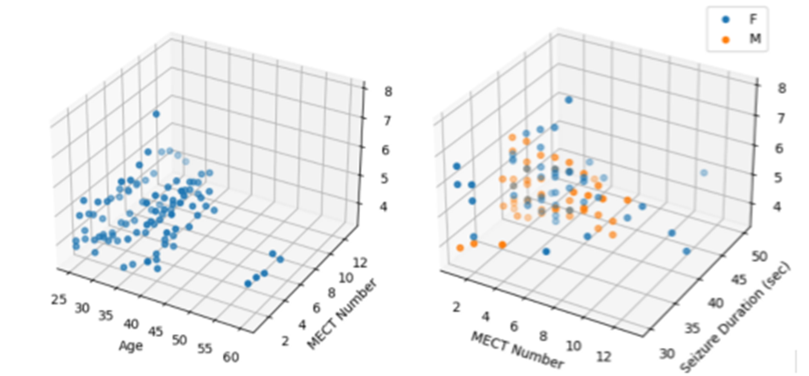
Figure 10: Lattice Plot between Age and MECT Repetition Number
Younger individuals and females needed more number of MECTs.
Scatter Plots between the Parameters along with Histogram
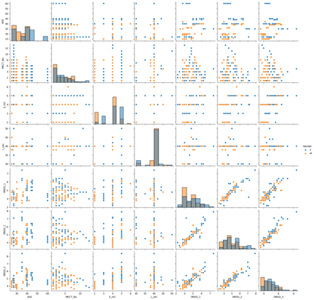
Figure 11: Scatter Plots between the Parameters along with Histogram
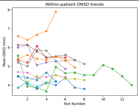
Figure 12: Patient ONSD Trends
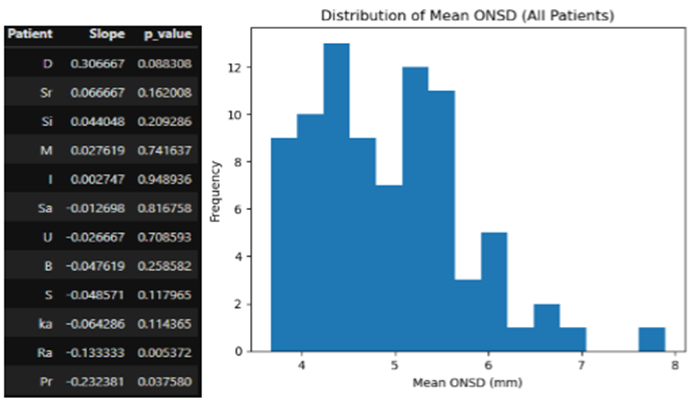
Figure 13: Distribution of Mean ONSD
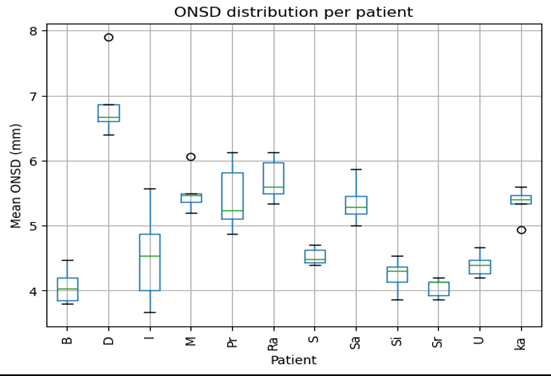
Figure 14: ONSD Distribution Per Patient
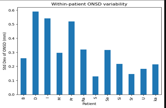
Figure 15: Patient ONSD Variability
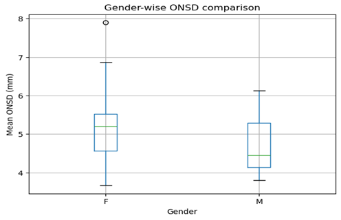
Figure 16: Gender Wise ONSD Comparison
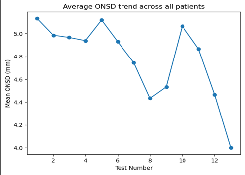
Figure 17: Average ONSD Trends
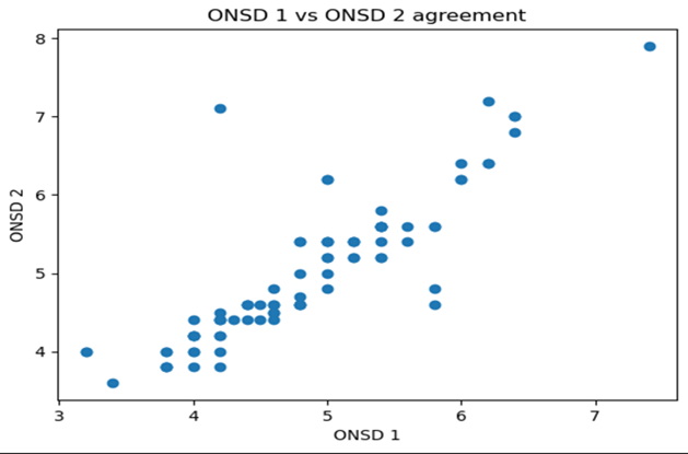
Figure 18: ONSD 1 VS ONSD 2
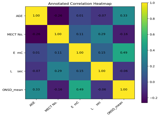
Figure 19: Annotated Correlation Heatmap
Discussion:
Treatment for psychiatric illnesses was limited until 1930s. Old somatic therapies included, hydrotherapy, sleep therapy, insulin coma and scotch douches were all attempted as various means to treat patients,[3] next came Barbiturates, bromides, chloral hydrate and paraldehyde until 1950s. In 1934, a Hungarian neuropsychiatrist Ladislas Joseph Von Meduna experimented repeated seizures as treatment for schizophrenia. Fink in 2001 and later in 2004 reaffirmed theory of biological antagonism which involved the idea of inducing one illness as treatment for another.[4] Induction of malarial fever as a cure for neurosyphilis yielded Nobel prize in medicine in 1928 for prof.Julcus Wagener Juaregg of Vienna. Thus convulsive therapy became acceptable and spread throughout Europe with clinical successful trials.
After camphor and pentylene fetoaxol electricity was used to induce seizures. By mid 1950s negative portrayals of ECT as a cruel therapy in the media declined the usage of ECT. Major innovations including use of anesthesia, oxygenation, muscle relaxation and seizure monitoring made ECT being increasingly recognized as a proven, effective intervention especially in pharmacologically refractive psychiatric disorders.[5]
The discussion on this paper is elaborated on the following topics:
1. Electroconvulsive therapy and its indications
2. Schizophrenia and the depressive psychosis-the two indications taken up in this study
3. Outline of anaesthesia principles for MECT
4. Neuro and sono anatomy of optic nerve sheath
5. Optic nerve sheath diameter and allied measurements
6. Results and conclusions
7. Limitations and summary
8. Recommendations and future research
Electroconvulsive Therapy and its Indications
Mood disorders and schizophrenia constitute the major and common conditions proving good response to electroconvulsive therapy.
Major depressive disorder or depressive phase of bipolar disorder and schizophrenia, along with catatonia see to improve with ECT when compared to using pharmacotherapy alone, bipolar disorder is frequently associated with cognitive and functional deterioration posing treatment challenges.[6] electroconvulsive therapy has a unique place in therapeutic armamentarium for bipolar, especially in highly suicidal patients catatonia and drug refractory disease.[7]
ECT is generally classified as UECT and MECT. UECT or unmodified ECT involves bilateral stimulation with a sine wave voltage sources at the powerline frequency without anaesthesia.
MECT maybe Uni or bilateral stimulation with individually titrated trains of ultra brief or brief rectangular constant current pulses under general anaesthesia. The impetus for these optimisations has been to prevent physical and cognitive side-effects. Low GABAminergic function is a subset of mood disorder. Over the course of treatment ECT itself results In decreased neural metabolic activity[8] older age; higher ECT dose and increased repetitive number were associated with shorter sizures. Enhanced activity of inhibitory circuits of human Motor Cortex and pharmacological therapy augumented treatment outcomes.
Mechanism of ECT is further explained by neurophysiological principles. Repeated electrically induced seizures increase seizure threshold and therefore mood stability by increasing activity of GABAergic neurons that regulate the functions of Neuro circuits relieving aspects of neuro vegetative depression such as rumination, anxious, distress, mania and catatonia.[9]
Serum proactive levels increase to 10 to 50 fold in response to seizures induced by ECT along with short increases in oestrogen stimulating neurohypophysis and nicotine stimulating neurohypophysin, atleast for 6 minutes post ECT.[10] Banki and colleagues observed reduced levels of corticotropin releasing factor in cerebrospinal fluid to correlate with clinical improvement following ECT.
Following electrical, electrophysiological and hormonal changes evident after ECT, molecular subtrates involving kynurenine pathway, and its metabolic have emerged as potential mediators in pathophysiology of depression. Tryptophan is the basic amino acid of serotonin that degrades along this pathway. Tryptophan also consist of QUIN and KYNA, NMDA receptor agonist and antagonist respectively. Schwieler et al found that after ECT, QUIN plasma levels and QUIN/KYNA ratios were decreased in 80% of patients.[11]
While treating severe depression with ECT glutamate and glutamine increased significantly, suggesting excitatory transmitter enhancement after ECT in major depression.
Magnetic resonance imaging studies after ECT shows volumetric, changes across cortical and sub cortical regions. Approximately 5% increase in grey matter subfields of hippocampus and basolateral nuclei of amygdala, anterior cingulate, post central fusiform Gyri and medial prefrontal cortex.[12]
Schizophrenia
Pathophysiology, structural changes in the brain and correlation with intra cranial pressure
Schizophrenia is one of the most devastating of mental illnesses, often strikes early in live and may signal an irreversible vulnerability towards psychosis creating lifetime burden for the patients and their loved ones. Psychodynamic and Neuro dynamic theories have been proposed to explain the disorder. Environmental, social, genetic and familial factors have been identified as significant contributors. Symptoms of schizophrenia occur due to shifts and conflicts in the hardware of mentation as well as conflicts and warps in the software of psychology.
Neuro dynamic theories concentrate on biologically programmed neuronal networks in dynamic communication via chemical and electrical connections.[13,14] neuroradiological postmortem studies have suggested reduced hippocampal volume and disarray of its pyramidal cells, suggesting insufficient number of synaptic connections that link the neurons of the system. Wide range of input information and co-erced into single memory activation pattern, explaining the term parasitic memory.[14]
Parasitic memories are products of biophysical energy processes
Subcortical dopamine dysfunction is the key mechanism underlying in Patho physiology of schizophrenia.
Neuro anatomical changes are seen in prefrontal, medial and superior temporal lobes as reduced Gray matter volume[15] resulting in ventricular dilatation. The enlarged cerebral ventricles could explain a borderline increase in the intracranial pressure in schizophrenics. CT imaging has demonstrated generalized brain tissue Loss along with ventricular enlargement in schizophrenic when compared to controls.[16] Negative symptoms, executive function impairment have been found to be associated with structural abnormalities in a range of brain networks.[17]
The ventricular dilatation correlates with the baseline Increase In optic nerve sheath diameter denoting a rise in intracranial pressure. The Administration of ECT only marginal raised ICP according to our study which then settled to baseline levels on recovery from MECT. In schizophrenics the rise was noted in spite of the sleep dose of thiopentone sodium as induction agent.
In two cases of schizophrenia, the clinical recovery correlated with baseline normalization of ONSD/ ICP. Late onset schizophrenia was associated with increased ventricle-to-brain ratio.[18]
Bipolar Disorder: (BD)
Bipolar disorder is a severe and chronic psychiatric illness that affects 1-4% of world population.[19] It is characterized by changes in mood that alternate between mania and hypomania or depression and mixed states often associated with functional impairment.[20] environmental, neurochemical and genetic factors contribute to etiology of BD. Neurotrophic factors a group of proteins responsible for regulating neuronal survival processes, neuronal growth, synaptic information and Neuroplasticity. Of these brain desired Neurotrophic factor BDNF has been found to be affected in BD. Decreased levels of BDNF have been noted along with that of few other factors in depressive like behaviors in animal models. Antidepressants and mood stabilizers could act on these molecules modulating their signaling pathways in BD.
BD has heterogeneous symptomatology and diagnosis is purely based on clinical criteria, history taking, interviewing and behavioural observation. Brain imaging studies have shown evidence of change in regional activity, functional connectivity and bioenergetics associated with BD. No structural changes signifying raised ICP or change in ventricular size has been found in BD In contrast to schizophrenia. MECT has been applied as treatment modality based on clinical judgement to supplement a pharmacotherapy that yielded reduced outcome.
In our study of serial changes of ONSD in individual patients during recurring settings of MECT, no significant rise in ONSD was noted In contrast to schizophrenic patients.
Optic Nerve Sheath Diameter
The changes in the diameter of the optic nerve reflects real time. Changes in intracranial pressure. This acts as a useful adjunct to a standard diagnostic modalities. The eyeball rests in a membranous sac called tenon capsule, surrounded by Periocular fat, soft tissue and orbital walls. It attaches via corneoscleral junction and optic nerve with extra ocular muscle tendons piercing the Tenon sac inserting the sclera and allowing eyeball mobility on multiple axis. Two fluid filled structures, the anterior and posterior chamber are separated by the lens and both harbour anechoic fluid. Posterior to the posterior chamber is the retina with hyper echoic nervous membrane.[21] optic nerve is identified by its anechoic core posterior to the retina and optic disc. This is flanked by a hyper echoic sheath on the medial and lateral border in the transverse plane.
The optic nerve can be defined as an outpouching of intact brain tissue with the intra orbital component fully encapsulated by Dura, pia and arachnoid matter, allowing optic nerve sheath to transmit cerebrospinal fluid and fluctuate in size based on changes in intracranial pressure.[22] the meningeal cover of the optic nerve is the continuation of dural and subarachnoid Space. The bulbous portion of the optic nerve approximately 3MM posterior to the globe, appears more distensible and sensitive to changes in ICP.[23]
The upper limit of the normal ONSD includes: 4MM in infants, 5MM in children and 5MM in adults. ONSD shows a linear increment until 7.5 MM after which it plateaus.[24] ONSD is the best non-invasive modality for dynamic estimation of ICP.[22] ICP can be calculated using the formula:
ONSD: n ICP ONSD = 5.00 x ONSD – 13.92.[25]
The calculation has been helpful in evaluating intracranial hypertension, intracranial, hypotension, encephalopathies, perioperative neurosurgical patients and head injury apart from haemorrhagic and ischemic strokes.
In this study ONSD was measured in electrical induction of seizures that could produce changes in ICP in addition to the antipsychotic drugs and anaesthetic drugs during MECT.
Bipolar disorder and schizophrenia cases who presented for MECT alone were taken up for the study. While schizophrenics had a pre-procedural ONSD with wide variations, BPD patients showed close to normal ONSD.
Ultrasound Image-ONSD
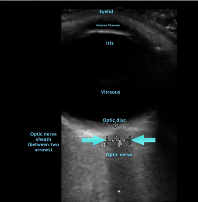
Figure 20: Ultrasound Image-ONSD
Summary
Optic nerve sheath diameter is an indirect measurement of intracranial pressure. Ultrasound measurement of ONSD appears to be non-invasive easy Real assessment which is also sensitive. In this study measurement of ONSD was performed on the left eye following sleep dose of induction agent to make them cooperative before MECT (1) after the seizure response, following MECT and after recover of spontaneous breathing. Patients diagnosed as schizophrenia and bipolar disorder were included in the study. Several studies, confirming structural changes in the brain with both the above disorders as well as MECT which seem to reverse the raised ONSD following clinical recovery, especially in schizophrenia. Dilated ventricular systems and reduced grey matter volume in schizophrenic cause a baseline Pre MECT rise in contrast to the readings in bipolar disorders.
Mean ONSD was plotted against several variables, taking all readings collectively and analysed. 84 observations from 12 patients were analysed with mean age of 36 years. The mean ONSD from the three readings was used for downstream analysis.
Excellent inter measurement agreement was demonstrated by the ONSD measurements with Pearson coefficient of 0.84 to 0.92 across the three readings. The mean coefficient of variation was 4.7% supporting the use of averaged ONSD as a reliable outcome variable.
Energy dose demonstrated strong association with ONSD spearman p= 0.52 indicating dose response relationship.
ONSD variability decreased modestly with increasing test number p= 0.039 suggesting positive physiological adaptation or measurement, stabilization overtime. Significant between patient variability with group variance of 0.67.
Conclusion:
Optic nerve sheath diameter appears to be a reliable real time non-invasive modality to measure ICP with strong associations to age and energy dose. The ONSD measurements even pre ECT are higher in schizophrenics than in patients with bipolar disorder, explained by structural changes in the brain in schizophrenia explained by dilatation of ventricular system and reduced gray matter in schizophrenia.
ONSD may be a useful tool in indicating recovery from schizophrenia, as our findings, suggesting normalization of ONSD with clinical improvement.
Limitations
Operator variability and inability to measure in an awake patient due to emotionally challenged situation.
More numbers and a better high resolution equipment with high frequency probe may play a better role in authenticating our findings.
References:
1. WHO global health estimates 2020: disease burden by cause, age, sex, country and region 2000 – 2019. Who.int/date/m.nb-ch-ad.ageing/advisory groups.
2. Fetahovic E, Janjic V, Muric M, et al. Neurobiological Mechanisms of Electroconvulsive Therapy: Molecular Perspectives of Brain Stimulation. International Journal of Molecular Sciences 2025;26(12):5905.
3. Beyer JL. History of electroconvulsive therapy. In: Mankad MV, Beyer JL, Weiner RD, et al, eds. Clinical manual of electroconvulsive therapy. 1st edn. Inc. Washington D.C.: American Psychiatric Publishing 2010: P. 3.
4. Fink M. Meduna and the origins of convulsive therapy. The American journal of psychiatry 1984;141(9):1034-41.
5. Beyer JL. History and indications for ECT. In: Mankad MV, Beyer JL, Weiner RD, et al, eds. Clinical manual of electroconvulsive therapy. 1st edn. American Psychiatric Publishing Inc 2010.
6. Ketter TA. Diagnostic features, prevalence, and impact of bipolar disorder. The Journal of clinical Psychiatry 2010;71(6):27652.
7. Dragovic M, Allet L, Janca A. Electroconvulsive therapy and determination of cerebral dominance. Annals of General Hospital Psychiatry 2004;3(1):14.
8. Duthie AC, Perrin JS, Bennett DM, et al. Anticonvulsant mechanisms of electroconvulsive therapy and relation to therapeutic efficacy. The Journal of ECT 2015;31(3):173-8.
9. Sackeim HA. The anticonvulsant hypothesis of the mechanisms of action of ECT: current status. The Journal of ECT 1999;15(1):5-26.
10. Whalley L, Dick H, Watts A, et al. Immediate increases in plasma prolactin and neurophysin but not other hormones after electroconvulsive therapy. The Lancet 1982;320(8307):1064-8.
11. Schwieler L, Samuelsson M, Frye MA, et al. Electroconvulsive therapy suppresses the neurotoxic branch of the kynurenine pathway in treatment-resistant depressed patients. Journal of neuroinflammation 2016;13(1):51.
12. Ousdal OT, Argyelan M, Narr KL, et al. Brain changes induced by electroconvulsive therapy are broadly distributed. Biological Psychiatry 2020;87(5):451-61.
13. Hoffman RE, McGlashan TH. Neurodynamics and schizophrenia research: editors' introduction. Schizophrenia Bulletin 1993;19(1):15-9.
14. Sadock BJ, Sadock VA, eds. Kaplan and Sadock's pocket handbook of clinical psychiatry. 7th edn. Lippincott William & Wilkins Publishers 2000.
15. Karlsgodt KH, Sun D, Cannon TD. Structural and functional brain abnormalities in schizophrenia. Current Directions in Psychological Science 2010;19(4):226-31.
16. Turner JA, Smyth P, Macciardi F, et al. Imaging phenotypes and genotypes in schizophrenia. Neuroinformatics 2006;4(1):21-49.
17. Dietsche B, Kircher T, Falkenberg I. Structural brain changes in schizophrenia at different stages of the illness: A selective review of longitudinal magnetic resonance imaging studies. Australian & New Zealand Journal of Psychiatry 2017;51(5):500-8.
18. Luvsannyam E, Jain MS, Pormento MK, et al. Neurobiology of schizophrenia: a comprehensive review. Cureus 2022;14(4):e23959.
19. Grande I, Berk M, Birmaher B, et al. Bipolar disorder. The Lancet 2016;387(10027):1561-72.
20. Zarate CA, Tohen M, Land M, et al. Functional impairment and cognition in bipolar disorder. Psychiatric Quarterly 2000;71(4):309-29.
21. Roque PJ, Hatch N, Barr L, et al. Bedside ocular ultrasound. Critical Care Clinics 2013;30(2):227-41.
22. Helmke K, Hansen HC. Fundamentals of transorbital sonographic evaluation of optic nerve sheath expansion under intracranial hypertension: II. Patient study. Pediatric Radiology 1996;26(10):706-10.
23. Liu D, Kahn M. Measurement of relationship of subarachnoid pressure of the optic nerve to intracranial pressures in fresh cadavers. Am J Ophthal 1993:116(5):548-56.
24. Hylkema C. Optic nerve sheath diameter ultrasound and the diagnosis of increased intracranial pressure. Critical care nursing clinics of North America 2016;28(1):95-9.
25. Hanafi MG, Verki MM, Parei SN. Ultrasonic assessment of optic nerve sheath to detect increased intracranial pressure. Journal of Medical Ultrasound 2019;27(2):69-74.