Role of Magnetic Resonance Imaging (MRI) In Evaluation of Posterior Reversible Encephalopathy Syndrome in Eclampsia Patients – An Observational Study in a Tertiary Care Hospital
- Dr. Abhishek Sunil Pingalkar , Senior Resident, Department of Radiodiagnosis, Sir JJ Group of Hospitals and Grant GMC Mumbai, India
- Dr. Amol Sakharam Jaitalkar , Assistant Professor, Department of Radio Diagnosis/ Radiology, GMC, Hingoli, India
- Dr. Jagannath Jare , Assistant Professor, General Medicine GMC Hingoli, India
Article Information:
Abstract:
Background: Posterior Reversible Encephalopathy Syndrome (PRES) is a well-recognized clinico-radiological condition commonly associated with hypertensive disorders of pregnancy, particularly eclampsia. It presents with acute neurological manifestations such as seizures, headache, visual disturbances, and altered sensorium, along with characteristic magnetic resonance imaging (MRI) findings of vasogenic edema. Early identification is crucial, as prompt management can lead to complete clinical and radiological reversibility. MRI remains the imaging modality of choice due to its high sensitivity in detecting both typical and atypical patterns of brain involvement. Objectives: To evaluate the role of MRI in diagnosing PRES among eclampsia patients, to describe the spectrum of typical and atypical MRI patterns, and to correlate imaging findings with clinical presentation and radiological outcomes. Materials and Methods: This ambispective observational study was conducted in the Department of Radiology of a tertiary care teaching hospital over a period of 12 months. A total of 57 antenatal and postnatal women aged ≥18 years, clinically diagnosed with eclampsia and suspected to have PRES, were included. All patients underwent MRI brain imaging. Clinical features, blood pressure parameters, gestational age, MRI lesion patterns, and follow-up imaging outcomes were recorded and analyzed using descriptive statistics. Results: The mean age of patients was 28.0 ± 5.8 years, with the majority belonging to the 21–30-year age group. Seizures were the most common presenting symptom (89.5%), followed by headache (49.1%), visual disturbances (35.1%), and altered sensorium (31.6%). MRI demonstrated classical parieto-occipital involvement in 24.6% of patients, atypical patterns in 36.8%, and combined typical and atypical patterns in 38.6%. Superior frontal sulcus and holohemispheric involvement were the most frequent atypical variants. Follow-up MRI showed complete reversibility in 84.2% of patients, while 15.8% exhibited persistent changes. Conclusion: MRI plays a pivotal role in the early diagnosis, characterization, and prognostication of PRES in eclampsia patients. Awareness of atypical imaging patterns is essential to prevent misdiagnosis and to facilitate timely intervention, thereby improving maternal neurological outcomes.
Keywords:
Article :
Introduction:
Hypertensive disorders of pregnancy are among the leading causes of maternal and perinatal morbidity and mortality worldwide. Eclampsia represents the most severe manifestation of preeclampsia and is defined by the occurrence of new-onset generalized tonic–clonic seizures in a woman with preeclampsia. It is a medical emergency and remains a significant contributor to maternal and fetal deaths, particularly in low- and middle-income countries.[1] Globally, hypertensive disorders of pregnancy account for nearly 10% of maternal mortality, with eclampsia contributing substantially to this burden. In India, the reported incidence of eclampsia ranges from 0.2% to 5% of all pregnancies, with a disproportionately high number of maternal deaths attributed to the condition.[2]
With advances in neuroimaging, particularly Magnetic Resonance Imaging (MRI), there has been a better understanding of the neurological mechanisms underlying eclamptic seizures. Increasing evidence has linked the neurological manifestations of eclampsia to a distinct clinico-radiological entity known as Posterior Reversible Encephalopathy Syndrome (PRES). PRES is characterized by acute neurological symptoms such as seizures, headache, visual disturbances, and altered mental status, accompanied by characteristic MRI findings of vasogenic edema predominantly involving the parieto-occipital regions of the cerebral hemispheres.[1,2]
The syndrome was first described in 1996 by Hinchey et al., who reported reversible posterior white matter changes in patients presenting with acute neurological symptoms, commonly in association with hypertensive encephalopathy or immunosuppressive therapy[3]. PRES has since been identified in various clinical settings, including renal failure, autoimmune diseases, cytotoxic drug exposure, and notably preeclampsia and eclampsia. In eclampsia, the development of PRES is believed to be multifactorial, involving sudden elevation of blood pressure, endothelial dysfunction, and disruption of cerebral autoregulation, resulting in leakage of fluid into the interstitial space and the formation of vasogenic edema.
The posterior cerebral circulation, particularly the parieto-occipital regions, is especially vulnerable due to relatively sparse sympathetic innervation, which limits its ability to respond to acute increases in blood pressure. This explains the characteristic posterior distribution of edema seen in PRES. Although PRES is described as a reversible condition, delayed diagnosis or inadequate management may lead to permanent neurological deficits or death. Studies have reported MRI evidence of PRES in up to 90% of women presenting with eclampsia, and in a significant proportion of patients with severe preeclampsia even in the absence of seizures.[4]
MRI plays a crucial role in the diagnosis, evaluation, and management of PRES. Typical MRI findings include bilateral, symmetrical hyperintensities on T2-weighted and fluid-attenuated inversion recovery (FLAIR) images involving the posterior white matter, usually without diffusion restriction on diffusion-weighted imaging (DWI), which helps differentiate PRES from acute ischemic infarction[5]. Atypical patterns may involve the frontal lobes, basal ganglia, cerebellum, or brainstem. Advanced MRI sequences such as susceptibility-weighted imaging or gradient-recalled echo can detect associated hemorrhagic changes, while contrast-enhanced imaging may show patchy enhancement in some cases. These imaging features assist in confirming the diagnosis, assessing severity, and monitoring resolution or complications.
Despite the high incidence of eclampsia in India, there is a relative paucity of observational studies focusing on the neuroimaging spectrum of PRES in this population. Most available literature originates from Western countries, where the prevalence and management protocols of eclampsia differ significantly. This highlights the need for region-specific studies evaluating MRI findings of PRES in Indian patients, particularly in tertiary care settings where advanced neuroimaging facilities are available.[6]
Preeclampsia is a pregnancy-specific disorder occurring after 20 weeks of gestation in previously normotensive women and is clinically characterized by hypertension (blood pressure ≥140/90 mmHg) and proteinuria (≥300 mg in a 24-hour urine collection) [7]. Preeclampsia and its variants remain major contributors to maternal and fetal morbidity and mortality worldwide[2], affecting approximately 5% of pregnancies. About 0.5% of women with mild preeclampsia progress to eclampsia. In developed countries, the incidence of eclampsia ranges from 1 in 2000 to 3000 deliveries [8]. Severe preeclampsia is defined by systolic blood pressure ≥160 mmHg, diastolic blood pressure ≥100 mmHg, and nephrotic-range proteinuria [7].
Douglas and Redman [9] reported that prodromal symptoms such as headache, visual disturbances, and epigastric pain frequently precede eclamptic seizures. Eclampsia affects pregnant or recently delivered women at a rate of 3–9 per 1000 pregnancies[7,8], and despite advances in antenatal care, early diagnosis remains a major challenge[9]. PRES is the most common neuroimaging abnormality associated with eclampsia and preeclampsia, though its exact pathophysiology remains debated. Diffusion-weighted imaging plays a key role in distinguishing vasogenic from cytotoxic edema and reflects dynamic pathophysiological changes in PRES [6].
The present study aims to evaluate the role of MRI in assessing PRES among eclampsia patients in a tertiary care hospital, focusing on incidence, imaging characteristics, clinical correlation, and outcomes.
AIMS AND OBJECTIVE
AIM
To evaluate role of Magnetic resonance imaging (MRI) in evaluation of posterior reversible encephalopathy syndrome in eclampsia patients.
OBJECTIVES
1. To determine the role of Magnetic resonance imaging (MRI) scan in diagnosis of posterior reversible encephalopathy syndrome in eclampsia patients.
2. To assess the neuroimaging abnormalities in a series of patients treated for eclampsia.
3. To assess the degree and type of brain edema in eclampsia patients with (PRES) posterior reversible encephalopathy syndrome.
4. To assess the incidence of atypical and typical regions of involvement and unusual imaging manifestations.
5. To assess the sequelae of ― (PRES) posterior reversible encephalopathy syndrome.
Materials and Methods:
Study Design: A tertiary care hospital–based ambispective observational study.
Study Setting: Department of Radiology, a tertiary care teaching hospital in a metropolitan city.
Study Duration: The study will be conducted for a period of 12 months.
Sample Size: A total of 57 patients.
Sample size was calculated using the formula:
n = Z² × p × (1−p) / E²
where p = 18% probability of PRES in pregnant women, confidence level = 95%.
Study Population: Antenatal and postnatal women admitted with clinical features of eclampsia or preeclampsia with neurological symptoms.
Inclusion Criteria
· Clinical suspicion or confirmed diagnosis of PRES
· MRI findings consistent with PRES
· Age ≥18 years
· History of eclampsia or preeclampsia
· New-onset seizures during antenatal or postnatal period
· Informed consent obtained
Exclusion Criteria
· Intracranial space-occupying lesions
· Pre-existing seizure disorders
· MRI contraindications (pacemakers, metallic implants)
· Claustrophobia
Study Procedure
All eligible patients underwent MRI brain imaging after informed consent. MRI findings were analyzed for lesion distribution, pattern type, and reversibility on follow-up imaging.
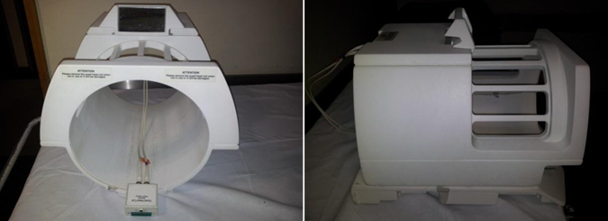
Figure 1: Brain coil used in MRI brain
Statistical Analysis
Data were analyzed using descriptive statistics. Continuous variables were expressed as mean ± standard deviation, and categorical variables as frequencies and percentages.
Results:
Table 1: Age-wise Distribution of Patients (n = 57)
|
Age Group (years) |
Frequency (n) |
Percentage (%) |
|
16–20 |
4 |
7.0 |
|
21–25 |
19 |
33.3 |
|
26–30 |
16 |
28.1 |
|
31–35 |
10 |
17.5 |
|
36–40 |
7 |
12.3 |
|
41–45 |
1 |
1.8 |
|
Total |
57 |
100.0 |
Mean age: 28.0 ± 5.8 years (Range: 18–41 years)
Table 1 depicts the age-wise distribution of the study population comprising 57 eclampsia patients with PRES. The patients ranged in age from 18 to 41 years, with a mean age of 28.0 ± 5.8 years. The highest proportion of patients belonged to the 21–25 years age group (33.3%), followed by the 26–30 years group (28.1%). A smaller proportion of patients were aged above 35 years, and only one patient was above 40 years of age. This distribution indicates that PRES in eclampsia predominantly affects young women in their reproductive years.
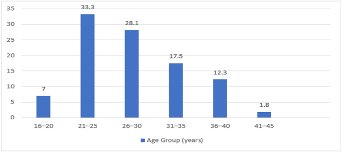
Figure 2: Age-wise Distribution of Patients
Table 2: Clinical Presentation of Eclampsia Patients
|
Clinical Feature |
Number of Patients (n) |
Percentage (%) |
|
Seizures |
51 |
89.5 |
|
Headache |
28 |
49.1 |
|
Visual disturbances |
20 |
35.1 |
|
Altered sensorium |
18 |
31.6 |
|
Focal neurological deficit |
10 |
17.5 |
Table 2 summarizes the clinical features observed in the study population. Seizures were the most common presenting symptom, occurring in 89.5% of patients. Headache was the next most frequent symptom (49.1%), followed by visual disturbances (35.1%) and altered sensorium (31.6%). Focal neurological deficits were observed in 17.5% of patients. These findings reflect the typical neurological manifestations of PRES in eclampsia, with seizures being the predominant feature.
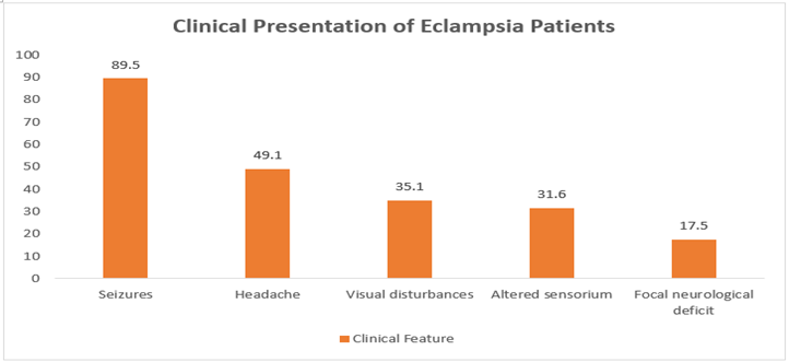
Figure 3: Clinical Presentation of Eclampsia Patients
Table 3: Blood Pressure and Gestational Age Distribution
Descriptive Statistics
|
Parameter |
Mean |
Standard Deviation |
Minimum |
Maximum |
|
Age (years) |
27.0 |
5.8 |
18 |
41 |
|
Systolic BP (mmHg) |
164.0 |
26.5 |
110 |
210 |
|
Diastolic BP (mmHg) |
94.9 |
11.9 |
70 |
116 |
|
Gestational age (days)* |
234.2 |
26.3 |
171 |
277 |
*Gestational age data available for 36 antepartum patients Mean gestational age ≈ 33.5 weeks (Range: 24.4–39 weeks)
Table 3 presents the descriptive statistics of age, blood pressure parameters, and gestational age among the study participants. The mean systolic blood pressure was 164.0 ± 26.5 mmHg, and the mean diastolic blood pressure was 94.9 ± 11.9 mmHg, indicating severe hypertension in most patients. Gestational age data, available for 36 antepartum patients, showed a mean gestational age of 234.2 ± 26.3 days (approximately 33.5 weeks), with a range of 24.4 to 39 weeks. These findings suggest that PRES in eclampsia commonly occurs in the late second or third trimester and is associated with markedly elevated blood pressure levels.
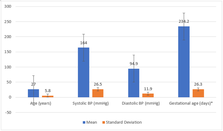
Figure 4: Blood Pressure and Gestational Age Distribution
Table 4: MRI Pattern Classification in PRES Patients
Final MRI Pattern Distribution (n = 57)
|
MRI Pattern Type |
Frequency (n) |
Percentage (%) |
|
Combination (Typical + Atypical) |
22 |
38.6 |
|
Atypical |
21 |
36.8 |
|
Typical (Parieto-occipital) |
14 |
24.6 |
|
Total |
57 |
100.0 |
Table 4B: Distribution of Atypical PRES Variants
|
Atypical Variant |
Number of Patients (n) |
Percentage (%) |
|
Superior frontal sulcus (SFS) |
10 |
17.5 |
|
Holohemispheric pattern |
8 |
14.0 |
|
Other atypical findings |
4 |
7.0 |
Table 4 illustrates the MRI pattern distribution among patients diagnosed with PRES. The most common imaging pattern was a combination of typical and atypical features, observed in 38.6% of patients. Atypical patterns alone were seen in 36.8% of patients, while classical parieto-occipital involvement was present in 24.6%. Among atypical variants, superior frontal sulcus involvement was the most frequent (17.5%), followed by holohemispheric patterns (14.0%) and other atypical findings (7.0%). These results highlight the heterogeneous MRI spectrum of PRES in eclampsia patients.
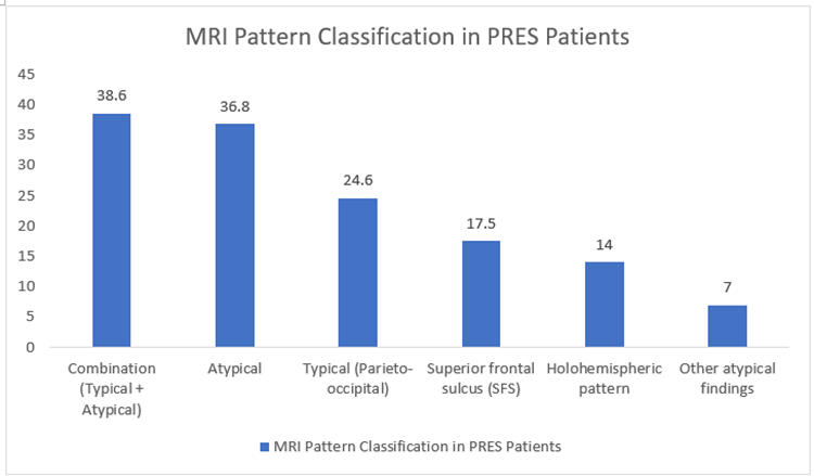
Figure 5: MRI Pattern Classification in PRES Patients
Table 6: Outcome on Follow-up MRI
|
Outcome |
Number of Patients (n) |
Percentage (%) |
|
Reversible MRI changes |
48 |
84.2 |
|
Irreversible MRI changes |
9 |
15.8 |
|
Total |
57 |
100.0 |
Table 5 demonstrates the radiological outcomes of patients on follow-up MRI. The majority of patients (84.2%) showed complete reversibility of MRI changes, indicating a favorable prognosis with appropriate management. However, 15.8% of patients exhibited persistent or irreversible MRI findings. This table emphasizes the generally reversible nature of PRES while also underscoring the importance of early diagnosis and timely intervention to prevent permanent neurological damage.
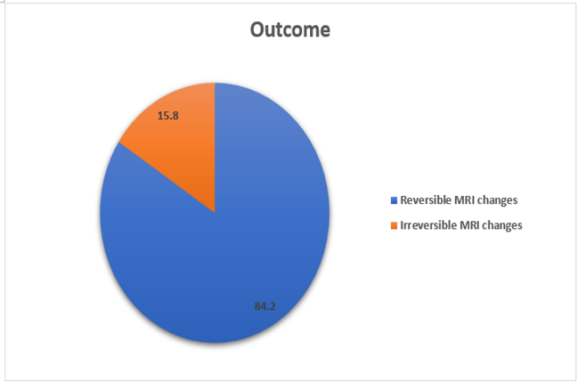
Figure 6: Outcome on Follow-up MRI
Discussion:
The present observational study evaluated the clinical, radiological, and outcome characteristics of Posterior Reversible Encephalopathy Syndrome (PRES) in eclampsia patients using magnetic resonance imaging. The mean age of 28.0 ± 5.8 years, with the majority of patients clustered between 21 and 30 years, reflects the well-known predilection of eclampsia and PRES for young women of reproductive age. Similar age distributions have been reported in earlier studies, where PRES associated with hypertensive disorders of pregnancy predominantly affected women in their third decade of life Wu Y, et al.[6]. This age trend underscores the vulnerability of younger pregnant women, particularly primigravida, to hypertensive cerebrovascular complications.
Seizures were the most common presenting symptom in the present study, observed in 89.5% of patients, followed by headache, visual disturbances, and altered sensorium. This clinical profile is consistent with the classical presentation of PRES originally described by Hinchey et al., who identified seizures, headache, visual symptoms, and encephalopathy as hallmark manifestations of the syndrome[3]. Subsequent studies have confirmed that seizures are the predominant presenting feature of PRES in eclampsia, often serving as the initial trigger for neuroimaging evaluation Garg RK et al.[5]. The high frequency of visual disturbances and altered sensorium in this cohort further supports posterior cerebral involvement, particularly of the parieto-occipital lobes, which are responsible for visual processing.
Severe hypertension was a prominent finding in this study, with a mean systolic blood pressure of 164.0 ± 26.5 mmHg and mean diastolic blood pressure of 94.9 ± 11.9 mmHg. These values exceed the upper limits of cerebral autoregulation, supporting the widely accepted hypothesis that acute elevations in blood pressure contribute to endothelial dysfunction, breakdown of the blood–brain barrier, and development of vasogenic edema Bartynski WS et al.[10]. Several authors have emphasized that severe hypertension is a major precipitating factor for PRES in preeclampsia and eclampsia, although it may not be present in all cases Fischer M et al.[11]. The mean gestational age of approximately 33.5 weeks observed in this study is also consistent with prior reports, indicating that PRES commonly manifests during the late second or third trimester when preeclampsia tends to progress to severe forms Jindal M et al.[12].
MRI pattern analysis revealed that combined typical and atypical patterns were the most frequent (38.6%), followed closely by atypical patterns alone (36.8%), while classical parieto-occipital involvement was observed in only 24.6% of patients. These findings highlight the evolving understanding that PRES is not confined to the posterior regions of the brain. Bartynski and Boardman described multiple imaging patterns of PRES, including superior frontal sulcus and holohemispheric watershed patterns, emphasizing that atypical distributions are common and should not exclude the diagnosis Sudulagunta SR et al.[7]. The relatively high proportion of atypical and combined patterns in this study may reflect the severity of disease, postpartum physiological changes, or early imaging during the disease course.
Among atypical variants, superior frontal sulcus involvement (17.5%) and holohemispheric patterns (14.0%) were the most frequently observed. Similar atypical patterns have been reported in obstetric PRES cohorts, where involvement of frontal lobes and diffuse hemispheric edema was noted, particularly in patients with severe hypertension or delayed presentation Liman T Get al.[13]. Recognition of these atypical patterns is critical, as they may mimic ischemic stroke, encephalitis, or demyelinating disorders, potentially leading to misdiagnosis and delayed treatment.
One of the defining features of PRES is its potential reversibility. In the present study, 84.2% of patients demonstrated complete radiological reversal on follow-up MRI, while 15.8% showed persistent or irreversible changes. These findings are in agreement with prior studies reporting favorable outcomes in the majority of eclampsia-associated PRES cases when timely blood pressure control and seizure management are instituted Frontiers in Neurology[14]. Tawati DA et al. reported that delayed diagnosis, presence of hemorrhage, and extensive cytotoxic edema were associated with poorer outcomes and incomplete reversibility[4]. The relatively high reversibility rate in this study likely reflects early MRI evaluation and management in a tertiary care setting. McKinney AM et al.[15]
Overall, the findings of this study reinforce the strong association between eclampsia and PRES and emphasize the critical role of MRI in early diagnosis. The high prevalence of atypical imaging patterns highlights the need for heightened awareness among clinicians and radiologists. Early recognition of PRES and prompt management of hypertension and seizures are essential to prevent irreversible neurological injury and improve maternal outcomes.
Conclusion:
This observational study demonstrates that MRI is an indispensable diagnostic and prognostic tool in evaluating Posterior Reversible Encephalopathy Syndrome in eclampsia patients. In addition to confirming the diagnosis, MRI shows the range of lesion distribution, including typical, atypical, and mixed patterns. For most patients, early detection of PRES by MRI enables timely blood pressure control and supportive care, resulting in positive clinical and radiological results. It is especially crucial to identify abnormal MRI patterns in order to avoid incorrect diagnoses and needless treatments. To enhance maternal outcomes, routine MRI scanning should be taken into consideration for eclamptic patients who report with neurological symptoms.
LIMITATIONS OF THE STUDY
Despite providing valuable insights into the MRI spectrum of PRES in eclampsia, this study has certain limitations. The sample size was relatively small, which may limit the generalizability of the findings to a broader population. Being a single-center study, the results may reflect institutional practices and referral patterns rather than population-wide trends. In order to establish greater correlations between clinical factors, imaging patterns, and outcomes, the study mainly relied on descriptive statistical analysis rather than sophisticated inferential studies. The lack of long-term neurological and cognitive patient follow-up restricts the evaluation of subtle or delayed consequences.
Furthermore, objective grading of illness severity and comparability with other research were hampered by the lack of a standardised PRES severity score system. Even while skilled radiologists evaluated the pictures, inter-observer variability in MRI interpretation may have potentially affected pattern categorisation. Lastly, not all patients had regular contrast-enhanced MRI and sophisticated perfusion procedures, which may have limited the ability to detect minor problems such microhemorrhages or infarction.
References:
1. World Health Organization. Pre eclampsia. WHO fact sheet. 2025. Available from: https://www.who.int/news room/fact sheets/detail/pre eclampsia
2. Abalos E, Duley L, Steyn DW, et al. Global and regional estimates of preeclampsia and eclampsia: a systematic review. PLoS One. 2013;8(1):e54856. doi:10.1371/journal.pone.0054856.
3. Hinchey J, Chaves C, Appignani B, et al. A reversible posterior leukoencephalopathy syndrome. N Engl J Med. 1996;334(8):494–500.
4. Tawati DA, Chan W S. A systematic review of posterior reversible encephalopathy syndrome in pregnant women with severe preeclampsia and eclampsia. Obstet Med. 2023;16(4):236 241. doi:10.1177/1753495X221150302
5. Garg RK, Kumar N, Malhotra HS. Posterior reversible encephalopathy syndrome in eclampsia. Neurol India. 2018;66:1316–23.
6. Wu Y, Wu L F, Li Q J, et al. MRI characteristics of brain edema in preeclampsia/eclampsia patients with posterior reversible encephalopathy syndrome. BMC Pregnancy Childbirth. 2021;21(1):536. doi:10.1186/s12884 021 04145 1
7. Sudulagunta SR, Sodalage S, Maheshwari A. Posterior reversible encephalopathy syndrome (PRES): clinical and imaging features. J Clin Imaging Sci. 2017;7:9. doi:10.4103/jcis.JCIS_21_17
8. Shandilya V. Prevalence, risk factors and impact on mother and fetus. Int J Clin Dev World Health. 2023;3(3):88–93. doi:10.12345/ijcdw.2023.03.03.88
9. Douglas KA, Redman CW. Eclampsia in the United Kingdom. BMJ. 1994;309(6966):1395–400.
10. Bartynski WS. Posterior reversible encephalopathy syndrome, part 1: fundamental imaging and clinical features. AJNR Am J Neuroradiol. 2008;29(6):1036-1042. doi:10.3174/ajnr.A0928.
11. Fischer M, Schmutzhard E. Posterior reversible encephalopathy syndrome. J Neurol. 2017;264(8):1608-1616. doi:10.1007/s00415-016-8377-8.
12. Jindal M, Singh P, Arora S. MRI spectrum of posterior reversible encephalopathy syndrome in pregnancy-related hypertension. J Obstet Gynaecol India. 2016;66(Suppl 1):460-466. doi:10.1007/s13224-015-0739-6.
13. Liman TG, Siebert E, Endres M. Posterior reversible encephalopathy syndrome. Curr Opin Neurol. 2019;32(1):25-35. doi:10.1097/WCO.0000000000000640.
14. Frontiers in Neurology. Clinical and imaging outcomes in PRES. Front Neurol. 2020;11:71. doi:10.3389/fneur.2020.00071.
15. McKinney AM, Short J, Truwit CL, et al. Posterior reversible encephalopathy syndrome: incidence of hemorrhage. AJNR Am J Neuroradiol. 2007;28(7):1370-1376. doi:10.3174/ajnr.A0525.