A Prospective Study of Cardiovascular Autonomic Neuropathy Assessment By Clinical Methods In Newly Diagnosed Type 2 Diabetes Mellitus Patients Before And After Acheiving Euglycemia In A Tertiary Care Centre
- Dr. Manjunatha K R , MBBS, MD, Department General Medicine, MGMMC & MYH, Indore, MP
- Dr. Rajesh Verma , Associate Professor MBBS, MD, DM Endocrinology, Department General Medicine, MGMMC & MYH, Indore, MP
- Dr. Pooja Sounr , Assistant Professor MBBS, MD , Department General Medicine, MGMMC & MYH, Indore, MP
Article Information:
Abstract:
Background: Cardiovascular autonomic neuropathy (CAN) is a serious yet often underdiagnosed complication of diabetes mellitus and is associated with increased cardiovascular morbidity and mortality. Although traditionally considered a late manifestation, emerging evidence suggests that autonomic dysfunction may occur early in the course of type 2 diabetes mellitus (T2DM) and may be reversible with timely metabolic intervention. The present study aimed to evaluate cardiovascular autonomic neuropathy using simple, non-invasive clinical methods in newly diagnosed T2DM patients and to assess changes in autonomic function before and after achieving euglycemia over a one-year period. Methods: This prospective study was conducted at a tertiary care center and included 100 newly diagnosed T2DM patients aged over 18 years. Cardiovascular autonomic function was assessed at baseline and after achieving euglycemia using standard bedside tests, including resting heart rate, beat-to-beat heart rate variation, heart rate response to standing, systolic blood pressure response to standing, and diastolic blood pressure response to isometric exercise. Electrocardiographic QT/QTc intervals were also analyzed. Repeated-measures ANOVA was used for longitudinal comparisons. Results: Significant improvements were observed across multiple autonomic parameters over one year. Resting heart rate showed a marked reduction, beat-to-beat heart rate variability improved, and baroreflex-mediated responses demonstrated favorable trends. Both resting and stress-induced blood pressure responses indicated improved sympathetic regulation. QT/QTc intervals shortened significantly, suggesting enhanced myocardial electrical stability. All changes were statistically significant (p < 0.0001). Conclusion: Newly diagnosed T2DM patients exhibit early cardiovascular autonomic dysfunction that is significantly reversible with sustained glycemic control. Early identification and intervention using simple clinical tests may help prevent progression of CAN and reduce long-term cardiovascular risk.
Keywords:
Article :
Introduction:
Diabetes mellitus (DM) is a chronic metabolic disorder characterized by persistent hyperglycemia due to defects in insulin secretion, insulin action, or both. The global burden of diabetes continues to rise, with type 2 diabetes mellitus (T2DM) accounting for the majority of cases. According to the International Diabetes Federation, nearly 500 million individuals worldwide are currently living with diabetes, and this number is expected to increase by nearly 30% by 2045 [1]. Diabetes and its complications are major contributors to morbidity, impaired quality of life, and premature mortality, with almost 10% of global all-cause deaths in adults aged 20–99 years attributed to diabetes [2].
Cardiovascular autonomic neuropathy (CAN) is one of the most serious and clinically relevant complications of diabetes. It results from damage to autonomic nerve fibers innervating the heart and blood vessels, leading to impaired heart rate control and abnormal vascular responses. The presence of CAN is strongly associated with increased cardiovascular events, silent myocardial ischemia, and sudden cardiac death, thereby significantly increasing mortality risk in diabetic patients [3]. Despite its importance, CAN often remains underdiagnosed, especially in the early stages of T2DM, and its risk factors and natural history in newly diagnosed patients are not fully defined [4].
The prevalence of CAN varies widely, from approximately 2% in newly diagnosed or well-controlled diabetes to as high as 60% in patients with long-standing T2DM, depending on the population studied and the diagnostic methods used [5,6]. This wide variability reflects heterogeneity in assessment techniques and diagnostic criteria. Several factors, including increasing age, longer duration of diabetes, and poor glycemic control, are known to increase the likelihood of CAN [7]. Although traditionally considered a late complication, emerging evidence indicates that subclinical autonomic dysfunction may be present at or soon after the diagnosis of T2DM. This early involvement is characterized by reduced parasympathetic activity with relative sympathetic predominance [8–11].
Large clinical trials, including the Diabetes Control and Complications Trial and observations from the ACCORD study, have demonstrated that improved glycemic control can favorably influence autonomic function and may reduce the progression of CAN [12,13]. Given the high mortality associated with diabetic autonomic neuropathy and evidence suggesting potential reversibility in early stages, timely identification is crucial. [14,15]
In this context, the present prospective study was designed to evaluate cardiovascular autonomic neuropathy using simple, non-invasive clinical methods in patients newly diagnosed with T2DM. The study aims to assess the frequency of cardiovascular autonomic neuropathic features at diagnosis and to compare autonomic function before and after the achievement of euglycemia, thereby exploring the potential impact of early glycemic control on autonomic dysfunction in a tertiary care centre.
Materials and Methods:
This prospective study was conducted over a period of one year after obtaining approval from the Institutional Ethical Committee. The study was carried out in the Outpatient Department (OPD) and Inpatient Department (IPD) of Maharaja Yeshwant Rao Hospital, a tertiary care government hospital in Central India. The study population consisted of newly diagnosed patients with type 2 diabetes mellitus (T2DM) who were evaluated for the presence of cardiovascular autonomic neuropathy (CAN).
A total of 100 newly diagnosed T2DM patients fulfilling the predefined inclusion and exclusion criteria were enrolled in the study. Patients aged more than 18 years with a recent diagnosis of T2DM were included. Patients with a prior diagnosis of diabetes mellitus and those already on antidiabetic treatment were excluded. Additional exclusion criteria included the presence of structural heart disease, ischemic heart disease, malignancy, or any other systemic illness known to affect cardiac function. Patients with hypertension and those receiving beta-blocker therapy were also excluded to avoid confounding effects on autonomic function.
Data Collection and Clinical Assessment
After obtaining written informed consent, data were collected using a prevalidated proforma. Demographic details including age, sex, and place of residence were recorded. All participants underwent a detailed clinical examination with particular emphasis on cardiovascular autonomic function assessment. Resting heart rate was recorded after adequate rest. Beat-to-beat heart rate variation was assessed using standard bedside methods. Heart rate response to standing and systolic blood pressure (SBP) response to standing were measured to evaluate parasympathetic and sympathetic function. Diastolic blood pressure (DBP) response to sustained isometric exercise was recorded as a measure of sympathetic integrity.
Relevant investigations were performed in all patients, including glycated hemoglobin (HbA1c), oral glucose tolerance test (OGTT), and resting electrocardiography (ECG). Routine laboratory tests such as complete blood count (CBC), renal function tests (RFT), and lipid profile were also obtained to assess baseline metabolic and systemic status.
Method of Assessment and Follow-up
All enrolled patients were evaluated for CAN at baseline, prior to achieving euglycemia
. Following standard diabetes management protocols, patients were treated and followed until euglycemic status was achieved, after which cardiovascular autonomic function tests were repeated to assess changes in autonomic parameters.
Statistical Analysis
Data were analyzed using Statistical Package for the Social Sciences (SPSS) software, version 27.0. Categorical variables were expressed as frequencies and percentages, while continuous variables were presented as mean and standard deviation. Repeated measures analysis of variance (ANOVA) was used to compare continuous variables at different time points. A p-value of less than 0.05 was considered statistically significant.
Ethical Considerations and Funding
The study protocol was approved by the Institutional Ethical Committee. Written informed consent was obtained from all participants prior to enrollment. As the study was conducted in a government hospital where investigations and treatment are provided free of cost, there was no financial burden on patients. The study did not receive funding from any pharmaceutical company or external institution.
Results:
A total of 100 newly diagnosed type 2 diabetes mellitus patients were included in the final analysis. All participants completed the planned follow-up. Normality testing using the Kolmogorov–Smirnov test confirmed that the continuous variables were approximately normally distributed. Continuous data are expressed as mean ± standard deviation, while categorical variables are presented as frequency and percentage. Longitudinal changes in cardiovascular autonomic parameters were assessed using repeated-measures ANOVA, with a p-value < 0.05 considered statistically significant.
The study population had a mean age of 53.51 ± 6.53 years, with ages ranging from 41 to 72 years. The majority of participants were middle-aged, with nearly half belonging to the 51–60-year age group, followed by those aged 41–50 years. Participants above 60 years constituted a smaller proportion of the cohort. Gender distribution was relatively balanced, with a slight female predominance. This demographic profile reflects the typical presentation of newly diagnosed type 2 diabetes mellitus in a tertiary care setting (Table 1).
Table 1. Demographic Characteristics of the Study Population (n = 100)
|
Variable |
Category |
n (%) / Mean ± SD |
|
Age (years) |
Mean ± SD |
53.51 ± 6.53 |
|
41–50 |
36 (36.0%) |
|
|
51–60 |
49 (49.0%) |
|
|
61–70 |
13 (13.0%) |
|
|
71–80 |
2 (2.0%) |
|
|
Gender |
Male |
46 (46.0%) |
|
Female |
54 (54.0%) |
Resting heart rate demonstrated a statistically significant change over the course of follow-up. After a mild increase at three months, a steady and sustained decline was observed, with mean values decreasing from 110.94 ± 9.74 bpm at baseline to 90.98 ± 8.18 bpm at one year. This progressive reduction suggests improvement in autonomic balance, likely reflecting reduced sympathetic overactivity following achievement of euglycemia. Beat-to-beat heart rate variation (>15 bpm) also showed a significant decline over time, with values stabilizing after six months, indicating improved heart rate stability and autonomic regulation. Similarly, heart rate response to standing showed a small but consistent reduction during follow-up, suggesting normalization of parasympathetic function (Table 2, Figure 1).
Table 2. Changes in Heart Rate and Parasympathetic Function Over Time
|
Parameter |
Baseline |
3 months |
6 months |
9 months |
1 year |
F value |
p value |
|
Resting HR (bpm) |
110.94 ± 9.74 |
112.63 ± 10.22 |
104.91 ± 11.48 |
95.48 ± 9.50 |
90.98 ± 8.18 |
48044.162 |
<0.0001 |
|
Beat-to-beat HR variation (>15 bpm) |
21.24 ± 3.92 |
19.63 ± 3.36 |
17.53 ± 3.01 |
18.51 ± 2.37 |
18.63 ± 2.16 |
18491.730 |
<0.0001 |
|
HR response to standing |
1.1848 ± 0.106 |
1.1750 ± 0.104 |
1.1662 ± 0.027 |
1.1680 ± 0.070 |
1.1687 ± 0.072 |
59673.047 |
<0.0001 |
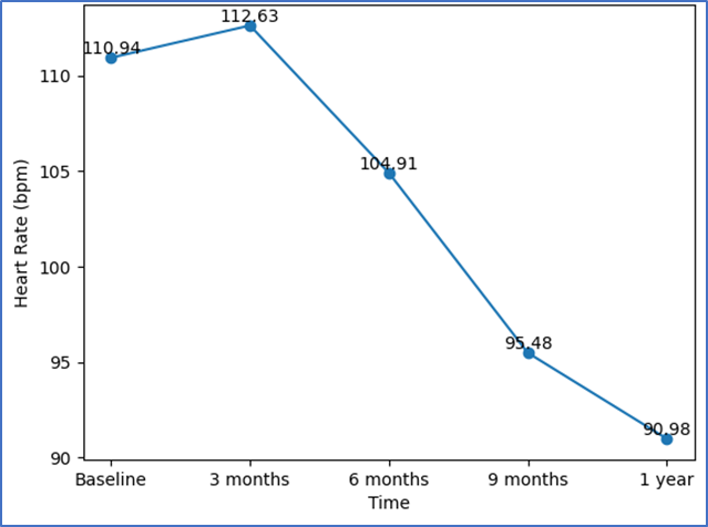
Figure 1. Change in Heart Rate Over Time
Resting systolic blood pressure showed a significant and clinically meaningful reduction over time, with the greatest decline observed by nine months, followed by a mild rebound at one year. This pattern suggests improved vascular tone and sympathetic modulation with glycemic control. Resting diastolic blood pressure exhibited a more variable trend, with an initial rise at three months followed by a decline at six and nine months and a return toward baseline by one year. The systolic blood pressure response to standing decreased progressively during follow-up, indicating improved orthostatic blood pressure regulation and baroreflex sensitivity. In addition, the diastolic blood pressure response to isometric exercise showed a significant reduction by one year, reflecting attenuation of exaggerated sympathetic responses to stress (Table 3).
Table 3. Blood Pressure and Sympathetic Function Parameters Over Time
|
Parameter |
Baseline |
3 months |
6 months |
9 months |
1 year |
F value |
p value |
|
Resting SBP (mmHg) |
132.42 ± 7.03 |
130.08 ± 7.69 |
126.93 ± 9.50 |
123.56 ± 9.12 |
126.50 ± 10.52 |
103796 |
<0.0001 |
|
Resting DBP (mmHg) |
80.51 ± 6.07 |
83.23 ± 3.07 |
79.00 ± 3.06 |
78.45 ± 5.12 |
81.64 ± 8.36 |
141042.8 |
<0.0001 |
|
SBP response to standing (mmHg) |
14.10 ± 1.78 |
14.84 ± 1.32 |
13.98 ± 2.01 |
13.45 ± 2.12 |
12.28 ± 2.12 |
25182.536 |
<0.0001 |
|
DBP response to isometric exercise (mmHg) |
11.54 ± 2.57 |
12.82 ± 3.55 |
10.62 ± 1.79 |
10.73 ± 2.09 |
9.56 ± 1.70 |
10567.895 |
<0.0001 |
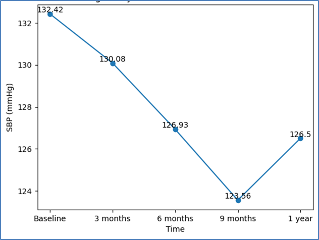
Figure 2. Change in Systolic Blood Pressure Over Time
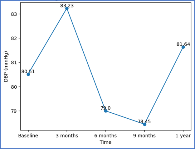
Figure 3. Change in Diastolic Blood Pressure Over Time
Electrocardiographic analysis revealed significant changes in QT/QTc intervals over time. After a slight prolongation at three months, a progressive and sustained shortening was observed from six months onward, with a reduction of more than 24 ms at one year compared to baseline. This improvement in ventricular repolarization suggests reduced arrhythmogenic risk and recovery of autonomic and metabolic stability with sustained euglycemia (Table 4).
Table 4. QT/QTc Interval Changes Over Time
|
Time Point |
QT/QTc Interval (ms) Mean ± SD |
F value |
p value |
|
Baseline |
474.74 ± 16.50 |
643007 |
<0.0001 |
|
3 months |
477.87 ± 14.62 |
||
|
6 months |
464.55 ± 11.45 |
||
|
9 months |
450.54 ± 12.95 |
||
|
1 year |
450.14 ± 12.18 |
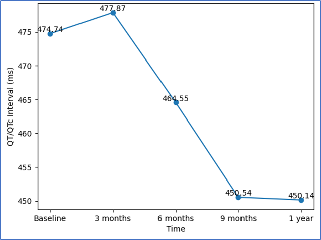
Figure 4. Change in QT/QTc Interval Over Time
Overall, the results demonstrate that newly diagnosed type 2 diabetes mellitus patients exhibit early cardiovascular autonomic dysfunction at presentation. Importantly, significant improvement in both parasympathetic and sympathetic autonomic parameters was observed following achievement and maintenance of euglycemia over one year, highlighting the potential reversibility of early cardiovascular autonomic neuropathy with timely intervention.
Discussion:
The present prospective study demonstrates statistically significant improvement across multiple cardiovascular autonomic parameters following one year of glycemic control in patients with newly diagnosed type 2 diabetes mellitus. These findings strongly support the concept that early cardiac autonomic neuropathy is not a fixed or irreversible complication but rather a dynamic process that can improve with timely metabolic correction.
Resting heart rate, a well-recognized surrogate marker of sympathetic overactivity and reduced parasympathetic tone, showed a marked and progressive decline over the study period. The reduction from a baseline mean of 110.94 bpm to 90.98 bpm at one year reflects restoration of autonomic balance, with attenuation of sympathetic dominance and improvement in vagal tone. This observation is clinically meaningful, as elevated resting heart rate has been consistently associated with increased cardiovascular morbidity and mortality in diabetic populations. Our findings are in agreement with Esmaeillou et al., [16] who reported significantly higher resting heart rates in patients with long-standing diabetes compared to newly diagnosed cases, suggesting that autonomic dysfunction worsens with disease duration. In contrast to the relatively static autonomic impairment seen in chronic diabetes, the present study highlights the potential for heart rate normalization when glycemic control is achieved early in the disease course.
Beat-to-beat heart rate variation, reflecting parasympathetic reactivity, also showed statistically significant improvement over time. Although absolute values remained below optimal physiological thresholds in some patients, the consistent reduction from baseline indicates enhanced vagal modulation. These findings align with those of Sukla et al. [17], who identified heart rate variability during deep breathing as one of the most sensitive indicators of parasympathetic dysfunction in diabetic patients. The longitudinal improvement observed in our cohort adds important evidence that early parasympathetic abnormalities are modifiable and reinforces the value of simple bedside tests in detecting and monitoring autonomic recovery.
Heart rate response to standing, representing baroreflex sensitivity and integrated autonomic control, demonstrated a small but statistically significant attenuation over the follow-up period. While the absolute changes were modest, their consistency across a large sample resulted in high statistical significance. Reduced 30:15 ratios have been associated with impaired insulin secretion and lower fasting C-peptide levels, as demonstrated by Yang et al.[18], emphasizing the close link between glycemic physiology and autonomic function. The stabilization of this parameter in our study suggests gradual normalization of baroreceptor reflexes, likely driven by sustained improvement in glycemic status.
Blood pressure parameters further support the concept of reversible autonomic vascular dysfunction. Resting systolic blood pressure showed a clinically meaningful decline, particularly during the first nine months, reflecting improved vascular compliance and sympathetic tone regulation. Esmaeillou et al. [16] reported higher systolic blood pressure values in chronic diabetic patients, often associated with increased body mass index and longer disease duration. Similarly, Bhuyan et al. [19] linked cardiovascular autonomic neuropathy to prolonged diabetes duration and renal dysfunction. The favorable systolic blood pressure trend in our cohort underscores the importance of early intervention before structural vascular changes become established.
Diastolic blood pressure exhibited a biphasic pattern, with an initial rise followed by subsequent decline and partial normalization. This fluctuation may represent early vascular tone adaptation and endothelial recovery in response to improved metabolic control. The transient rise at three months could reflect compensatory vascular responses before stabilization occurs. Bhuyan et al. highlighted the association between declining renal function and autonomic neuropathy, suggesting that improvements in diastolic blood pressure may also indirectly indicate recovery of renal–autonomic interactions in early diabetes.
The systolic blood pressure response to standing declined progressively over time, indicating improved orthostatic regulation and enhanced baroreflex sensitivity. This finding is particularly relevant, as exaggerated orthostatic responses are a hallmark of sympathetic overactivity in early autonomic dysfunction. Sukla et al. [17] identified postural blood pressure changes as reliable noninvasive markers of sympathetic integrity, especially in resource-limited settings. Our results reinforce the utility of this test and demonstrate that sympathetic hyperresponsiveness is amenable to reversal with metabolic stabilization.
Similarly, the diastolic blood pressure response to isometric exercise showed a clear attenuation by one year, reflecting reduced sympathetic efferent activity during stress. The exaggerated pressor response observed early in the study gradually normalized, suggesting improved cardiovascular adaptability. Handgrip testing has been emphasized by Sukla et al. [17] as a sensitive and practical tool for evaluating sympathetic function, and our findings validate its relevance in longitudinal follow-up of newly diagnosed diabetic patients.
Electrocardiographic QT/QTc interval shortening was one of the most striking findings of this study. Prolonged QT intervals are associated with autonomic imbalance, myocardial repolarization abnormalities, and increased risk of malignant arrhythmias. The significant reduction observed over time suggests improvement in autonomic regulation of ventricular repolarization and a potential reduction in arrhythmogenic risk. Esmaeillou et al. [16] reported longer QT intervals in patients with chronic diabetes, supporting the notion that QT prolongation reflects cumulative autonomic damage. Our findings demonstrate that such electrical abnormalities are not necessarily permanent and may improve with sustained glycemic control.
Collectively, the improvement in heart rate variability, blood pressure responses, and QT/QTc intervals parallels observations by Liu et al., who linked reduced heart rate variability parameters to more severe coronary atherosclerosis. Their work suggests that cardiovascular autonomic neuropathy may serve as an early marker of macrovascular risk. By demonstrating improvement in these surrogate markers, the present study supports the hypothesis that early correction of autonomic dysfunction may have broader cardiovascular protective implications.
Despite its strengths, this study has certain limitations. Being a single-center study conducted in a tertiary care hospital, the findings may not be fully generalizable to community-based populations. The absence of a control group limits causal inference. The one-year follow-up, although sufficient to demonstrate early reversibility, does not provide insight into long-term sustainability of autonomic improvement. In addition, lifestyle factors and medication adherence were not standardized, and biochemical markers such as serial HbA1c levels were not analyzed in parallel with autonomic changes. Finally, advanced autonomic testing modalities were not employed. Future multicenter studies with longer follow-up, control groups, and comprehensive autonomic profiling are warranted to further elucidate the long-term clinical implications of early autonomic recovery in type 2 diabetes mellitus.
Conclusion:
Early cardiovascular autonomic neuropathy in newly diagnosed type 2 diabetes mellitus emerges as a dynamic and potentially reversible condition rather than a fixed complication. Assessment with simple, non-invasive autonomic function tests demonstrated meaningful restoration of autonomic balance after one year of sustained glycemic control. Improvements in heart rate behavior, blood pressure responses, and QT/QTc intervals indicate recovery of parasympathetic activity, reduction in sympathetic overdrive, and improved myocardial electrical stability. These findings highlight the importance of early identification and intensive metabolic optimization, supporting routine autonomic screening at diagnosis to prevent progression and reduce long-term cardiovascular risk in patients with type 2 diabetes mellitus.
References:
1. American Diabetes Association. Diagnosis and classification of diabetes mellitus. Diabetes Care. 2009;32(Suppl 1):S62-S67.
2. Cho NH, Shaw JE, Karuranga S, Huang Y, da Rocha Fernandes JD, Ohlrogge AW, et al. IDF Diabetes Atlas: Global estimates of diabetes prevalence for 2017 and projections for 2045. Diabetes Res Clin Pract. 2018;138:271-281.
3. Tesfaye S, Boulton AJ, Dyck PJ, Freeman R, Horowitz M, Kempler P, et al. Diabetic neuropathies: update on definitions, diagnostic criteria, estimation of severity, and treatments. Diabetes Care. 2010;33:2285-2293.
4. Vinik AI, Ziegler D. Diabetic cardiovascular autonomic neuropathy. Circulation. 2007;115:387-397.
5. Vinik AI, Erbas T, Casellini CM. Diabetic cardiac autonomic neuropathy, inflammation and cardiovascular disease. J Diabetes Investig. 2013;4:4-18.
6. Pop-Busui R. What do we know and we do not know about cardiovascular autonomic neuropathy in diabetes. J Cardiovasc Transl Res. 2012;5:463-478.
7. Agashe S, Petak S. Cardiac Autonomic Neuropathy in Diabetes Mellitus. Methodist Debakey Cardiovasc J. 2018 Oct-Dec;14(4):251-256.
8. Singh JP, Larson MG, O’Donnell CJ, et al. Association of hyperglycemia with reduced heart rate variability (The Framingham Heart Study). Am J Cardiol. 2000;86:309-312.
9. Pierson CR, Zhang W, Murakawa Y, Sima AA. Insulin deficiency rather than hyperglycemia accounts for impaired neurotrophic responses and nerve fiber regeneration in type 1 diabetic neuropathy. J Neuropathol Exp Neurol. 2003;62:260-271.
10. Pierson CR, Zhang W, Sima AA. Proinsulin C-peptide replacement in type 1 diabetic BB/Wor-rats prevents deficits in nerve fiber regeneration. J Neuropathol Exp Neurol. 2003;62:765-779.
11. Najem D, Bamji-Mirza M, Yang Z, Zhang W. Abeta-induced insulin resistance and the effects of insulin on the cholesterol synthesis pathway and Abeta secretion in neural cells. Neurosci Bull. 2016;32:227-238
12. The Diabetes Control and Complications Trial Research Group. The effect of intensive diabetes therapy on measures of autonomic nervous system function in the Diabetes Control and Complications Trial (DCCT). Diabetologia. 1998;41(4):416-423.
13. Zoppini G, Cacciatori V, Raimondo D, Gemma M, Trombetta M, Dauriz M, et al. Prevalence of cardiovascular autonomic neuropathy in a cohort of patients with newly diagnosed type 2 diabetes: the Verona newly diagnosed type 2 diabetes study (VNDS). Diabetes Care. 2015;38(8):1487-1493.
14. Jeyaraman K. Autonomic neuropathy. In: Thomas N, editor. A Practical Guide to Diabetes Mellitus. 6th ed. Vellore: Jaypee; 2012. p. 234-239.
15. Rao MS. Noncoronary cardiac complications in diabetes. In: Tripathy BB, editor. RSSDI Textbook of Diabetes Mellitus. 2nd ed. New Delhi: Jaypee; 2012. p. 798-801
16. Esmaeillou M, Houshyar J, Akbarzadeh F, Aliasgarzadeh A. Evaluation of autonomic neuropathy in newly diagnosed and chronic type 2 diabetic patients: A retrospective cross-sectional study. J Res Clin Med. 2020;8:49:1-6.
17. Shrivastava S, Sukla P, Shrivastava P, Rao N. Assessment of the cardiac autonomic neuropathy among the known diabetics and age-matched controls using noninvasive cardiovascular reflex tests in a South-Indian population. Avicenna J Med. 2016;6(3):81–85.
18. Yang X, Lu J, Wu H, et al. Decreased β-cell function is associated with cardiovascular autonomic neuropathy in Chinese patients newly diagnosed with type 2 diabetes. Neurosci Bull. 2019;35(1):25-33.
19. Bhuyan AK, Baro A, Sarma D, Choudhury B. A study of cardiac autonomic neuropathy in patients with type 2 diabetes mellitus: A Northeast India experience. Indian J Endocrinol Metab. 2019;23(2):246-250.
20. Liu J, Zhang Y, Yang Y, Du J, Li J, Wang J. Reduced heart rate variability is associated with the severity of coronary artery disease. PLoS One. 2013;8(10):e77245.