Morphometrical Analysis of Femoral Proximal end in Telangana Population- Regional variations and Implications in Prosthesis
- MS. Hima Sarika K , (PhD. Scholar), MSc. medical Anatomy, Department of Anatomy, Shadan Institute of Medical Sciences, Hyderabad, Telangana. (Assistant Professor)
- Dr. Sreekanth T , MD. partment of Anatomy, Shadan Institute of Medical Sciences, Hyderabad, Telangana (Professor)
- Dr. Somavathi , MD, Assistant Professor, Department of Anatomy, Shadan institute of Medical Sciences, Hyderabad, Telangana
- Dr. Taqiuddin , MD, Assistant professor, Department of Anatomy, Shadan Institute of Medical Sciences, Hyderabad, Telangana
Article Information:
Abstract:
Background: The understanding of bone structural alterations and dimensions is enhanced by utilizing data from morphometric analysis. Morphometry plays an essential role, especially in orthopedics. In the current research, the authors have evaluated the morphometric elements of the proximal part of the femur. This evaluation holds significant value, specifically in terms of total hip replacement surgical procedures. Aim: To study the morphometry of femoral proximal end and assess regional variations to imply its Clinical relevance. Materials and Methods: The study was undertaken at the Shadan Institute of Medical Sciences, Anatomy department in Hyderabad, Telangana. 90 dry femora of both right limb 46 and left limb 44, regardless of gender, were evaluated by taking 7 parameters. The study lasted a full year i.e., 2023–2024. For this study, completely ossified adult long bones were taken into account. Bones that were broken or damaged were not allowed in the study. Results: The authors of the current study found significant variance in NSA i.e, 142.8 +4.47 in left femur and FIL i.e , 53.86+5.2 in right femur exhibiting higher values when compared with the other studies invariably. Conclusion: This study is a valuable contribution to the field of orthopedics and biomedical engineers to construct prostheses according to regional variations. The morphometry of the femur directly implies the surgical methods, implant designs, and therapeutic outcomes.
Keywords:
Article :
INTRODUCTION:
The femur stands as the longest and most robust bone within the human skeletal framework. Its proximal segment is characterized by prominent anatomical structures, including the femoral head, neck, and the greater and lesser trochanters, alongside the intertrochanteric line and crest. The head of the femur is predominantly sheathed in hyaline cartilage, with the exception of a central indentation, the fovea capitis, which provides a critical anchoring site for ligament attachment. The greater trochanter represents a prominent protrusion, extending both upwards and laterally, whereas the lesser trochanter is situated in a most posterior and medial position, nestled between the neck and the shaft. Bridging the space between the two trochanters is the intertrochanteric line. The femoral neck, approximately 5 cm in length, serves as the crucial link between the femoral head and shaft. This connection forms an angle with the shaft, typically ranging from 120° to 140° in adults. However, this angle is subject to variation, with deviations classified as Coxa Vara when reduced and Coxa Valga when increased. The neck-shaft angle plays a pivotal role in facilitating the movement of the lower limb at the hip joint and has a significant impact on an individual’s height and gait. [3]
The detailed morphometric analysis of the proximal end of femur plays a crucial role in orthopedics and biomedical engineering for designing prosthesis and regional investigations are necessary, particularly in the context of total hip replacement procedures.[6]
Aims & Objectives:
To study morphometric parameters of the proximal end of the femur.
Compare our results with previous studies and find out regional variations to imply its Clinical relevance.
MATERIALS AND METHODS:
In this investigation, 90 dry femora—comprising 46 from the right limb and 44 from the left—were meticulously analyzed at the Department of Anatomy, Shadan Institute of Medical Sciences, Hyderabad, Telangana, over the span of one year from 2023 to 2024. The study, classified as observational in nature, aimed to identify any regional anatomical variations. Excluded from the study were bones that are broken, malformed, or from infants, while only fully ossified femora were included in the analysis. The primary objective was to assess seven distinct parameters to determine the proximal femoral morphology, thereby facilitating the development of appropriately designed implants.
A digital vernier caliper, characterized by a precision of 0.01 mm, was utilized to obtain the measurement of the femoral head diameter (FHD) at its broadest transverse expanse. The femoral neck diameter (FND) was meticulously measured at the midpoint of the neck, situated between the femoral head and the intertrochanteric line. To determine the femoral neck length (FNL), measurements were conducted from the lowest extremity of the femoral head to the apex of the lesser trochanter. The femoral intertrochanteric line (FIL) was quantified by measuring from the highest point of the greater trochanter to the lowest point of the lesser trochanter. Furthermore, the fovea capitis width (WFC) was gauged at its maximum transverse span, while its depth (DFC) was ascertained by recording its greatest vertical depth.
Furthermore, the femoral neck-shaft angle (FNA) was assessed using a goniometer, with the axis placed at the midpoint of the femoral head and the midpoint of the intertrochanteric line.
Approval from the institutional ethical committee was obtained before the commencement of this study.
Statistical Analysis:
The data underwent collection, organization, and statistical evaluation utilizing both Microsoft Excel and Jamovi 2.3.28 software. Key statistical measures, including the mean, standard deviation, and P-value of the proximal femur measurements, were derived. A Chi-square test was conducted, with a P-value of less than 0.05 deemed statistically significant.
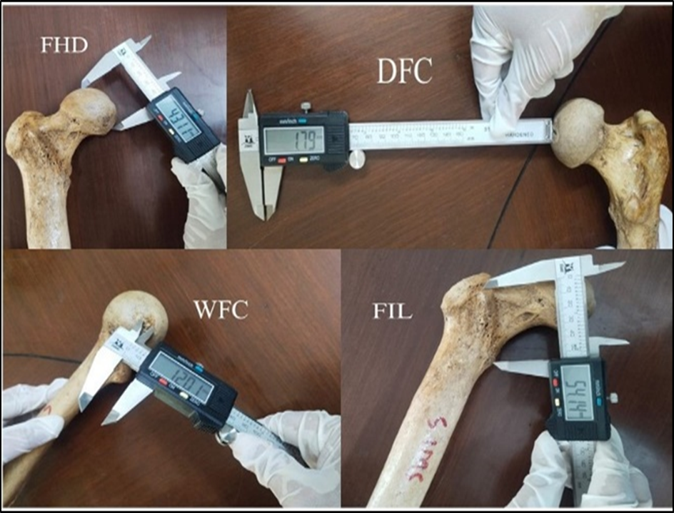
Figure 1: Shows measurements of FHD (Femoral Head Diameter),DFC(Depth of Fovea Capitis),WFC(Width of Fovea Capitis), FIL(Femoral Intertrochanteric line Length) taken by Digital vernier caliper.
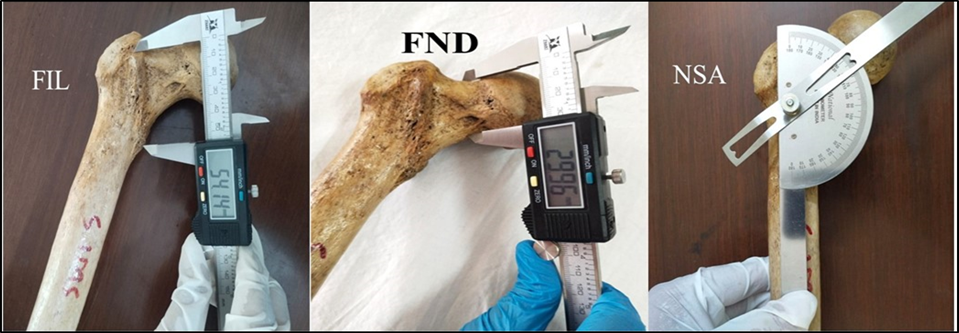
Figure 2: Shows measurements of FNL (Femoral Neck Length) FND (Femoral Neck Diameter) with a Digital vernier caliper and NSA (Neck Shaft Angle) with a Goniometer,
RESULTS:
In the course of this investigation, we identified notable disparities in the intertrochanteric line lengths and neck shaft angles between the right and left femora. Specifically, the neck shaft angle (NSA) for the left femur was measured at 142.8±4.47 mm, contrasting with 135.06±4.17 mm for the right. Regarding the femoral intertrochanteric line (FIL) the left exhibited a measure of 48.46±5.1 mm, while the right side slightly exceeding presented a length of 53.86+5.2 mm. Furthermore, the femoral head diameter (FHD) on the right was observed to be 40.28±3.26 mm, compared to 41.12±4.33 mm on the left. Measurements of the width of the fovea capitis (WFC) showed a right-side value of 12.76±2.53 mm, while the left side measured 13.42±1.96 mm. The depth of fovea capitis (DFC) measurements revealed a right-side reading of 2.95±1.18 mm and a left-side reading of 3.46±1.09 mm. Additionally, the femoral neck length (FNL) for the left side was recorded at 35.62±6.63 mm, in comparison to 33.15 cm±7.07 for the right. The left femoral neck diameter (FND)) was 31.20±5.53 mm, while the right side was again recorded at 31.20±4.42 mm. Table 1 outlines the proximal measurements of the left and right femora, encompassing both maximum and minimum values. Table 2 presents the mean, standard deviation and P-value, indicating the statistical significance of the differences observed between the two sides.
Table -1
|
S.no |
parameters |
Max (Rt) |
Min (Rt) |
Max (Lt) |
Min (Lt) |
|
1. |
FHD (mm) |
45.66 |
33.96 |
51.28 |
32.64 |
|
2. |
FND (mm) |
43.68 |
24.02 |
44.14 |
22.56 |
|
3. |
DFC (mm) |
6.26 |
1.17 |
5.46 |
1.82 |
|
4. |
WFC (mm) |
17.19 |
7.57 |
16.45 |
9.51 |
|
5. |
NSA(degrees) |
145 |
128 |
150 |
133 |
|
6. |
FNL (mm) |
44.75 |
22.62 |
46.03 |
24.74 |
|
7. |
FIL (mm) |
63.96 |
42.13 |
58.78 |
40.77 |
TABLE – 1: Shows maximum and minimum measurements of the proximal end of the femur in right & left dry bones
TABLE – 2
|
S.No |
Parameters |
Mean ± SD (Rt) |
Mean ± SD (Lt) |
P-Value |
|
1 |
FHD (mm) |
40.28 ± 3.26 |
41.12 ± 4.33 |
0.318 |
|
2 |
FND (mm) |
31.24 ± 4.42 |
31.20 ± 5.53 |
0.975 |
|
3 |
DFC (mm) |
2.95 ± 1.18 |
3.46 ± 1.09 |
0.027 |
|
4 |
WFC (mm) |
12.76 ± 2.53 |
13.42 ± 1.96 |
0.180 |
|
5 |
NSA (°) |
135.06 ± 4.17 |
142.80 ± 4.47 |
0.001 |
|
6 |
FNL (mm) |
33.15 ± 7.07 |
35.62 ± 6.63 |
0.054 |
|
7 |
FIL (mm) |
53.86 ± 5.20 |
48.46 ± 5.11 |
0.001 |
TABLE – 2: Shows statistical analysis, Significant co-relation is studied as p-value <0.05
Table -3
|
S. no |
Author, & year |
Sample size |
Side of femur |
FHD (mm) |
DFC (mm) |
WFC (mm) |
|
1. |
Manjunath et al.,2023. |
346 |
Rt |
41.1+5.6 |
------ |
------- |
|
Lt |
41.8+4.8 |
------ |
------- |
|||
|
2. |
Minakshi verma et al., 2017. |
91 |
Rt |
42.51+3.7 |
------ |
------- |
|
Lt |
42.11+4.5 |
------ |
------- |
|||
|
3. |
Prabhanand et al., 2017. |
100
|
Rt |
------ |
------- |
------- |
|
Lt |
------ |
------- |
------- |
|||
|
4. |
Ravi G.O et al., 2016. |
592 |
Rt |
------ |
------ |
------- |
|
Lt |
------- |
------- |
------- |
|||
|
5. |
Rathija et al., 2023 |
140
|
Rt |
33.3+3.7 |
------ |
------- |
|
Lt |
33.8+2.4 |
------ |
------- |
|||
|
6. |
Mayank Gupta et al., 2022 |
96 |
Total |
41.5+3.2 |
||
|
7. |
Present study, 2024 |
90 |
Rt |
40.2+3.2 |
2.9+1.1 |
12.7+2.5 |
|
Lt |
41.1+4.3 |
3.4+1.1 |
13.4+1.9 |
Table -3 : Comparison of morphometry of Femur Head Diameter (FHD), Depth of Fovea Cpitis (DFC), Width of Fovea Capitis (WFC) with the other studies
Table -4
|
S.no |
Author, & year |
Sample size |
Side of femur |
FND (mm) |
NSA (Degrees) |
FNL (mm) |
FIL (mm) |
|
1. |
Manjunath et al., 2023. |
346 |
Rt |
34.4+4.6 |
137.6+3.2 |
34.9+6 |
-------- |
|
Lt |
32.1+5.2 |
137.2+3.8 |
35.2+4 |
-------- |
|||
|
2. |
Minakshi verma et al., 2017. |
91 |
Rt |
127.5+4.6 |
44.8+9 |
-------- |
|
|
Lt |
31.7+3.6 |
130.3+3.8 |
44.6+6 |
-------- |
|||
|
3. |
Prabhanand et al., 2017. |
100
|
Rt |
-------- |
126.7+3.3 |
28.8+3.3 |
-------- |
|
Lt |
-------- |
127.6+4.1 |
28.8+4 |
-------- |
|||
|
4. |
592 |
Rt |
------- |
36.4+5.2 |
-------- |
||
|
Lt |
------ |
36.1+5.6 |
-------- |
||||
|
5. |
Rathija et al., 2023 |
140
|
Rt |
17.49+2 |
------ |
------- |
-------- |
|
Lt |
18.11+2 |
------- |
------- |
-------- |
|||
|
6. |
Mayank Gupta et al., 2022 |
96 |
Total |
29.4+3.3
|
119+5.1 |
36+4.9
|
|
|
7. |
Present study, 2024 |
90 |
Rt |
31.2+4.4 |
135+4.1 |
33.1+7 |
|
|
Lt |
31.2+5.5 |
142+4.4 |
35.6+6.6 |
Table 4 – Comparison of morphometry of Femur Neck Diameter (FND), Neck Shaft Angle (NSA), Femur Neck Length (FNL), Femur Intertrochanteric Line (FIL) with other studies.
DISCUSSION:
The total hip replacement surgeries has typically increased globally and in India. Specifically, it is observed in postmenopausal women suffering from osteoarthritis, a condition that leads to increased susceptibility to bone deterioration, joint stiffness, and fractures.
The femur head dislocation is predominantly raised in age, gender and fractures. Currently oversized heads are used in total hip arthroplasty to decrease the chances of dislocation but they can cause chronic complication of wear [8]. Therefore an accurate measuring of femoral head considering regional variations is predominantly raised in past years for preventing instability and dislocation [6]. Our findings indicated the femoral head diameter (FHD) to be 40.28±3.26 mm on the right side and 41.12±4.33 mm on the left. Our findings align with those reported by Manjunath et al as 41.12 ± 5.6 mm on the right side and 41.84 ± 4.8 mm on the left, closely resembling the results of our measurements.
Fovea capitis is the site of attachment of ligamentum teres which maintains the joint stability. Blood vessels to the head of femur is traversed through the ligament of head of femur. In case of avascular necrosis of head of femur it is highly necessary to measure the geometry of fovea capitis[6]. It is observed that the depth of fovea capitis (DFC) 2.95+1.18 mm on the right side and 3.46+1.09 mm on the left side and width of fovea capitis (WFC) 12.76+2.53 mm on the right and 13.42+1.96 on the left side values are greater than the values obtained by the Mayank Gupta et al DFC as 2.9+0.7 mm and WFC as 11.3+2.3 mm . Table 3 explains the comparison of femoral head variables with other studies.
Femur neck play crucial role in the Hip fractures. In retrospective study it is observed that, there is increase in risk of fractures by 24% for every one millimeter increase in femoral neck width [2]. Therefore it is significant to measure the femur neck diameter (FND) in our study we found right as 31.2+4.4mm and left as 31.2+5.5mm. our findings are comparatively nearby with values of Minakshi verma et al studies obtained right as 34.2+4.3mm and left as 31.7+3.6mm.
Neck – shaft angle (NSA) plays an important role in diagnosis of hip joint alignment. It varies with age, and is influenced by various factors like, occupation, race, ethnicity and sedentary lifestyle [6].Notably, we observed NSA values in our study 135.06 ± 4.17 degrees on the right and 142.8 ± 4.47 degrees on the left. In Manjunath et al.'s [1] study, the NSA values were 137.6 ± 3.2 degrees on the right and 137.2 ± 3.8 degrees on the left was nearly close to our findings. In another study Ravi G.O et al[4] reported NSA as 136.9+4.4 degrees on the right and 136.7+4.4 degrees on the left which was again close to our findings. In comparison with other studies we observed NSA values are higher in our study.
Femur intertrochanteric line (FIL) statistical data is significant in non union of femoral neck fractures after internal fixation. In patients of young and middle age the intertrochanteric osteotomy alters the anatomy of proximal end of femur therefore inspite of avascular necrosis of femur the early diagnosis and treatment is worth in exploring [14]. Notably, in our study (FIL) was 53.8+5.2mm on the right and 48.4+5.1mm on the left side. Whereas Mayank Gupta et al [6] finds FIL as 41.9+5.1mm and comparatively we found higher values of FIL . Table 4 provides a detailed breakdown of the proximal femur morphometry considering femoral neck variables. The regional variations observed in our study offer valuable insights for optimizing implant design.
Limitation:
There is a limitation in measuring gender and age differences in our study.
CONCLUSION:
There are various investigations on morphometrical analysis of proximal end of femur in India and globally. In the present study we found regional differences when compared with parameters of other studies. The necessity of considering such variations will provide valuable insight for biomedical engineers and orthopedics in the design and development of prosthetic devices . Further study is required in case of gender and age of the femur.
REFERENCES:
1. Manjunath TH , Santhosh CS. Morphometric Study of Adult Dry Femur and its Forensic Importance. DOI: 10.47009/jamp.2023.5.5.245
2. Minakshi Verma, Sheetal Joshi, Anita Tuli, Shashi Raheja, Priyanka Jain, Priyanka Srivastava et al. Morphometry of Proximal Femur in Indian Population. Journal of Clinical and Diagnostic Research. 2017;11(2):AC01-AC04.
3. Chaudhary PN, Shirol VS, Virupaxi RD. A morphometric study of femoral length, anterior neck length, and neck-shaft angle in dry femora: A cross-sectional study. Indian J Health Sci Biomed Res 2017;10:331-4.
4. Ravi G.O, Shaik Hussain Saheb,Abraham Ratna Joseph N. A Morphometric Study of Femur and its Clinical Importance. Int J Intg MedSci. 2016; 3(7):341-44.
5. Rathija Sreekumar, Morphometric Analysis of Proximal End of Femur. International Journal of Anatomy, Radiology and Surgery. 2023 Nov, Vol-12(6): AO01-AO04.
6. Gupta M, Devadas D, Sahni C, et al. (September 04, 2022) Morphometric Analysis of the Proximal Femur with its Clinical Correlation in Eastern Uttar Pradesh Region. Cureus 14(9): e28780. DOI 10.7759/cureus.28780
7. Sajan Skaria, Manoj Kulkarni, Hetal Vaishnani. Morphometric Study of Proximal Femur and its Applications in Prosthesis Designing: A Cross-sectional study from Western India. Journal of Clinical and Diagnostic Research. 2022; 16(5): AC01-AC04
8. Cho M, Choi WK, Kim JJ. Current Concepts of Using Large Femoral Heads in Total Hip Arthroplasty. H&P 2016;28:134-141. https://doi.org/10.5371/hp.2016.28.3.134
9. Rajendran HSR, Raamabarathi K, Sundaramurthi I, Gnanasundaram V, Balaji T. [3] Anthropometric analysis of femur in south Indian population. Biomed and Pharm J. 2020;13(1):167-73.
10. Vinay G, Kumar NB, Thondapu K. Morphometric study of proximal end of femur in [5] Telungana population. Int J Anat Res. 2020;8:7247-50. Available at: Doi: 10.16965/ijar.2019.350.
11. S Dhivya, V Nandhini. A Study of Certain Femoral Metrics in South Indian Population and its Clinical Importance International Journal of Scientific Study.2015; 3(7):132-135.
12. Siwach RC: Anthropometric study of proximal femur geometry and its clinical application. Ann Natl Acad Med Sci (India). 2018, 54:203-215. 10.1055/s-0040-1712831
13. Yadav SK, Arun Chandra Singh S, Yadav R. Determining the angle of femoral torsion and association with shaft length and mid circumference. Asian Journal of Medical Sciences. 2020;11(3):58
14. Zhou QJ, Peng XQ, Fei ZG, Zhu XH, Sun CG. Treatment of nonunion after femoral neck fracture with valgus intertrochanteric osteotomy: a case report with 18-year follow-up. J Int Med Res. 2021 May;49(5):3000605211012662. doi: 10.1177/03000605211012662. PMID: 34034563; PMCID: PMC8161879.