Spectrum of benign and malignant lesions of male urinary tract in a tertiary care hospital of Jammu- A one-year retrospective study
- Aanchal , MBBS, MD Pathology. Department of Pathology, Govt. Medical College Jammu
- Akhtar Un Nisa Salaria , Assistant Professor, Department of Pathology, Govt. Medical College Jammu
- Anu Radha , Assistant Professor, Department of Radiodiagnosis, Rama Medical College Hospital & Research Centre, Hapur. (Corresponding Author)
- Subash Bhardwaj , Professor. & Ex. Head Department of Pathology, Govt. Medical College Jammu
Article Information:
Abstract:
Background: Lesions of the male urinary tract constitute a major proportion of the surgical pathology workload in tertiary care hospitals and include a wide spectrum of benign, premalignant, and malignant conditions. Accurate histopathological diagnosis is essential for appropriate clinical management, particularly for prostatic enlargement-related lesions and urothelial neoplasms of the urinary bladder. Materials & Methods: This retrospective observational study was carried out in the Department of Pathology of a tertiary care hospital in Jammu over one year. All male urinary tract specimens received for histopathological examination during the study period were included (N = 61). Data regarding patient age, organ/site, type of procedure, and final histopathological diagnosis were retrieved from departmental records. Specimens were processed routinely and stained with hematoxylin and eosin. Diagnoses were grouped into clinically relevant categories. Bladder tumors were further assessed for histologic grade and invasion status as mentioned in reporting notes. Descriptive statistics were used to summarize the findings. Results: The study population predominantly comprised older adults with a mean age of 63.30 ± 12.70 years (median 66.50; range 22–85). Prostate specimens were most frequent (33/61; 54.10%), followed by bladder specimens (24/61; 39.30%), with few kidney (3/61; 4.90%) and testis (1/61; 1.60%) specimens. TURP/prostate chips were the commonest specimen type (19/61; 31.10%), followed by TURBT (14/61; 23.00%). Benign prostatic pathology was the largest diagnostic category (27/61; 44.30%). Urothelial neoplasms constituted a significant proportion of cases, with low-grade (12/61; 19.70%) and high-grade (10/61; 16.40%) tumors. Among bladder cases (n = 24), low-grade tumors were slightly more common than high-grade tumors, and invasion was documented in a minority of cases. Conclusion: The male urinary tract lesion spectrum in this tertiary care hospital was dominated by prostate and bladder pathology, with benign prostatic lesions forming the largest group and urothelial neoplasms representing a major malignant burden. Histopathological evaluation remains crucial for definitive diagnosis, tumor grading, and invasion assessment.
Keywords:
Article :
INTRODUCTION :
Diseases of the male urinary tract account for a substantial share of outpatient visits, emergency presentations, and operative workload in tertiary care hospitals. The urinary tract is continuously exposed to metabolic, infectious, obstructive, and carcinogenic influences, and lesions may range from non-neoplastic inflammatory/reactive changes to premalignant conditions and overt malignancies. In routine practice, clinical and radiological impressions often overlap—hematuria, lower urinary tract symptoms, dysuria, flank pain, or obstructive uropathy can be produced by both benign and malignant processes—making tissue diagnosis a cornerstone for accurate classification, prognostication, and treatment planning. At the population level, the burden of genitourinary cancers is rising with aging demographics, and prostate and bladder cancers remain among the leading malignancies affecting men globally.¹ The male urinary tract also shows strong age-related clustering of disease. Obstructive lower urinary tract symptoms and benign prostatic enlargement typically increase with advancing age, while malignant lesions such as prostate carcinoma and urothelial carcinoma frequently present in later decades. These epidemiologic features shape the histopathology workload at tertiary centres, where tissue is commonly obtained via endoscopic resections and targeted biopsies. In parallel, global projections indicate that the absolute number of prostate cancer cases will rise sharply in the coming decades, largely driven by population ageing and increased life expectancy, highlighting the growing importance of hospital-based audits that describe local patterns of disease and specimen flow.2,3 Bladder lesions represent another major component of uropathology practice, particularly in men. Hematuria is a key symptom prompting evaluation for urothelial malignancy, but it can also be due to benign conditions such as cystitis, stones, or instrumentation-related injury. Risk-based approaches to hematuria assessment emphasize identifying patients who warrant timely cystoscopy and imaging to exclude malignancy, reflecting the diagnostic priority of early detection for urothelial tumors.⁴ From a pathology standpoint, bladder resections and biopsies must address not only the presence of neoplasia but also clinically decisive parameters such as histologic grade and evidence of invasion, because these variables are central to stratifying recurrence and progression risk and to selecting intravesical therapy versus more aggressive interventions. Globally, bladder cancer continues to impose a considerable mortality burden, and the disparity between men and women is consistent across regions, reflecting differences in exposure to risk factors such as tobacco smoke and occupational carcinogens. Contemporary analyses of worldwide mortality patterns show that bladder cancer is strongly influenced by preventable exposures, and that deaths occur disproportionately in men, underlining the relevance of studying urothelial lesions within male urinary-tract specimen series.5 Modern classification systems have increasingly emphasized reproducibility and clinical relevance in diagnosing urinary tract tumors. The 5th edition of the World Health Organization (WHO) classification integrates morphologic criteria with evolving molecular insights, while still relying on histopathology as the diagnostic gold standard for most settings. Updates in tumor terminology, grading practices, and recognition of histologic variants aim to improve consistency across laboratories and to align pathology reporting with clinical decision-making.⁶ For urinary bladder tumors in particular, refinements in defining papillary lesions, grading thresholds, and the interpretation of early stromal invasion directly affect patient management. Consequently, hospital-based studies that report the spectrum of lesions using contemporary frameworks can help harmonize local reporting with international standards, especially in regions where resource constraints may limit extensive ancillary testing. Renal lesions, although less frequent in specimen-based audits compared with prostate and bladder submissions, are clinically important because many renal masses are malignant and may require nephron-sparing or radical surgery. The epidemiology of renal cell carcinoma (RCC) varies by geography and risk-factor prevalence, and evidence continues to highlight potentially modifiable contributors such as hypertension, obesity, and smoking.
MATERIALS AND METHODS:
This retrospective observational study was conducted in the Department of Pathology of a tertiary care hospital in Jammu over a period of one year. The study aimed to analyze the spectrum of benign and malignant lesions of the male urinary tract received for histopathological examination during the study period and to describe their distribution with respect to age, organ involved, type of procedure, and final diagnosis.
All histopathology specimens from the male urinary tract received in the pathology department during the one year were included. The organs evaluated included the prostate, urinary bladder, kidney, and testis. Specimens were obtained through routine urological procedures such as transurethral resection of prostate (TURP), transurethral resection of bladder tumor (TURBT), TRUS-guided prostate biopsies, nephrectomies (partial or radical), orchidectomy specimens, and other biopsy samples. Repeat entries were considered as separate cases when received as separate specimens during the study period, consistent with a specimen-based audit approach.
Patient demographic details were retrieved from the histopathology requisition forms and departmental records. Age, organ/site of specimen, procedure performed, and histopathological diagnosis were recorded. Patient identifiers such as names and hospital registration numbers were excluded to maintain confidentiality. The diagnoses were grouped into clinically relevant categories such as benign prostatic lesions (including benign prostatic hyperplasia and associated lesions), prostate adenocarcinoma, urothelial neoplasms of urinary bladder (further classified as low grade, high grade, or grade not specified), renal tumors (including renal cell carcinoma and other renal tumors), premalignant lesions such as prostatic intraepithelial neoplasia (PIN), testicular germ cell tumor, and benign bladder papilloma. For bladder tumors, additional documentation of invasion status was extracted from the reporting notes wherever mentioned (e.g., lamina propria invasion, muscle invasion, pT1, pTa), and cases were categorized as “invasion noted” or “no invasion mentioned.”
All specimens were processed routinely. Tissue was fixed in 10% neutral buffered formalin, processed through graded alcohols, cleared, and embedded in paraffin. Sections of approximately 3–5 µm thickness were stained with hematoxylin and eosin (H&E) and examined under light microscopy. The final diagnosis was recorded as per standard histopathological criteria, and tumor grading and invasion details were documented as available in the reports.
Data were entered in a structured format and analyzed using descriptive and inferential statistics. Continuous variables such as age were summarized as mean, standard deviation, median, and range. Categorical variables were summarized as frequencies and percentages. Where appropriate, chi-square (χ²) goodness-of-fit tests were applied to evaluate whether the observed categorical distributions were significantly non-uniform. For the bladder invasion variable, a binomial test was applied to assess whether the proportion of “invasion noted” differed significantly from “no invasion mentioned.” A p-value of <0.05 was considered statistically significant. Ethical considerations were maintained by anonymizing patient information, and the study was based solely on archived records and histopathology materials from routine clinical practice.
RESULTS:
Table 1 (Age of patients, N = 61) shows that the study population was predominantly older adults. The mean
age was 63.30 ± 12.70 years, with a median of 66.50 years, indicating that most cases clustered in the sixth to seventh decade. The age range was wide (22.00 to 85.00 years), suggesting that urological specimens were received across a broad adult age spectrum, although the central tendency clearly reflects an older population.
Table 1. Age of the patients (N = 61)
|
n |
Mean age (years) |
SD |
Median |
Min |
Max |
p-value |
|
61 |
63.30 |
12.70 |
66.50 |
22.00 |
85.00 |
NA |
Table 2 (Specimen distribution by organ, N = 61) demonstrates that the majority of specimens were from the prostate (33 cases, 54.10%), followed by the bladder (24 cases, 39.30%). Specimens from the kidney (3 cases, 4.90%) and testis (1 case, 1.60%) were relatively infrequent. The overall distribution was statistically non-uniform (χ² test p < 0.001), meaning the specimens were not evenly distributed across organs; instead, the case load was clearly dominated by prostate and bladder specimens.
Table 2. Specimen / Entry distribution by organ (N = 61)
|
Organ |
n |
% |
p-value |
Organ |
n |
|
Prostate |
33 |
54.10 |
|
Prostate |
33 |
|
Bladder |
24 |
39.30 |
|
Bladder |
24 |
|
Kidney |
3 |
4.90 |
|
Kidney |
3 |
Table 3 (Procedure/specimen type distribution, N = 61) reflects the types of urological procedures contributing to the pathology workload. The commonest specimen type was TURP/prostate chips (19 cases, 31.10%), consistent with frequent evaluation for benign prostatic enlargement and related conditions. The next most common was TURBT/bladder resection (14 cases, 23.00%), highlighting a substantial burden of bladder lesions requiring endoscopic resection. Other/unclear procedures (12 cases, 19.70%) and biopsies other than TRUS (9 cases, 14.80%) formed a notable proportion, suggesting some heterogeneity in referral patterns or documentation. Major resections such as nephrectomy (3 cases, 4.90%), TRUS prostate biopsy (3 cases, 4.90%), and orchidectomy (1 case, 1.60%) were comparatively uncommon, indicating that large oncologic resections were a smaller part of the total specimen load during the study period.
Table 3. Procedure / specimen type distribution (N = 61)
|
Procedure / specimen type |
n |
% |
|
TURP / prostate chips |
19 |
31.10 |
|
TURBT / bladder resection |
14 |
23.00 |
|
Other / unclear |
12 |
19.70 |
|
Biopsy (other) |
9 |
14.80 |
|
Nephrectomy (partial/radical) |
3 |
4.90 |
|
TRUS / prostate biopsy |
3 |
4.90 |
|
Orchidectomy |
1 |
1.60 |
Table 4 (Diagnosis categories, N = 61) shows that benign prostatic pathology was the single largest diagnostic category, with Prostate: Benign (BPH/ALMH) accounting for 27 cases (44.30%), confirming that non-malignant prostate enlargement-related pathology formed a major proportion of the workload. Among bladder lesions, urothelial neoplasms were prominent, with low-grade tumors in 12 cases (19.70%) and high-grade tumors in 10 cases (16.40%), indicating a substantial malignant/pre-malignant burden in bladder specimens. Prostate adenocarcinoma comprised 5 cases (8.20%), showing that malignant prostate disease was less frequent than benign prostate conditions in this set. Renal malignancies were relatively rare, with RCC in 2 cases (3.30%) and one other renal tumor (1.60%). Single-case categories included PIN (1.60%), testicular germ cell tumor (1.60%), benign bladder papilloma (1.60%), and grade-unclear urothelial neoplasm (1.60%). The overall diagnostic distribution was significantly non-uniform (χ² test p < 0.001), meaning that a few diagnostic categories (notably benign prostate disease and urothelial tumors) contributed disproportionately to the overall case mix.
Table 4. Diagnosis categories (N = 61)
|
Diagnosis category |
n |
% |
p-value |
|
Prostate: Benign (BPH / ALMH) |
27 |
44.30 |
|
|
Bladder: Urothelial neoplasm – Low grade |
12 |
19.70 |
|
|
Bladder: Urothelial neoplasm – High grade |
10 |
16.40 |
|
|
Prostate: Adenocarcinoma |
5 |
8.20 |
|
|
Kidney: RCC |
2 |
3.30 |
|
|
Bladder: Urothelial neoplasm – Grade unclear |
1 |
1.60 |
|
|
Kidney: Other renal tumor |
1 |
1.60 |
|
|
Prostate: PIN |
1 |
1.60 |
|
|
Testis: Germ cell tumor |
1 |
1.60 |
|
|
Bladder: Benign papilloma |
1 |
1.60 |
|
|
Overall χ² test |
— |
— |
<0.001 |
Table 5 (Bladder cases only, n = 24) provides a focused view of bladder tumors by grade and invasion status. For tumor grade, low-grade urothelial neoplasms were slightly more common (12 cases, 50.00%) than high-grade tumors (10 cases, 41.70%), while grade-unclear (1 case, 4.20%) and benign papilloma (1 case, 4.20%) were uncommon. The grade distribution was statistically significant (χ² test p = 0.002), indicating that the observed frequencies across grade categories were not evenly distributed and were concentrated in low- and high-grade disease. Regarding invasion status as documented in the notes, no invasion was mentioned in 17 cases (70.80%), while invasion was noted in 7 cases (29.20%); this difference was statistically significant (binomial test p = 0.031), suggesting that the proportion of cases without documented invasion was meaningfully higher than those with invasion recorded. The invasion-noted group included descriptions such as lamina propria invasion, muscle-invasive disease, pT1, and pTa, indicating that a clinically important subset of bladder tumors demonstrated invasive or stage-assigned disease features.
Table 5. Bladder cases (n = 24): Tumor grade and invasion status
|
Variable |
Category |
n |
% |
p-value |
|
Tumor grade |
Low grade |
12 |
50.00 |
|
|
|
High grade |
10 |
41.70 |
|
|
|
Grade unclear |
1 |
4.20 |
|
|
|
Benign |
1 |
4.20 |
|
|
|
Overall χ² test |
— |
— |
0.002 |
|
Invasion status (as mentioned in notes) |
No invasion mentioned |
17 |
70.80 |
|
|
|
Invasion noted |
7 |
29.20 |
|
|
|
Binomial test |
— |
— |
0.031 |
DISCUSSION:
The present one-year audit from a tertiary care hospital in Jammu showed a predominantly elderly male urinary-tract specimen profile, with a mean age of 63.30 ± 12.70 years (median 66.50, range 22–85; N=61). This age pattern is comparable to operative TURP populations reported elsewhere; for example, Agrawal et al. (2019) reported mean ages of 62.1 ± 8.22 years (2006 cohort) and 66.94 ± 9.12 years (2016 cohort) among TURP patients, supporting that male lower urinary tract pathology requiring tissue diagnosis largely clusters in the 6th–7th decades.⁷
In this study, specimen contribution was organ-skewed, with prostate (33/61; 54.10%) and bladder (24/61; 39.30%) dominating, while kidney (3/61; 4.90%) and testis (1/61; 1.60%) were uncommon (χ² p < 0.001). This distribution aligns with the known epidemiology of male urological morbidity in older age groups, where benign prostatic enlargement and bladder neoplasia contribute heavily to clinical symptoms and procedural interventions; population-level summaries also show that BPH frequency rises steeply with age (e.g., high histological prevalence in older decades), which helps explain why prostate-related specimens tend to predominate in routine histopathology workloads.⁸
Procedure patterns in Jammu further reflect this clinical burden: TURP/prostate chips (19/61; 31.10%) were the most frequent, followed by TURBT (14/61; 23.00%), with fewer biopsies and major resections. These findings are consistent with specimen-based series of prostatic lesions where transurethral resections form the bulk of submissions and are largely driven by obstructive symptoms and suspected neoplasia; for instance, Aslam et al. (2013) reported that benign prostatic hyperplasia formed 87.5% (42/48) of prostatic lesions in their spectrum study, illustrating how TURP-type material commonly represents the largest procedural contribution in similar settings.⁹
On histopathology, the single largest diagnostic group in the current study was benign prostate pathology (BPH/associated lesions) at 27/61 (44.30%), reinforcing that non-malignant prostatic enlargement remains the principal reason for tissue evaluation in older men. This predominance mirrors the broad uropathology literature where BPH accounts for the majority of prostatic specimens, and age-linked increases in BPH burden translate directly into higher TURP volumes and benign diagnoses in tertiary-care pathology practice.⁸
Although benign lesions dominated, malignant prostate disease was also detected: prostate adenocarcinoma comprised 5/61 (8.20%) in this series (figure 1a &1b. This magnitude is comparable to the “incidental cancer” phenomenon reported in TURP materials across regions; for example, Janjua et al. (2021) observed 10.7% incidental prostate cancer among TURP specimens in their retrospective series, emphasizing the continued role of routine histopathology in identifying unsuspected carcinoma even when surgery is performed for presumed benign obstruction.¹⁰
Bladder pathology constituted a substantial fraction of the workload (24 cases), and within the total study cohort the bladder urothelial neoplasm categories were prominent (low grade 12/61; 19.70% and high grade 10/61; 16.40%). Comparable tertiary-care bladder biopsy data demonstrate that urothelial carcinoma is typically the predominant neoplastic diagnosis and that high-grade invasive disease often forms a major subset; Kundlia et al. (2024), for example, reported invasive high-grade urothelial neoplasm 36.7% ,(figure 2a&2b))and non-invasive low-grade papillary urothelial neoplasm 20.0% (figure3a&3b)(figure among their neoplastic bladder cases, supporting the clinical reality that both low-grade papillary and high-grade invasive tumors commonly contribute to tertiary-center case mixes.¹¹
When bladder cases were analyzed internally (n=24), low-grade tumors were slightly more frequent (12/24; 50.00%) than high-grade tumors (10/24; 41.70%), with few benign/unclear cases, and the grade distribution was statistically non-uniform (χ² p = 0.002). In contrast, some institutional series show a heavier high-grade burden; Agarwal et al. (2024) reported infiltrating urothelial carcinoma high-grade 58% and infiltrating low-grade 14% in their histopathology spectrum, indicating that grade proportions can vary widely by referral pattern, inclusion of cystectomy material, and the proportion of infiltrating tumors captured at a center.¹²
Invasion documentation in this study showed “invasion noted” in 7/24 (29.20%) and “no invasion mentioned” in 17/24 (70.80%) (binomial p = 0.031). This lower documented invasion proportion may reflect (i) true predominance of non-muscle-invasive disease in TURBT material, and/or (ii) incomplete representation of muscularis propria in some resections and reporting notes. In comparison, Agarwal et al. (2024) found deeper invasion to be common in infiltrating carcinomas, reporting muscularis propria invasion in 58.33% of infiltrating urothelial carcinoma cases, and muscle invasion was overwhelmingly associated with high-grade tumors in their dataset—highlighting how invasion rates rise markedly in cohorts enriched for infiltrating/high-grade disease and in series with consistent muscle assessment.¹²
Renal and testicular specimens were infrequent in the current one-year audit (kidney 3/61; 4.90% and testis 1/61; 1.60%), with RCC diagnosed in 2/61 (3.30%). Low renal specimen contribution is typical for specimen-mix audits because nephrectomies are fewer than TURP/TURBT; however, when nephrectomy/tumor nephrectomy cohorts are analyzed, malignancy dominates. Datta et al. (2016) reported that among adult renal tumors, 91.6% were malignant and the mean age was 54.5 years, with RCC being the most common malignant type—supporting that even though renal cases are few in general surgical pathology flow, they carry a high likelihood of malignancy when they do occur.¹³
Finally, only one testicular tumor (a germ cell tumor) was encountered (1/61; 1.60%), consistent with the relative rarity of orchidectomy specimens in a general one-year uropathology audit compared with prostate and bladder resections. Larger tertiary-care series confirm that germ cell tumors constitute the dominant histology among testicular neoplasms: Chakrabarti et al. (2016) reported germ cell tumors in 77.1% of testicular/paratesticular neoplasms (mean age 38.1 years), with most cases occurring in the 3rd–4th decades; this contrasts with the older age-skew of the overall Jammu urinary-tract specimen set and underscores that testicular tumors represent a distinct, younger-age clinical subgroup that may appear only sporadically in short-duration audits.14
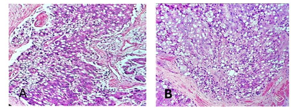
Figure1.Photomicrographs of Prostatic Adenocarcinoma (signet ring type), 20 X (A) and 40X (B)
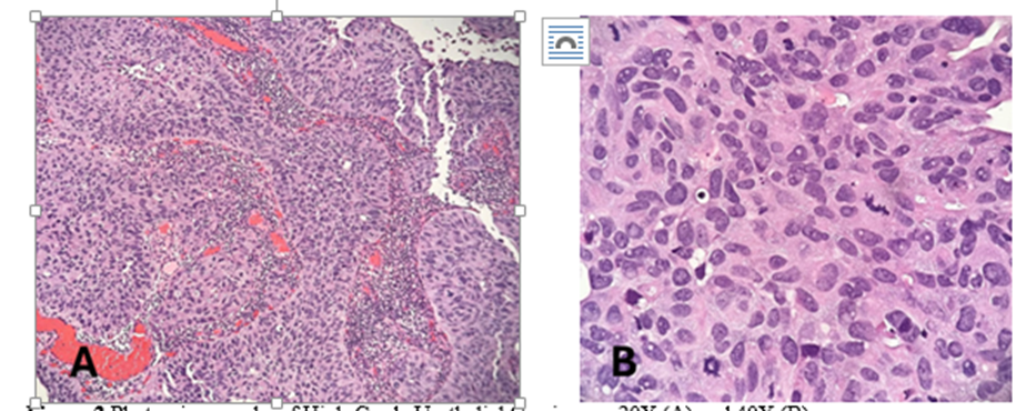
Figure 2 Photomicrographs of High-Grade Urothelial Carcinoma, 20X (A) and 40X (B).
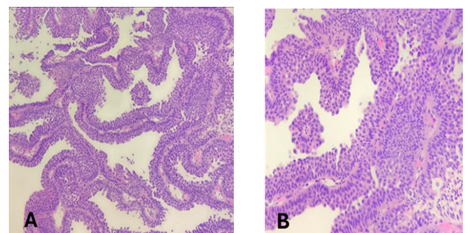
Figure3. Photomicrographs of Papillary urothelial neoplasm of low malignant potential (PUNLMP), 20 X (A) and 40X (B).
CONCLUSION:
We concluded that the spectrum of male urinary tract lesions in this tertiary care hospital over one year was dominated by prostate- and bladder-related specimens. Benign prostatic pathology constituted the largest diagnostic group, reflecting the high burden of obstructive prostatic disease in older men. Urothelial neoplasms formed a substantial proportion of bladder lesions, with both low- and high-grade tumors being commonly encountered. The findings highlight the importance of routine histopathological evaluation for accurate diagnosis, grading, and assessment of invasion to guide appropriate clinical management.
REFERENCES:
1. Bray F, Laversanne M, Sung H, Ferlay J, Siegel RL, Soerjomataram I, Jemal A. Global cancer statistics 2022: GLOBOCAN estimates of incidence and mortality worldwide for 36 cancers in 185 countries. CA Cancer J Clin. 2024 May–Jun;74(3):229-263.
2. James ND, Tannock I, N’Dow J, Feng F, Gillessen S, Ali SA, et al. The Lancet Commission on prostate cancer: planning for the surge in cases. Lancet. 2024 Apr 27;403(10437):1683-1722.
3. Wéber A, Vignat J, Shah R, Morgan E, Laversanne M, Nagy P, et al. Global burden of bladder cancer mortality in 2020 and 2040 according to GLOBOCAN estimates. World J Urol. 2024 Apr 16;42(1):237.
4. Barocas DA, Boorjian SA, Alvarez RD, Downs TM, Gross CP, Hamilton BD, et al. Microhematuria: AUA/SUFU Guideline. J Urol. 2020 Oct;204(4):778-786.
5. Moch H, Amin MB, Berney DM, Compérat EM, Gill AJ, Hartmann A, et al. The 2022 World Health Organization Classification of Tumours of the Urinary System and Male Genital Organs—Part A: Renal, penile, and testicular tumours. Eur Urol. 2022 Nov;82(5):458-468.
6. Guo CC, Zhang S, Shen SS, et al. Recent Advances in the Classification of Bladder Cancer—Updates from the 5th Edition of the World Health Organization Classification of the Urinary and Male Genital Tumors. Bladder Cancer. 2023;9(1):1-14.
7. Agrawal M, Kumar M, Pandey S, Aggarwal A, Sankhwar S. Changing profiles of patients undergoing transurethral resection of the prostate over a decade: A single-center experience. Urol Ann. 2019 Jul–Sep;11(3):270-275. doi:10.4103/UA.UA_198_17.
8. Ng M, Leslie SW, Baradhi KM. Benign Prostatic Hyperplasia. In: StatPearls [Internet]. Treasure Island (FL): StatPearls Publishing; 2025 Jan–. Updated 2024 Oct 20.
9. Aslam HM, Shahid N, Shaikh NA, Shaikh HA, Saleem S, Mughal A. Spectrum of prostatic lesions. Int Arch Med. 2013 Sep 25;6(1):36.
10. Janjua TK, Yousuf MA, Iqbal MT, Memon SM, Abdullah A, Faridi N, et al. Incidental finding of prostate cancer in transurethral resection of prostate (TURP) specimens: a retrospective analysis from a tertiary care hospital in Pakistan. Pan Afr Med J. 2021 May 7;39:20. doi:10.11604/pamj.2021.39.20.26931.
11. Kundlia A, Dharwadkar A, Gore C, Viswanathan V, Ingale A, et al. Exploring the Histopathological Landscape of Urinary Bladder Diseases: A Tertiary Care Center Study. Cureus. 2024 Jul 15;16(7):e64557.
12. Agarwal S, et al. A Clinicopathologic Study of Urinary Bladder Lesions Amongst North Indian Population: An Experience From a Tertiary Care Centre. Cureus. 2024 May 7;16(5):e59792.
13. Datta B, Giri A, Halder B. Histopathological evaluation of surgically treated adult renal tumors: Report from a tertiary care center in India. Indian J Cancer. 2016 Jan–Mar;53(1):124-126.
14. Chakrabarti PR, Dosi S, Varma A, Kiyawat P, Khare G, Matreja S. Histopathological Trends of Testicular Neoplasm: An Experience over a Decade in a Tertiary Care Centre in the Malwa Belt of Central India. J Clin Diagn Res. 2016 Jun;10(6):EC16-EC18.