Utility of trichoscopy in the evaluation of alopecia: A hospital based descriptive cross-sectional study.
- Dr Irfan Ahmed , Assistant professor, Department of Dermatology, Veneorology and Leprosy, Acharaya Shri Chander College of Medical Sciences and Hospital, Sidhra, Jammu, Jammu and Kashmir, India
- Dr. Reeta Sood , Professor and HOD, Department of Dermatology, Veneorology and Leprosy, Acharaya Shri Chander College of Medical Sciences and Hospital, Sidhra, Jammu, Jammu and Kashmir, India
- Dr. Ennakshee Sharma , Assistant professor, Department of Dermatology, Veneorology and Leprosy, Acharaya Shri Chander College of Medical Sciences and Hospital, Sidhra, Jammu, Jammu and Kashmir, India.
Article Information:
Abstract:
Background: Trichoscopy can be a valuable investigation in routine workup for alopecia as it is noninvasive and convenient however its usefulness is yet to be known. Objectives: To differentiate alopecia cases based on trichoscopic features, identify features specific to each alopecia type and determine the usefulness of trichoscopy in alopecia. Materials and methods: A descriptive cross-sectional study was done on alopecia patients. Each patient was subject to history and clinical examination before undergoing trichoscopy using a non-polarized hand held dermoscope with 10 times magnification. Results: Seventy-five patients were enrolled in the study. A clinical diagnosis was made based on history and examination and patients were divided in cicatricial (68 patients) and non-cicatricial (7 patients) alopecia. Trichoscopy was performed on each patients. Hair diameter diversity was the most common finding in both male and female androgenic alopecia (92.3%, 86.6% respectively), followed by vellus hair. Yellow dots (76.47%) and broken hair (76.47%) were the most common finding in alopecia areata. Hair diameter diversity >20% a feature seen in most cases of androgenic alopecia (89.3%) was not seen in any of the telogen effluvium patients. Patients with tinea capitis had corkscrew hairs and barcode hair. Among patients with scarring alopecia loss of hair follicles and white dots were seen in all cases. Patients with lichen planopilaris had blue grey perifollicular pigmentation, perifollicular scaling and hair cast. Patients with frontal fibrosis alopecia had single hair follicular units and none had blue grey pigmentation. Discoid lupus erythematosus patient had pinkish white background with loss of follicles along with red arborising patterns, follicular plugging. Conclusion: Trichoscopy was found useful in diagnosing alopecia.
Keywords:
Article :
INTRODUCTION:
Alopecia is a very common yet problematic condition for many. It is broadly classified into non-scarring and scarring alopecia depending upon the cause. Diagnosis of specific type of alopecia relies on history, examination using naked eye or hand lens and laboratory investigation. Finding the exact cause is often difficult and might require biopsy which is an invasive procedure and is often time consuming. Handheld dermoscope is a valuable addition to existing clinical practice to arrive at a diagnosis with reliable accuracy.
MATERIALS AND METHODS:
All patients presenting with alopecia at Department of Dermatology ASCOMS hospital Sidhra, Jammu were subjected to clinical evaluation during a six-month period between August 2024 to January 2025. A hand held dermoscope with 10times magnification was used for trichoscopy and scalp dermoscopy. Patients were classified into non-scarring alopecia which included Telogen effluvium, androgenic alopecia, alopecia areata, and scarring alopecia including lichen planopilaris, discoid lupus erythematosus. Data was collected on hair diameter variability, black dots, yellow dots, plugged follicles, exclamation mark hair etc and arranged in tables. Means and proportions were calculated. Hair diameter diversity was calculated by counting the number of thin and thick hairs and deriving percentage of thin hair in each trichoscopic field.
Statistical analysis: Data so collected was tabulated in an excel sheet, under the guidance of statistician. The means and standard deviations of the measurements per group were used for statistical analysis (SPSS 22.00 for windows; SPSS inc, Chicago, USA). For each assessment point, data were statistically analyzed using chi square test. The level of significance was set at p < 0.05.
RESULTS:
A total of 75 patients with alopecia were evaluated with trichoscopy. Out of 75 patients 54 were females and 21 males. Mean age of presentation was 27.73 years (range: 4-73 years). 28 patients had androgenic alopecia (15 females and 13 males), 21 had telogen effluvium, 17 had alopecia areata, 7 had scarring alopecia, 2 had tinea capitis. The demography of non-scarring and scarring alopecia is listed in table 1.
Hair diameter diversity/anisotrichosis (92.3%), vellus hair (84.6%), thin hair (84.6%) were the most common findings in male androgenic alopecia [Figure 1]. In female androgenic alopecia, anisotrichosis (86.6%), vellus hair (66.6%), honeycomb pigmentation (60%) were the most common features [Figure 2].
In telogen effluvium focal atrichia was seen in 66% patients and hair diameter variability <10% was present in 28.57%. Vellus hairs inter follicular scaling was seen in chronic telogen effluvium and none in acute telogen effluvium [Figure 3].
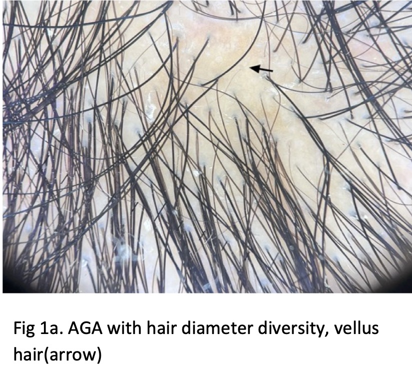
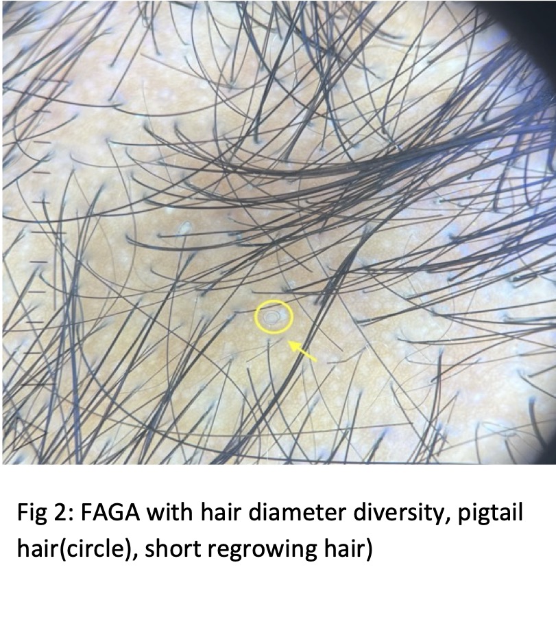
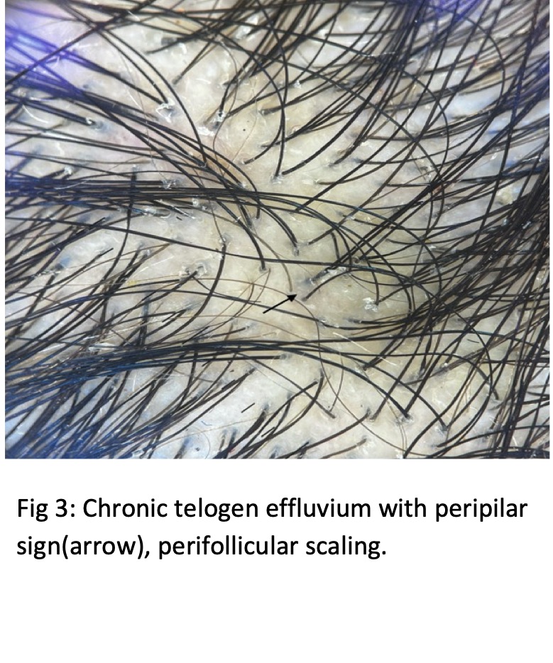
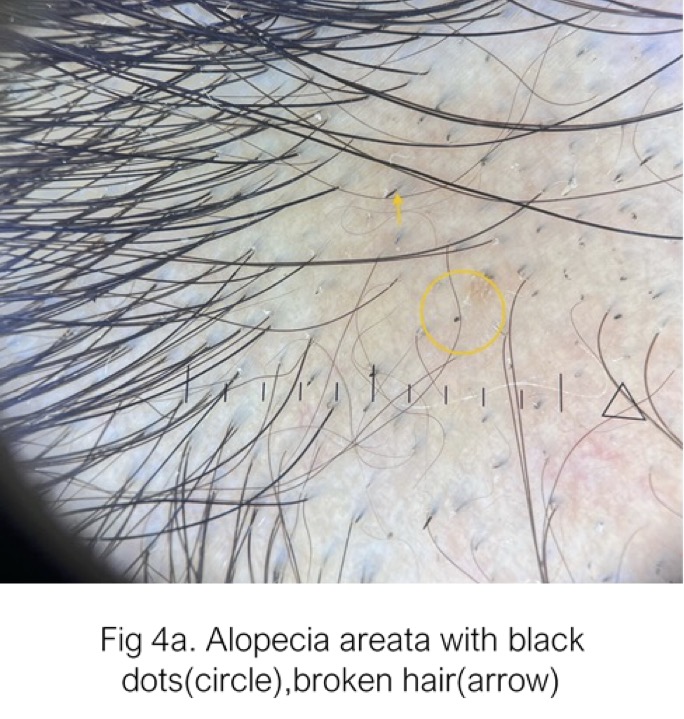
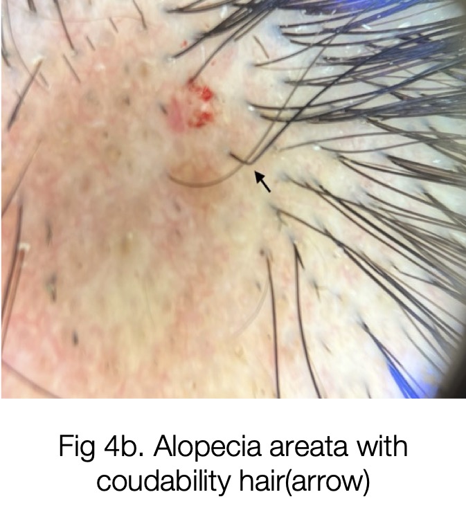
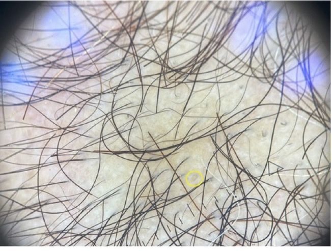
Fig 4c. Diffuse Alopecia areata yellow dots (circle), blackdots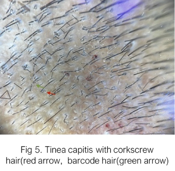
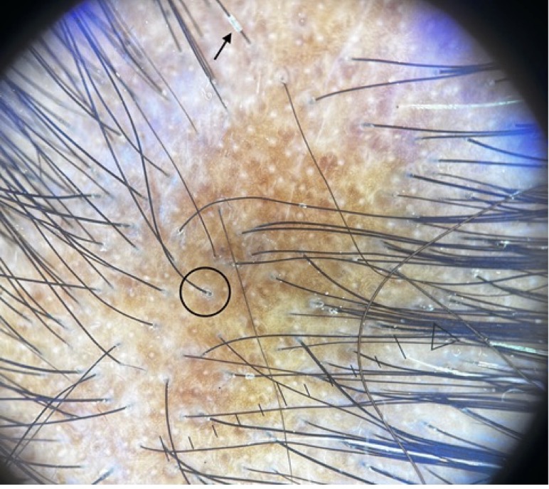 In alopecia areata, yellow dots, broken hair, short vellus hair were the most common finding seen in 75% patients [Figure 4]. Black dots were seen in 56% patients.
Both patient with tinea capitis had cork screw hair, morse code/bar code hair perifollicular scaling and loop hairs [Figure 5].
Table 1: Demography of non-scarring and scarring alopecia in this study
|
Non-scarring Alopecia |
No. of Males |
Mean age (years) |
No. of females |
Mean age (Years) |
Total |
Percentage of total cases |
|
Androgenic alopecia |
13 |
24.79 |
0 |
|
13 |
17.3% |
|
Female pattern hair loss |
0 |
0 |
15 |
32.9 |
15 |
20% |
|
Alopecia areata |
7 |
28.4 |
10 |
19.6 |
17 |
22.6% |
|
Chronic telogen effluvium |
0 |
0 |
16 |
27.5 |
16 |
21.3% |
|
Acute telogen effluvium |
0 |
0 |
5 |
21 |
5 |
6.6% |
|
Tinea capitis |
1 |
6 |
1 |
4 |
2 |
2.6% |
|
Scarring Alopecia |
|
|
|
|
|
|
|
Lichen planopilaris |
0 |
0 |
4 |
45 |
4 |
5.3% |
|
Frontal fibrosing alopecia |
0 |
0 |
2 |
40.5 |
2 |
2.6% |
|
Discoid lupus erythematous |
0 |
0 |
1 |
54 |
1 |
1.3% |
Table 2: Trichoscopic features comparing different non scarring alopecia
|
Trichoscopic finding |
Androgenic alopecia |
Alopecia areata |
Telogen effluvium |
P value |
|||
|
No. |
% |
No. |
% |
No. |
% |
||
|
Hair diameter variability ≥20% |
25 |
89.3 |
0 |
0 |
0 |
0 |
<0.01* |
|
Hair diameter variability <20% |
3 |
10.7 |
0 |
0 |
6 |
28.57 |
0.001* |
|
Yellow dots |
9 |
32.1 |
13 |
76.47 |
1 |
4.7 |
<0.01* |
|
Black dots |
0 |
0 |
10 |
58.8 |
0 |
0 |
<0.01* |
|
White dots |
12 |
42.8 |
2 |
12.5 |
5 |
23.8 |
0.006* |
|
Exclamation mark hair |
0 |
0 |
7 |
43.7 |
0 |
0 |
<0.01* |
|
Broken hair |
0 |
0 |
13 |
76.47 |
0 |
0 |
<0.01* |
|
Coudability sign |
0 |
0 |
2 |
12.5 |
0 |
0 |
0.019* |
|
Short vellus hair |
21 |
75 |
12 |
75 |
3 |
14.3 |
0.027* |
|
Peripilar sign |
11 |
39.3 |
1 |
6.2 |
4 |
19 |
0.008* |
|
Coiled hair |
7 |
25 |
7 |
43.7 |
6 |
28.6 |
0.012* |
|
Thin short hair |
16 |
57.1 |
3 |
18.7 |
12 |
57.1 |
0.036* |
|
Single hair FU |
11 |
39.3 |
2 |
12.5 |
10 |
47.6 |
0.011* |
|
Honey comb pigmentation |
11 |
39.3 |
3 |
18.7 |
5 |
23.8 |
0.015* |
*: statistically significant
Table 3: Comparison between MAGA and FAGA based on trichoscopic findings
|
Trichoscopic finding |
Male Androgenic alopecia |
Female pattern hairloss |
P value |
||
|
|
No. |
% |
No. |
% |
|
|
Hair diameter variability |
12 |
92.3 |
13 |
86.6 |
0.23 |
|
Vellus hair |
11 |
84.6 |
10 |
66.6 |
0.016* |
|
Yellow dots |
7 |
53.8 |
2 |
13.3 |
<0.01* |
|
White dots |
4 |
30.7 |
8 |
53.3 |
0.017* |
|
Perifollicular brown sign |
6 |
46.1 |
5 |
33.3 |
0.048* |
|
Single hair FU |
5 |
38.4 |
6 |
40 |
0.81 |
|
Thin short hair |
11 |
84.6 |
5 |
33.3 |
<0.01* |
|
Honeycomb pigmentation |
2 |
15.4 |
9 |
60 |
<0.01* |
|
Coiled hair |
4 |
30.7 |
3 |
20 |
0.10 |
Patients with scarring alopecia had loss of follicles and white dots in all patients. All patients with lichen planopilaris had peripilar scaling amorphous white areas and honey comb pigmentation [Figure 6]. Follicular plugging was seen in 3 (75%), bluish grey pigmentation in 2(50%) patients and hair casts in 1(25%) patient.
2 patients with frontal fibrosing alopecia had single hair follicular unit, white dots, white amorphous areas in both and peripilar scaling in one patient.
Patient with discoid lupus erythematosus had follicular plugging, arborizing red lines starburst patterns, red dots pinkish white structureless areas follicular scales and honeycomb pigmentation.
Comparative analysis of androgenic alopecia, alopecia areata and telogen effluvium based on trichoscopic findings of statistical significance is presented in table 2. Trichoscopic features comparing MAGA and FAGA is presented in table 3.
DISCUSSION:
This study was done to establish the usefulness of a handheld dermoscope in routine evaluation of alopecia. The study involved 75 alopecia (54 females, 21 males) patients attending outpatient department. A female preponderance (2.57:1) was seen owing to increased cosmetic concern among females in this population.
Androgenic alopecia: In this study hair diameter diversity >20% was seen overall 89.3% of patients and < 20% in rest patients with androgenic alopecia, establishing the hair diameter diversity as a major sign of androgenic alopecia. Inui et al, Hu et al demonstrated hair diameter diversity of >20 % and >10% respectively in all of their patients of AGA1,2.
Vellus hair results from the abnormal sensitivity of hair follicles to androgens and irregularities in the arrector pili muscles3. Vellus hair were present in 80% patients with AGA in a study by Wang Y et al4. In this study vellus hair were seen in 75% of the patients.
Peripilar brownish discolouration indicates perifollicular inflammation seen in early stages of AGA5. Peripilar sign was seen in 39.3% of patients with androgenic alopecia in current study similar to Inui et al and Hu et al who demonstrated peripilar sign in 35% and 44% of their patients respectively1,2.
Yellow dots result from sebaceous hypertrophy and lagooning in glands as a result of end-organ hypersensitivity6. Yellow dots were observed in 32.14% patients (FAGA>MAGA) in this study compared to 23% and 51% by Inui et al and Kebar et al respectively1,7.
White dots were present in 42.85% AGA patients in this study. Pinpoint white dots represent eccrine sweat duct openings and are indicative of hypertrophic sebaceous glands8.
Honey comb pigmentation results from sun exposure on thinning or complete hair loss area 8. It is associated with more advanced MAGA according to Kebar et al7. In the study HCP was seen in 39.28% patients and more common in FAGA.
Alopecia Areata: Yellow dots are the most common and most sensitive trichoscopic finding in alopecia areata as initially proposed by Ross et al9.
Yellow dots are polycyclic yellow to pink dots devoid of hair or contain a cadaverized or dystrophic hair 10. 76.47% patients of alopecia aerate had yellow dots in current study which was the commonest finding. Mane et al study found YD in 81.8% patients, Ross et al in 94.8% patients and Inui et in 63.7% of their AA patients9,11,12.
Broken hair are a marker of AA and a clinical marker of disease activity and severity12. Broken hair were also seen in 76.47% of AA patients in this study.
Short vellus hairs result from hair regrowth either with treatment or spontaneously12. 75% patients in the current study had SVH.SVH in combination with YD provide a sensitive clue for diagnosing alopecia aerate especially diffuse forms12. According to Alessandrini et al empty yellow dots, yellow dots with vellus hair, black dots were a feature of diffuse alopecia areata and presence of pigtail hairs was almost exclusively seen in Alopecia areata incognito13.
Black dots are a characteristic marker of AA but can also be seen in other conditions like TTM, Tinea capitis10.
Black dots were present in 58.8% of patients in this study. Tapering hair or exclamation mark hair are produced by hair shaft shortening towards the follicle during disease activity are seen in lesion periphery. Black dots, tapering hair and broken hair were the most specific signs of AA according to Inui et al12. 43.7% AA patients had tapering hair in current study.
Coudability hair are normal looking hair which can kink easily when bent or pushed14. 2 (12.5%) patients had coudability hair in current study. 1 case with diffuse alopecia areata in this study was initially clinically misdiagnosed as FAGA but trichoscopy revealed numerous yellow dots, vellus hairs, black dots and broken hair all over the scalp.
Telogen effluvium: Telogen effluvium is mainly a diagnosis of exclusion15. Trichoscopic features are not specific and may be detected in other forms of alopecia. The main difference between TE and AGA is that trichoscopic features are present on whole scalp and not just androgen dependent sites in latter 16.
Most common findings in cases of telogen effluvium were thin short hair (57.1%), single hair follicular unit (47.6%). A hair diameter variability of <20% was observed in 28.57% patients in our study. Dandruff was seen in 8(38%),and 5 had acute telogen effluvium with 4 having a history of preceding febrile illness 3 to 6 months back and 1 with h/o pregnancy 4 months back. Blood tests were not done in these cases to rule out deficiencies.
Scarring alopecia: Out of 7 patients in this study with scarring alopecia 4 had LPP, 2 had frontal fibrosing alopecia and 1 had discoid LE. Loss of hair follicles and white dots were present in all cases.
LPP cases all were found to have peripilar scaling, amorphous white areas, predominantly single hair follicular unit, and honeycomb pigmentation. Blue grey targeted pigmentation was seen in 3 patients 3 had follicular plugs and hair cast was seen in 1 patient.
In 2 FFA cases Single hair FU, white dots, loss of follicles were observed. Peripilar scaling was seen in 1 and none had blue grey pigmentation. 1 DLE case had pinkish white background with loss of hair follicles, follicular plugging red dots arborizing red lines and starburst pattern. There were 2 cases of tinea capitis in this study both had black dots, corkscrew hair, barcode hairs follicular scaling.
Limitations: The study was limited by the small sample size of the study which might have missed true prevalence of different conditions causing alopecia across the population.
CONCLUSION:
Dermoscopy of scalp can serve as a valuable non invasive tool in diagnosing alopecia in a clinical setup.In this study dermoscopy was useful in diagnosing conditions like diffuse alopecia areata, alopecia aerate incognito which can present with generalised hairloss and scarring alopecia like frontal fibrosing alopecia which can mimic AGA, although differentiating between early AGA and telogen effluvium was difficult in some cases.
REFERENCES:
1.Inui S, Nakajima T, Itami S. Scalp dermoscopy of androgenetic alopecia in Asian people. J Dermatol. 2009;36(2):82-5.
2.Hu, R.; Xu, F.; Han, Y.; Sheng, Y.; Qi, S.; Miao, Y.; Yang, Q. Trichoscopic findings of androgenetic alopecia and their association with disease severity. J. Dermatol. 2015, 42, 602–607. [CrossRef]
3.Martinez-Jacobo, L.; Villarreal-Villarreal, C.D.; Ortiz-Lopez, R.; Ocampo-Candiani, J.; Rojas-Martinez, A. Genetic and molecular aspects of androgenetic alopecia. Indian J. Dermatol. Venereol. Leprol. 2018, 84, 263–268. [CrossRef] [PubMed]
4.Wang Y, Ding W, Yao M, Li Y, Wang M, Wang L, et al. Diagnostic and grading criteria for androgenetic alopecia using dermoscopy. Skin Res Technol. 2024;30:e13649. https://doi.org/10.1111/srt.13649 .
5. Tosti A, Duque-Estrada B. Dermoscopy in hair disorders. J Egypt Women Dermatol Soc 2010;7(1):1–4.
6.Rakowska A, Slowinska M, Kowalska-Oledzka E, Olszewska M, Rudnicka L. Dermoscopy in female androgenic alopecia: method standardization and diagnostic criteria. Int J Trichology 2009;1:123-130.
7.Kibar, M.; Aktan, S.; Bilgin, M. Scalp dermatoscopic findings in androgenetic alopecia and their relations with disease severity. Ann. Dermatol. 2014, 26, 478–484. [CrossRef]
8.Khopkar, U.; Doshi, B.; Jain, N. Trichoscopy in alopecias: Diagnosis simplified. Int. J. Trichology 2013, 5, 170. [CrossRef]
9.Ross EK, Vincenzi C, Tosti A. Videodermoscopy in the evaluation of hair and scalp disorders. J Am Acad Dermatol. 2006;55(5):799-806.
10. Miteva M, Tosti A. Hair and scalp dermatoscopy. J Am Acad Dermatol. 2012;67(5):1040-1048.
11.Mane M, Nath AK, Thappa DM. Utility of dermoscopy in alopecia areata. Indian J Dermatol. 2011;56: 407–411.12.
12.Inui S, Nakajima T, Nakagawa K, ItamiS.Clinical significance of dermoscopy in alopecia areata: analysis of 300 cases. Int J Dermatol. 2008;47(7):688-693.
13.Alessandrini A, Starace M, Bruni F, Brandi N, Baraldi C, Misciali C, et al. Alopecia areata incognita and diffuse alopecia areata: clinical, trichoscopic, histopathological, and therapeutic features of a 5-year study. Dermatol Pract Concept. 2019;9(4):272–7.
14. Shuster S. ‘Coudability’: a new physical sign of alopecia areata. Br J Dermatol. 1984;111(5):629.Inui et al.21 found that the coudability score correlated positively with the disease activity but not with the severity of AA.
15.Jain N, Doshi B, Khopkar U. Trichoscopy in alopecias: diagnosis simplified. Int J Trichology. 2013;5(4):170–8.
16.Rakowska A, Olszewska M, Rudnicka L. Telogen effluvium. In: Rudnicka L, Olszewska M, Rakowska A, editors. Atlas of trichoscopy. Volume 18. London: Springer; 2012. p. 237–44.