A STUDY OF MORPHOMETRY AND MICROMETRY OF HUMAN SUPRARENAL GLAND AT VARIOUS GESTATIONAL AGE GROUP: AN OBSERVATIONAL STUDY
- Sagar M Nerpagar , Associate professor; Department of Anatomy, Parbhani Medical College, RP Hospital and research institute, Parbhani
- Gautam A Shroff , Professor; Department of Anatomy, MGM Medical College and Hospital, Chh.Sambhajinagar
- Geetanjali B Sudke , Associate professor; Department of Anatomy; PAH Government Medical College, Baramati
- Chhaya Diwan , Professor (Retd.) Department of Anatomy, GMC, Chh.Sambhajinagar.
Article Information:
Abstract:
Background: The suprarenal glands are distinctive endocrine organs characterized by continuous structural remodeling during both prenatal and postnatal life, ultimately attaining their definitive adult form. Objective: To study micrometry of suprarenal gland at various gestational age group. Materials and Methodology: The present observational study conducted at Department of Anatomy and Obstetrics and Gynecology of Government Medical College and Hospital, Aurangabad. The study was done on 40 spontaneous abortuses and stillborns ranging from 12th weeks to 36th weeks of gestation. The study was approved by the Ethical Committee of the Institute. Results: It was observed that weight of right and left suprarenal gland at 12th week was 0.808 and 0.828 respectively while the weight of right and left suprarenal gland at 36th week was 4.725 and 4.775 respectively. The average thickness of definitive cortex at 12 weeks is 114.4µm, while the thickness of fetal cortex is 1072.5µm. The total cortical thickness is 1186.9µm. Conclusion: The study concludes that, as gestational age advances, both the weight and linear dimensions of the suprarenal glands show a progressive and consistent increase.
Keywords:
Article :
INTRODUCTION:
The Anatomy of adrenal gland was described almost 450 years ago in 1563 by Bartholomeo Eustacius and zonation of gland and its distinction from medulla were elucidated shortly thereafter. (1)
The suprarenal glands, commonly known as the adrenal glands, are paired endocrine organs situated at the superior poles of the kidneys. Although small in size, they play a vital role in maintaining metabolic homeostasis and coordinating the mammalian stress response. The human suprarenal gland exhibits unique morphological, maturational, and functional characteristics that distinguish it from other endocrine glands. (2)
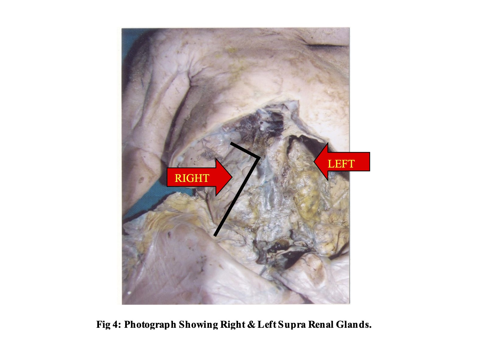
During early fetal life, both the right and left suprarenal glands typically present a tetrahedral shape. As development progresses, their morphology becomes asymmetrical. In adults, the right suprarenal gland is usually pyramidal in form, though it may appear triangular, tetrahedral, or inverted Y/V-shaped. (3,4) In contrast, the left gland gradually assumes a crescentic or semilunar configuration. These morphological transitions reflect the dynamic structural adaptations that occur throughout intrauterine life. A notable feature of the suprarenal gland is its ability to undergo significant structural modifications both prenatally and postnatally before attaining its definitive adult architecture. (4,5)
The pattern of prenatal growth of the suprarenal gland remains controversial. Some investigators report a steady increase in gland weight throughout fetal life, (6) whereas others describe rapid early growth followed by relative stabilization around the fourth fetal month.(7) Conversely, certain studies suggest slow initial growth with accelerated enlargement during later gestation.(8) These differing observations indicate that suprarenal development is influenced by complex regulatory mechanisms.
The present was conducted to study the histogenesis of fetal adrenal gland at different stages of development in intra uterine life in Marathwada region of Maharashtra.
OBJECTIVE: To study micrometry of suprarenal gland at various gestational age group.
MATERIALS AND METHODS:
The present observational study conducted at Department of Anatomy and Obstetrics and Gynecology of Government Medical College and Hospital, Aurangabad. The study was done on 40 spontaneous abortuses and stillborns ranging from 12th weeks to 36th weeks of gestation. Twins and fetuses with gross anomalies were excluded. The study was approved by the Ethical Committee of the Institute.
The gestational age, sex, weight crown-rump length was studied in detail. Weight of fetuses were measured in grams on double pan balance.
The fixation of fetuses was ensured by injecting them with 10 % formalin locally, at various sites with the help of a 10-ml. syringe and 20 number needle in cranial cavity, in the neck and subcutaneously in the upper and lower limbs.
The suprarenal glands were obtained from fetuses by a midline incision over the anterior abdominal wall. Both glands were carefully dissected out, immediately weighed, and initially fixed in 10% formalin. Micrometry is done to measure thickness of definitive zone and fetal zone, diameter and size of neuroblastic nodule. 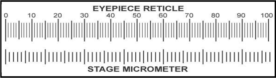
Fig 1: Diagrammatic representation of calculation of eye piece and stage micrometer.
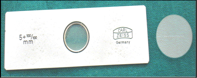
Fig 2: Micrometre.
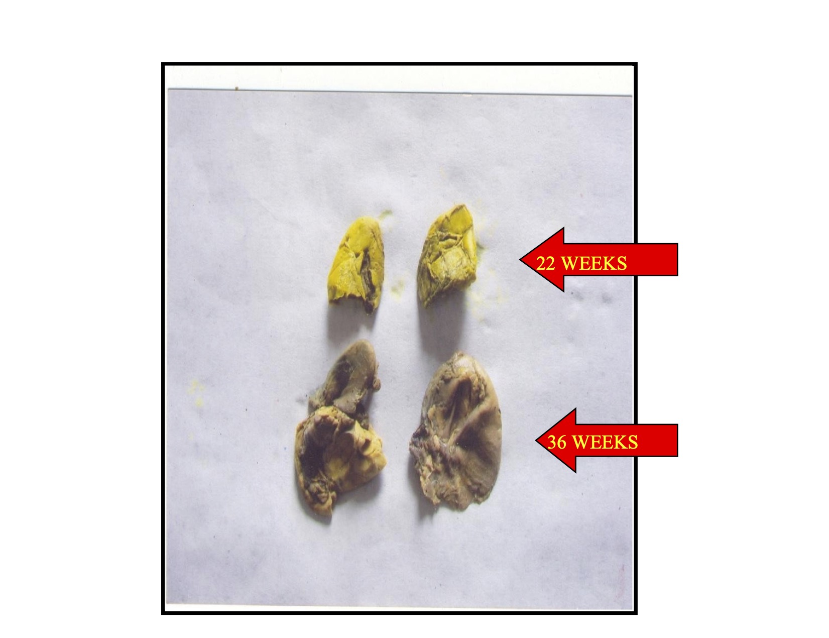
Fig 3: Photographs Showing Supra Renal Glands At 22 Weeks & 36 Weeks.
RESULTS:
In the present study, a total of 40 fetuses (19-males, 21-females) ranging from 12th weeks to 36th weeks of gestation were studied.
Table 1: Showing average weight and relative weight of Suprarenal Glands against weeks of gestational age:
|
Gestational Age (Weeks) |
Average Weight of Right Suprarenal (gram) |
Average Weight of Left Suprarenal (gram) |
Relative Weight of Right Suprarenal |
Relative Weight of Left Suprarenal |
|
12 |
0.808 |
0.828 |
0.641 |
0.657 |
|
14 |
0.945 |
0.96 |
0.675 |
0.685 |
|
16 |
1.409 |
1.491 |
0.728 |
0.770 |
|
18 |
1.787 |
1.999 |
0.560 |
0.627 |
|
20 |
2.099 |
2.308 |
0.387 |
0.426 |
|
22 |
2.326 |
2.397 |
0.363 |
0.378 |
|
24 |
2.455 |
2.497 |
0.339 |
0.345 |
|
26 |
2.621 |
2.685 |
0.329 |
0.337 |
|
28 |
3.403 |
3.649 |
0.269 |
0.288 |
|
34 |
4.175 |
4.333 |
0.225 |
0.234 |
|
36 |
4.725 |
4.775 |
0.192 |
0.194 |
It was observed that weight of right and left suprarenal gland at 12th week was 0.808 and 0.828 respectively while the weight of right and left suprarenal gland at 36th week was 4.725 and 4.775 respectively. It was observed that weight of both glands was increasing constantly. At 12th week of gestation, the percentage relative weight in grams. of right and left suprarenal was 0.641 and 0.657 respectively. At 36 weeks of gestation the right and left suprarenal relative weight was 0.192 grams. and 0.194 grams. respectively. (Table 1)
Table 2: Showing Micrometry of Suprarenal Gland across gestational ages:
|
Stage in weeks |
Definitive zone (A) (µm) |
Fetal zone (B) (µm) |
Total thickness of cortex (A+B) (µm) |
Proportion Definitive zone to Total cortex in % |
|
12 |
114.4 |
1072.5 |
1186.9 |
9.64 % |
|
16 |
128.7 |
1229.8 |
1358.5 |
9.48 % |
|
20 |
143 |
1401 |
1544 |
9.26 % |
|
28 |
171.6 |
1287 |
1458.6 |
11.77 % |
|
34 |
314.6 |
1215.5 |
1530.1 |
20.56 % |
|
36 |
314.6 |
1144.4 |
1458.6 |
21.57 % |
The average thickness of definitive cortex at 12 weeks is 114.4µm, while the thickness of fetal cortex is 1072.5µm. The total cortical thickness is 1186.9µm. The proportion of definitive cortex to total cortex is approximately 10.37%. At 36 weeks average thickness of definitive zone is 314.6µm, while thickness of fetal zone is 1144µm. The total thickness of cortex is 1458. 6µm.The proportion of total cortex to definitive cortex is 4.63%. (Table 2)
DISCUSSION:
Human fetal growth is a net result of a complex interplay of genetic, hormonal and growth factor effects. Suprarenal gland is essential for intrauterine development of fetus. The fetal adrenal cortex is involved in maintenance of intrauterine homeostasis and in the preparation of fetus for extra uterine life, by regulating the maturation of essential organ systems including lungs, liver and gut.(9)
The transition to extrauterine life requires rapid adaptation from the protected intrauterine environment supported by the placenta to the relatively hostile external environment, a process largely regulated by the fetal suprarenal gland. Near term, a cortisol surge from the transitional zone of the fetal adrenal augments surfactant synthesis, lung fluid absorption, T4–T3 conversion, ductus arteriosus closure, and maturation of intestinal enzymes and transport systems, while a concurrent catecholamine surge increases blood pressure, myocardial contractility, glucagon secretion, thermogenesis in brown adipose tissue, and plasma free fatty acid levels. (9)
In the present study, while studying the development of adrenal in antenatal period, different morphological parameters of adrenal glands were considered. The study of weight of adrenal gland at different stages of gestation and their proportion to the body weight. Each adrenal gland measures approximately 50 mm vertically, 30 mm transversely, and 10 mm in the anteroposterior dimension, with an average weight of about 5 g. At birth, the adrenal gland is roughly one-third the size of the kidney, whereas in adults it is about one-thirteenth the size of the kidney.
Increase in fetal weight is a good indicator of fetal growth in general. Rate of weight gain during first, second and third trimester differs considerably. Generally, organ size and histological maturation commensurate with total body size. The size and weight of supra renal gland is proportional to the body weight. The cortical portion of the suprarenal gland develops rapidly and is relatively more advanced during the first half of the pregnancy than are the other abdominal or thoracic organ, whereas the total mass of chromaffin tissue is proportionately much less in the fetus & newborn than it is in later life. The provisional cortex continues to grow as long as fetus is inside the uterus.
The weight of adrenal gland as reported by Potter and Craig (10) in an average 3400 gm in infant is 5 gm. each. They grow in proportion with body size. In some premature infants weighing 1200 to 1500 gms adrenal glands weighted as less as 2 gms showing a direct correlation of the body weight with the supra renal weight and size. The decrease in the weight of supra renal afterwards is mostly due to progressive degeneration of the inner zone which weigh proportionally more than the definitive cortex. Thus, causing a general shrinkage of the gland after birth.
Table No. 3: Showing comparisons of average percentage relative weight of suprarenal gland at different stages observed in present study with the findings of other workers.
|
Gestational Age (Weeks) |
Grunwald (11) (1960) |
Potter (10) (1961) |
Schultz (12) (1962) |
Present Study (2024) |
|
12 |
-- |
-- |
-- |
0.64 |
|
14 |
-- |
-- |
-- |
0.67 |
|
16 |
-- |
-- |
-- |
0.74 |
|
18 |
-- |
-- |
-- |
0.59 |
|
20 |
-- |
-- |
0.73 |
0.40 |
|
22 |
-- |
-- |
-- |
0.36 |
|
24 |
0.45 |
-- |
0.47 |
0.33 |
|
26 |
0.40 |
0.41 |
-- |
0.32 |
|
28 |
0.36 |
-- |
0.35 |
0.27 |
|
34 |
0.30 |
-- |
-- |
0.23 |
|
36 |
0.30 |
-- |
0.27 |
0.19 |
The comparison of relative weight of supra renal gland with other studies was done. In all studies it was found that, relative weight of supra renal gland decreases with gestational age. Present study also shows similar findings.
In the present study, at 12 weeks of gestation, the fetal cortex constitutes the major component of the gland, measuring 1072.5 µm in thickness, compared to 114.4 µm for the definitive cortex. The total cortical thickness at this stage is 1186.9 µm, with the definitive cortex contributing approximately 10.37% of the total cortical thickness. This marked predominance of the fetal zone in early gestation reflects its crucial endocrine role, particularly in the production of steroid precursors such as DHEA-S, which are essential for placental estrogen synthesis and fetal organ maturation.
By 36 weeks of gestation, substantial micrometric changes are evident. The definitive cortex increases in thickness to 314.6 µm, nearly three times its measurement at 12 weeks, indicating progressive maturation and differentiation. The fetal zone, although still the thicker component at 1144 µm, shows only a modest increase compared to earlier gestation. The total cortical thickness at 36 weeks measures 1458.6 µm. Importantly, the proportion of definitive cortex relative to total cortex increases, with the ratio of total cortex to definitive cortex being 4.63%, demonstrating a relative expansion of the definitive zone as term approaches.
These findings suggest that early gestation is characterized by rapid growth and functional dominance of the fetal zone, whereas late gestation shows progressive maturation and proportional enlargement of the definitive cortex. The shift in cortical proportions likely represents preparation for postnatal life, when the definitive cortex will differentiate into the adult cortical zones (zona glomerulosa, zona fasciculata, and zona reticularis), and the fetal zone will undergo rapid involution after birth.
Thus, micrometric evaluation across gestational ages highlights the dynamic growth pattern and structural reorganization of the suprarenal cortex. Such quantitative data contribute to a better understanding of normal fetal adrenal development and may serve as baseline reference values for identifying developmental deviations in pathological conditions.
CONCLUSION:
The study concludes that, as gestational age advances, both the weight and linear dimensions of the suprarenal glands show a progressive and consistent increase. Understanding the morphometric parameters, growth patterns, and weight changes of the fetal adrenal glands provides valuable insight into normal intrauterine development.
REFERENCES:
1. Cope CL. The adrenal cortex in internal medicine. I. British Medical Journal. 1966 Oct 8; 2(5518):847.
2. Ram KS, Sharma A, Sharma M. Histogenesis of suprarenal gland in fetuses of different gestational age groups. Int J Biol Med Res. 2013;4(1):2675-82.
3. Anand MK, Anand C, Choudhry R, Sabharwal A. Morphology of human suprarenal glands: a parameter for comparison. Surgical and radiologic anatomy. 1998 Sep;20(5):345-9.
4. Sangma GT, Ibochouba Y, Damayanti N. Development and maturation of suprarenal glands in human fetuses. Journal of the Anatomical Society of India. 2008 Jun 1;57(1):1-7.
5. Singh, P. R. Dimension and shape of human suprarenal gland. J. Anat Soc. India. 2002; 51,202–15.
6. Tanimura T, Nelson T, Hollingsworth RR, Shepard TH. Weight standards for organs from early human fetuses. The Anatomical Record. 1971 Oct;171(2):227-36.
7. Salmi HA, Puliciiinen M, Savola P. The Ponderal Growth of Human Organs in Early Fetal Life I. Glandular Organs. Acta Pædiatrica. 1962 May;51(3):375-9.
8. Carr BR, Casey ML. Growth of the adrenal gland of the normal human fetus during early gestation. Early Human Development. 1982 Apr 1;6(2):121-4.
9. Benirschke K, Bloch E, Hertig AT. Concerning the function of the fetal zone of the human adrenal gland. Endocrinology. 1956;58:598–625.
10. Potter EL. Pathology of the Fetus and Infant. Academic Medicine. 1962 Feb 1;37(2):160.
11. Schulz DM, Giordano DA, Schulz DH. Weights of organs of fetuses and infants. Archieves of pathology, vol. 74: 244-250.