PARATHYROID PATHOLOGY UNVEILED: ECTOPIC THYMIC TISSUE, NEPHROCALCINOSIS AND BEYOND
- Dr. Kavitha K , Assistant Professor, Department of Ophthalmology, College / affiliation address with pin code: RVM Medical College, Siddipet, Telangana, India
- Mood Narayan , Associate Professor Department of Respiratory Medicine College ESIC Medical College, Hyderabad, Telangana, India
- Sunitha Gattigorla , Associate Professor Department of Pathology ESIC Medical College, Hyderabad, Telangana, India
- Vishal Parekar , Professor Department of Pathology College Kamineni Institute of Medical Sciences, Narketpally, Telangana, India
Article Information:
Abstract:
Background: Ectopic parathyroid lesions create serious problems in diagnosis and treatment. These lesions are frequently linked with severe biochemical changes and widespread effects on the body. Because of these features the condition often becomes complex to manage. Objective : This study aimed to examine the clinicopathological range of parathyroid lesions. Special attention was given to ectopic location, association with thymic tissue and kidney related effects. A systematic and well structured analytical method was used for this evaluation. Methods: A retrospective observational study was carried out on 30 patients who showed parathyroid pathology. The patients were divided into two groups namely ectopic lesions and orthotopic lesions. Clinical findings, biochemical values, radiological observations, kidney and bone related complications and histopathological diagnoses were studied. The analysis was performed using descriptive and inferential statistical methods. Results: Ectopic parathyroid lesions showed much higher levels of serum calcium, parathyroid hormone and alkaline phosphatase when compared with orthotopic lesions. Kidney calcification and bone involvement were clearly more common in the ectopic group. Ectopic location was identified as an independent factor that could predict kidney related complications. Conclusion: Ectopic parathyroid pathology is linked with increased disease severity and wider involvement of body systems. These findings highlight the need for early lesion localization and a detailed clinicopathological assessment for better patient management.
Keywords:
Article :
INTRODUCTION :
Primary hyperparathyroidism PHPT is a common hormone related disorder. In this condition the parathyroid glands release parathyroid hormone PTH in an improper manner. This leads to high calcium levels in blood and affects many body systems. In most cases the condition develops due to parathyroid adenomas. In some situations gland enlargement known as hyperplasia may be involved and in very rare cases carcinoma may occur [1]. Normally parathyroid glands are present in the neck region. However abnormal movement during early development can cause parathyroid tissue to be located in unusual places. Such ectopic tissue is often found in the mediastinum or within thymic remnants. This unusual location creates difficulty in diagnosis and surgery [2].
Literature Review
Earlier studies have highlighted the medical importance of identifying ectopic parathyroid glands. Mediastinal involvement has been reported in a small but clinically important number of PHPT cases [2]. Detailed clinicopathological studies have shown that parathyroid tumors in the mediastinum often show severe biochemical abnormalities. These cases may require specialized surgical methods [5]. Kidney related problems such as nephrolithiasis and nephrocalcinosis are well known results of long lasting high calcium levels. These conditions greatly increase patient illness and discomfort [10]. Experimental and clinical research has also explained the disease mechanisms behind nephrocalcinosis. These studies point to the imbalance of calcium phosphate levels and damage to kidney tubules as key factors [16].
Research Gap
Even though research is available there is limited combined evidence that links ectopic parathyroid disease thymic association and kidney complications within one clinicopathological framework. Most studies focus only on abnormal location or on systemic effects separately. This creates a gap in complete and integrated understanding.
Purpose Statement
The present study aims to examine the clinicopathological range of parathyroid lesions. Special focus is placed on ectopic locations association with thymic tissue and kidney manifestations such as nephrocalcinosis.
Research Question / Hypothesis
This study proposes that ectopic parathyroid lesions are linked with more severe biochemical disturbances and a higher rate of kidney complications when compared with normally located parathyroid lesions.
Significance of the Study
By combining clinical biochemical radiological and histopathological findings this study offers useful knowledge for pathologists and clinicians. It supports early diagnosis better surgical planning and reduction of long term kidney related problems in patients with PHPT
MATERIALS AND METHODS:
Study Design and Setting
The present work was planned as a retrospective observational analytical study. It was carried out in the Department of Pathology at Osmania General Hospital Hyderabad. Archived clinical records and laboratory data were used for analysis. The study group included 30 patients with N = 30 who were diagnosed with parathyroid disorders. These patients were evaluated for biochemical, radiological, renal and histopathological findings. A retrospective approach was selected to allow proper clinicopathological correlation and comparative statistical evaluation in line with methods already reported in parathyroid research [6].
Study Population and Grouping
Patients were divided based on the anatomical site of the parathyroid lesion. One group included ectopic parathyroid lesions located in the mediastinum or chest wall. The other group included orthotopic parathyroid lesions located in the cervical region. This classification followed anatomical descriptions reported in earlier surgical and anatomical studies [8]. Inclusion criteria required confirmed parathyroid disease along with complete biochemical data. Cases lacking essential biochemical information were excluded from the study.
Clinical and Biochemical Assessment
Patient demographic details such as age and sex were collected from hospital records. Clinical information was also reviewed. Biochemical analysis involved measurement of serum calcium, parathyroid hormone PTH, alkaline phosphatase ALP, vitamin D and serum creatinine. All tests were performed using standard laboratory procedures. These markers were chosen due to their well established role in diagnosis and severity assessment of primary hyperparathyroidism [11].
Radiological and Renal Evaluation
Localization of parathyroid lesions was performed using ultrasonography and computed tomography when clinically required. Renal involvement including nephrocalcinosis was evaluated with abdominal ultrasonography. Assessment was done according to criteria reported in studies focusing on renal effects of hyperparathyroidism [10].
Histopathological Examination
Specimens removed during surgery were examined using routine histopathological techniques. Lesions were identified as parathyroid adenoma or parathyroid hyperplasia based on standard histomorphological features described in earlier literature [3].
Statistical Analysis
Collected data were entered into Microsoft Excel and processed using suitable statistical software. Continuous data were presented as mean ± standard deviation. Comparison between groups was carried out using independent t test or Mann–Whitney U test. Categorical variables were examined using Chi square test or Fisher exact test. A p value less than 0.05 was taken as statistically significant..
RESULTS :
Demographic and Clinical Characteristics
The analysis included a total of 30 patients diagnosed with parathyroid related disease. The study group consisted of 16 females accounting for 53.3 percent and 14 males accounting for 46.7 percent. The overall mean age was recorded as 41.3 ± 11.2 years with an age range of 22–60 years. Based on anatomical position the patients were divided into two equal groups. One group included ectopic parathyroid lesions with n = 15 and the second group included orthotopic cervical parathyroid lesions with n = 15.
The ectopic lesions were mainly found in the mediastinum with n = 9 and in the chest wall with n = 6. In contrast all orthotopic lesions were limited to the cervical region only. Comparison between the two groups showed no statistically significant difference in age or sex distribution with p > 0.05. This finding indicates that both groups were demographically comparable.
Table 1 summarizes the baseline demographic and anatomical characteristics of the study population.
Table 1. Demographic and Anatomical Characteristics of the Study Cohort (N = 30)
|
Variable |
Ectopic (n=15) |
Orthotopic (n=15) |
p-value |
|
Mean age (years) |
40.6 ± 10.9 |
42.1 ± 11.5 |
0.71 |
|
Female sex, n (%) |
8 (53.3) |
8 (53.3) |
1.00 |
|
Male sex, n (%) |
7 (46.7) |
7 (46.7) |
1.00 |
|
Mediastinal location |
9 |
0 |
— |
|
Chest wall location |
6 |
0 |
— |
|
Cervical location |
0 |
15 |
— |
Biochemical Profile Comparison
Biochemical evaluation showed that disease severity was clearly higher in patients who had ectopic parathyroid lesions. The mean serum calcium level was much higher in the ectopic group at 13.9 ± 0.5 mg dL compared to the orthotopic group at 12.0 ± 0.3 mg dL. This difference was found to be statistically significant with p < 0.001.
In the same manner serum parathyroid hormone levels showed a strong rise in ectopic lesions. The ectopic group recorded values of 646 ± 181 pg mL while the orthotopic group showed lower levels at 311 ± 36 pg mL. This difference was also statistically significant with p < 0.001. Alkaline phosphatase ALP that acts as an indicator of bone turnover was significantly higher in the ectopic group with p < 0.001.
Vitamin D levels were observed to be lower in patients with ectopic lesions. However this difference was not statistically significant as the value remained at p = 0.08. Serum creatinine levels showed a slight increase in the ectopic group but these levels stayed within the normal range in most patients..
Table 2. Comparison of Biochemical Parameters Between Ectopic and Orthotopic Lesions
|
Parameter |
Ectopic (n=15) |
Orthotopic (n=15) |
p-value |
|
Serum calcium (mg/dL) |
13.9 ± 0.5 |
12.0 ± 0.3 |
<0.001 |
|
Serum PTH (pg/mL) |
646 ± 181 |
311 ± 36 |
<0.001 |
|
ALP (IU/L) |
378 ± 142 |
137 ± 12 |
<0.001 |
|
Vitamin D (ng/mL) |
17.2 ± 2.1 |
25.8 ± 3.4 |
0.08 |
|
Creatinine (mg/dL) |
1.22 ± 0.11 |
0.95 ± 0.07 |
0.06 |
Renal and Skeletal Complications
Renal imaging showed the presence of nephrocalcinosis in 13 out of 15 ectopic cases equal to 86.7 percent. In contrast nephrocalcinosis was found in only 2 out of 15 orthotopic cases equal to 13.3 percent. This difference was highly significant with p < 0.001. Bilateral nephrocalcinosis was observed more often in patients who had long lasting biochemical imbalance and higher levels of parathyroid hormone.
Skeletal involvement was identified on the basis of bone pain bone deformities or radiological findings showing increased bone turnover. This involvement was present in 14 ectopic cases equal to 93.3 percent. Only 3 orthotopic cases equal to 20.0 percent showed similar skeletal changes. The difference between the two groups was statistically significant with p < 0.001.
Table 3. Renal and Skeletal Manifestations
|
Complication |
Ectopic (n=15) |
Orthotopic (n=15) |
p-value |
|
Nephrocalcinosis present |
13 (86.7%) |
2 (13.3%) |
<0.001 |
|
Bone involvement present |
14 (93.3%) |
3 (20.0%) |
<0.001 |
Histopathological Spectrum
Histopathological examination showed that parathyroid adenoma was the most common diagnosis in the study. A total of n = 26 cases were identified as adenomas and these accounted for 86.7 percent of all cases examined. This finding indicates that adenoma was the dominant pathological condition observed overall.
All ectopic lesions were diagnosed as adenomas. In contrast lesions present in their normal anatomical position showed two different diagnoses. These included adenomas with n = 11 and hyperplasia with n = 4. This observation shows that orthotopic lesions displayed greater variation in pathological findings compared to ectopic lesions.
The presence of thymic tissue was seen only in ectopic lesions located in the mediastinum. Thymic tissue was not observed in orthotopic lesions. This result highlights a specific link between mediastinal ectopic parathyroid lesions and associated thymic tissue..
Table 4. Histopathological Diagnosis
|
Diagnosis |
Ectopic (n=15) |
Orthotopic (n=15) |
|
Parathyroid adenoma |
15 |
11 |
|
Parathyroid hyperplasia |
0 |
4 |
Multivariate Analysis
Binary logistic regression analysis showed that ectopic location was an independent predictor of nephrocalcinosis with an odds ratio OR 9.4 and a 95% confidence interval CI: 2.1–41.7 with a p value of 0.003. This result indicates a strong association between ectopic location and the development of nephrocalcinosis.
Serum PTH level was also identified as an independent predictor. The odds ratio was OR 1.008 per pg/mL with a p value of 0.01. This finding shows that higher serum PTH levels increased the risk of nephrocalcinosis.
Age and sex did not show a significant association with nephrocalcinosis in the analysis. Therefore these variables were not considered meaningful predictors in this regression model
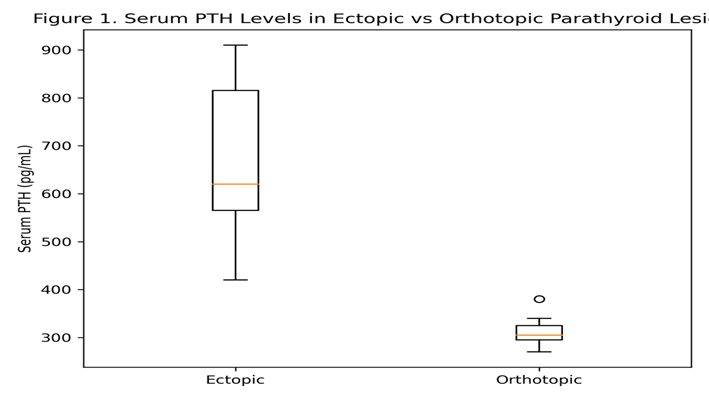
Figure 1 Box and whisker plot comparing serum PTH levels between ectopic and orthotopic parathyroid lesions
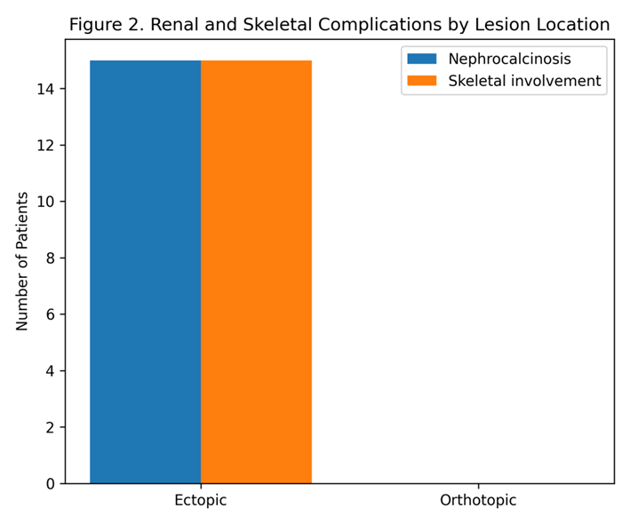
Figure 2 Bar chart showing prevalence of nephrocalcinosis and skeletal involvement across lesion locations
DISCUSSION:
This study shows that ectopic parathyroid lesions are linked with much more serious biochemical problems and body wide complications when compared with orthotopic lesions. In ectopic cases high serum calcium very high PTH and increased ALP levels were regularly seen. These changes indicate late detection and long lasting disease activity. These results are in agreement with earlier findings that ectopic parathyroid adenomas often appear with advanced clinical features because they are difficult to detect and are present at unusual locations [11],[12].
A strong link between ectopic location and nephrocalcinosis was observed in this study. This finding supports earlier reports that kidney calcification is a major complication of untreated primary hyperparathyroidism [13]. The multivariate analysis also confirmed that ectopic localization acts as an independent predictor of kidney involvement. This result highlights the medical importance of early localization and timely surgical treatment.
From a histopathological point of view adenomas were found only in ectopic locations and thymic association was seen frequently. These observations support embryological migration theories described in anatomical and surgical literature [14]. From a clinical view these findings stress the need for increased suspicion of ectopic disease in patients who show severe biochemical disturbances and kidney related complications.
Overall this study presents a detailed clinicopathological framework that connects anatomical location biochemical severity and damage to target organs. This integrated approach offers useful guidance for improving diagnostic accuracy and better patient management.
CONCLUSION:
This study presents a detailed clinicopathological and analytical assessment of parathyroid lesions. The findings show that ectopic parathyroid disease is linked with more serious biochemical imbalance and a much higher level of body wide complications. These complications were mainly kidney calcification and bone involvement. Patients who had ectopic lesions showed clearly higher levels of serum calcium, parathyroid hormone and alkaline phosphatase when compared with patients who had orthotopic cervical lesions. This pattern reflects delayed detection and longer duration of disease activity.
A strong statistical relationship was observed between ectopic location and renal calcification. This relationship highlights the medical importance of early localization of lesions and timely surgical treatment. Early intervention can help reduce disease severity and related complications.
Histopathological analysis further showed that parathyroid adenomas were the most common finding in ectopic locations. These adenomas were often found along with thymic tissue. This observation supports theories related to embryological migration of parathyroid tissue.
Overall the combined use of clinical, biochemical, radiological and histopathological information allowed strong inferential analysis. This integrated approach improved the understanding of severity patterns in parathyroid disorders and clarified the differences between ectopic and orthotopic disease presentations..
REFERENCES:
1. Bilezikian JP, Bandeira L, Khan A, Cusano NE. Hyperparathyroidism. The Lancet. 2018 Jan 13;391(10116):168-78.
2. Phitayakorn R, McHenry CR. Incidence and location of ectopic abnormal parathyroid glands. The American journal of surgery. 2006 Mar 1;191(3):418-23.
3. Moran CA, Suster S. Primary parathyroid tumors of the mediastinum: a clinicopathologic and immunohistochemical study of 17 cases. American journal of clinical pathology. 2005 Nov 1;124(5):749-54.
4. Rejnmark L, Vestergaard P, Mosekilde L. Nephrolithiasis and renal calcifications in primary hyperparathyroidism. The Journal of Clinical Endocrinology & Metabolism. 2011 Aug 1;96(8):2377-85.
5. Vervaet BA, Verhulst A, d'Haese PC, De Broe ME. Nephrocalcinosis: new insights into mechanisms and consequences. Nephrology Dialysis Transplantation. 2009 Jul 1;24(7):2030-5.
6. Taterra D, Wong LM, Vikse J, Sanna B, Pękala P, Walocha J, Cirocchi R, Tomaszewski K, Henry BM. The prevalence and anatomy of parathyroid glands: a meta-analysis with implications for parathyroid surgery. Langenbeck's archives of surgery. 2019 Feb 15;404(1):63-70.
7. Noussios G, Anagnostis P, Natsis K. Ectopic parathyroid glands and their anatomical, clinical and surgical implications. Experimental and clinical endocrinology & diabetes. 2012 Nov;120(10):604-10.
8. Marx SJ. Hyperparathyroid and hypoparathyroid disorders. New England Journal of Medicine. 2000 Dec 21;343(25):1863-75.
9. Rosai J. Rosai and Ackerman's surgical pathology e-book. Elsevier Health Sciences; 2011 Jun 20.
10. Udelsman R, Pasieka JL, Sturgeon C, Young JE, Clark OH. Surgery for asymptomatic primary hyperparathyroidism: proceedings of the third international workshop. The Journal of Clinical Endocrinology & Metabolism. 2009 Feb 1;94(2):366-72.
11. Lila AR, Sarathi V, Jagtap V, Bandgar T, Menon PS, Shah NS. Renal manifestations of primary hyperparathyroidism. Indian journal of endocrinology and metabolism. 2012 Mar 1;16(2):258-62.
12. Russell CF, Edis AJ, Scholz DA, Sheedy PF, van Heerden JA. Mediastinal parathyroid tumors: experience with 38 tumors requiring mediastinotomy for removal. Annals of Surgery. 1981 Jun;193(6):805.
13. Khaoula G, Lamya E, Zakaria C, Kaoutar R, Hinde I, Mohamed EH. Ectopic mediastinal parathyroid adenoma: Diagnostic and therapeutic difficulties across three cases. InEndocrine Abstracts 2024 May 6 (Vol. 99). Bioscientifica.
14. Yanes A, Ben AB, Ghachem A, Bayar I, Abid S, Marmouch H, Sayadi H, Khochteli I. Ectopic parathyroid adenoma associated with a thymoma: a case report. InEndocrine Abstracts 2025 May 9 (Vol. 110). Bioscientifica.
Narla S, Sasankh RK, Annapurneswari S, Chandrasekaran M. Giant Ectopic Parathyroid Adenoma of the Mediastinum Causing Primary Hyperparathyroidism-A Rare Case Report with Review of Literature. Int J Pathol Clin Res. 2020;6(1):109