Prevalence of Multidrug Resistant and Extended-Spectrum Beta-Lactamase Producing Gram- Negative Isolates from Exudate Samples
- Anu Mariya David , Student, Allied Health Sciences, Dr. Moopen’s Medical College, Meppadi, Kerala, India.
- Madhuri K.R. , Assistant Professor, Department of Microbiology, Dr. Moopen’s Medical College, Meppadi, Kerala, India.
- Jisha P , Associate Professor, Department of Microbiology, Dr. Moopen’s Medical College, Meppadi, Kerala, India.
- Ashitha P , Assistant Professor, Department of Microbiology, Dr. Moopen’s Medical College, Meppadi, Kerala, India.
Article Information:
Abstract:
Background: Pyogenic infections, caused by a range of pathogenic microorganisms, are significant contributors to morbidity and mortality globally. The rise of multidrug-resistant (MDR) and extended-spectrum beta-lactamase (ESBL)-producing Gram-negative bacteria poses serious therapeutic challenges, particularly in resource-limited settings. Objective: To determine the prevalence of MDR and ESBL-producing Gram-negative bacterial isolates from exudate samples collected at a tertiary care hospital in Wayanad. Methods A cross-sectional study was conducted over six months (October 2024 to March 2025) at DMMC, Wayanad. A total of 1513 exudate samples from inpatients and outpatients were analyzed. Samples were cultured and bacterial isolates identified using standard microbiological techniques. Antimicrobial susceptibility testing was performed using the Kirby-Bauer disk diffusion method. Isolates resistant to three or more classes of antibiotics were categorized as MDR. ESBL production was screened and confirmed using standard CLSI-recommended methods. Results :Out of 1513 samples, 1008 (66%) were culture positive. Among these, 107 isolates (62%) were identified as MDR and 65 isolates (37%) as ESBL producers. The predominant MDR organisms were Acinetobacter spp. (27%), E. coli (23%), and Klebsiella pneumoniae (24%). Among ESBL producers, E. coli accounted for 73%, followed by Klebsiella pneumoniae (9.2%) and Acinetobacter spp. (4.6%). Conclusion: This study highlights a high prevalence of MDR and ESBL-producing Gram-negative bacteria in exudate samples, underscoring the urgent need for routine antimicrobial resistance surveillance, robust infection control measures, and judicious antibiotic use. Though limited by the absence of molecular testing and clinical outcome data, these findings offer crucial insights into local resistance patterns and support the call for enhanced diagnostic and stewardship practices.
Keywords:
Article :
INTRODUCTION:
Pyogenic infections are characterized by local inflammation in the skin, soft tissues, and various bodily organs, primarily resulting from the invasion and multiplication of pathogenic microorganisms. These pathogens release cellular toxins, metabolites, and leukocidins that damage neutrophils, leading to the formation of abscesses and pus. Common examples of pyogenic infections include impetigo, osteomyelitis, septic arthritis, spondylodiscitis, otitis media, cystitis, meningitis, and surgical site infections. The primary pathogens responsible for these infections are Staphylococcus aureus, Streptococcus pyogenes, Escherichia coli, Klebsiella spp., Proteus spp., and Pseudomonas spp.[1] Pyogenic infections are not only a leading cause of morbidity and mortality but also contribute to prolonged hospital stays and long-term disability on a global scale.[2]
Prevalence of MDR and ESBL producing isolates has been reported to be on rise worldwide, and most of the studies showed increasing trend of prevalence in recent times.[3, 4] The emergence of multi drug-resistant (MDR) and extended-spectrum beta-lactamase (ESBL)- producing Gram-negative bacteria has become a major concern in clinical settings due to their association with poor treatment outcomes and limited therapeutic options. This study aimed to investigate the prevalence of MDR and ESBL-producing Gram-negative isolates from exudate samples.
MATERIALS AND METHODS:
A hospital-based cross-sectional study was conducted for 6 months (October 2024 to March 2025) at DMMC, Wayanad. Exudates samples taken from patients admitted in the hospital and from visiting outpatient departments of the hospital were included. In the case of exudates proper information about sample collection was given. Improperly collected samples or those lacking proper labeling were excluded from the study.
The specimens were cultured on MacConkey agar, Chocolate agar and blood agar. The isolates were identified based on colony morphology, Gram’s stain result, and conventional biochemical methods. Antimicrobial susceptibility testing (AST) was performed using the standardized Kirby- Bauer disk diffusion method.
Antimicrobial susceptibility testing (AST) was done by the Kirby-Bauer disk diffusion technique using Muller Hinton agar (MHA) [CLSI; 2010, Al-Ouqaili et al; 2018]]. The antibiotics used were amikacin (30 μg), amoxyclav (30 μg), amoxicillin (10 μg), gentamicin (10 μg), ceftriaxone (30 μg), cotrimoxazole (25 μg), ciprofloxacin (5 μg), cefixime (30 μg), gentamicin (10 μg), ofloxacin (5 μg), meropenem (10 μg), piperacillin/tazobactam (100/10), polymyxin B (100 IU), imipenem (10 μg), and cefepime (30 μg). The bacterial isolates showing resistance towards three or more different antibiotics classes were considered multidrug-resistant (MDR) bacteria.
Screening and confirmation of ESBL producers:
Ceftazidime, cefpodoxime, ceftriaxone, and cefotaxime were included in the primary panel for screening potential ESBL producers. Isolates showing resistance to any of these antibiotics were suspected as potential ESBL producers.
RESULTS:
A total of 1513 exudate samples received in the laboratory were included for this study. Of them 43% were from females and 57% from males. Out of 1513 samples, 1008 (66%) were culture positive and in that 107 (62%) bacterial isolates were identified as MDR, 65(37%) bacterial isolates were identified as ESBL producers. Out of the 107(62%) MDR producers, Acinetobacter spp were 29(27%), E.coli 25(23%), Klebsiella pneumoniae 26(24.%), Pseudomonas aeruginosa 8(4.7%), Enterobacter spp 5(2.9%), Pseudomonas spp 4(3.7%), Morganella spp 3(2.8%), Klebsiella oxytoca 2(1.8%), Klebsiella spp 2(1.8%), Citrobacter spp 1(0.9%), Citrobacter freundii 1(0.9%), Proteus vulgaris 1(0.9%). Out of the 65(37%) ESBL producers, E.coli were 48(73%), Klebsiella pneumoniae 6(9.2%). Acinetobacter spp 3(4.6%), Pseudomonas aeruginosa 3(4.6%), Enterobacter spp 2(3.0%), Morganella spp 1(1.5%), Klebsiella oxytoca 1(1.5%), Citrobacter koseri 1(1.5%).
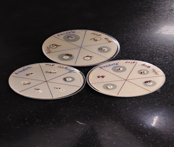
Figure 1 showing Mueller Hinton agar plate with antibiotic susceptibility pattern of MDR Klebsiella pneumoniae isolated from exudate sample of a patient.
DISCUSSION:
The present study underscores a concerning prevalence of multidrug-resistant (MDR) and extended-spectrum beta-lactamase (ESBL)-producing Gram-negative bacteria isolated from exudate samples. Among the 1008 culture-positive samples, 62% were identified as MDR and 37% as ESBL producers. These findings are consistent with previous studies reporting a global surge in MDR and ESBL-producing pathogens, particularly in nosocomial settings where antibiotic pressure is high and infection control practices may be inconsistent.[5,6]
The predominance of Acinetobacter spp. (27%) among MDR isolates in our study aligns with trends observed in similar tertiary healthcare centers, where Acinetobacter has emerged as a leading MDR pathogen due to its high adaptability and ability to survive under diverse environmental conditions.[7] Likewise, E. coli (23%) and Klebsiella pneumoniae (24%) also featured prominently, reflecting their widespread involvement in pyogenic infections and their propensity for acquiring resistance mechanisms, including ESBL production.[8,9]
Among ESBL producers, E. coli accounted for the majority (73%), a finding in concordance with numerous regional and international studies which report E. coli as the most common ESBL-producing pathogen in both community and hospital settings.[10,11] The detection of other ESBL producers, such as Klebsiella pneumoniae (9.2%), Acinetobacter spp. (4.6%), and Pseudomonas aeruginosa (4.6%), further highlights the widespread dissemination of resistance genes across multiple genera.[12].
The rising trend of ESBL production severely limits therapeutic options, as many of these isolates exhibit co-resistance to multiple non-beta-lactam antibiotics, including fluoroquinolones and aminoglycosides. This phenomenon was clearly evident in our study where resistance to three or more antibiotic classes defined the MDR phenotype. Similar resistance patterns have been reported in South Asia and other low-to-middle-income countries (LMICs), where overuse and misuse of antibiotics are prevalent.[13, 14].
SUMMARY AND CONCLUSION :
The prevalence of multi drug-resistant (MDR) and extended-spectrum beta-lactamase (ESBL)- producing organisms is rapidly increasing, presenting a significant public health concern. The detection of these enzymes using simple disk diffusion methods should be routinely implemented in clinical laboratories. Such testing is crucial for guiding appropriate antibiotic use, understanding resistance patterns, and preventing the spread of these resistant organisms, ultimately reducing therapeutic failures. The study underscores the alarming prevalence of MDR and ESBL-producing Gram-negative bacteria in exudate samples, posing a serious challenge to effective infection management. Surveillance, strict infection control practices, and appropriate antibiotic stewardship are essential to mitigate the spread of these resistant pathogens in clinical settings.
Limitations of the study include the lack of molecular confirmation of ESBL genes and the absence of clinical outcome data, which could provide insight into treatment efficacy and morbidity. Nevertheless, the findings offer valuable epidemiological data and reinforce the need for ongoing surveillance, particularly in high-burden regions.
REFERENCES:
1. Singh S, Khare M, Patidar RK, Bagde S, Sahare K, Dwivedi D, et al. Antibacteria activity against pyogenic pathogens. Int J Pharm Sci Res. 2013;4:2974–9.
2. Lozano R, Naghavi M, Foreman K, Lim S, Shibuya K, Aboyans V, et al. Global and regional mortality from 235 causes of death for 20 age groups in 1990 and 2010: A systematic analysis for the global burden of disease study 2010. Lancet. 2012 Dec 15;380(9859):2095–128. Doi: 10.1016/S0140-6736(12)61728-0.
3. Fennell J, Vellinga A, Hanahoe B, Morris D, Boyle F, Higgins F, Lyons M, O’Connell K, Keady D, Cormican M. Increasing prevalence of ESBL production among Irish clinical Enterobacteriaceae from 2004 to 2008: an observational study.BMC Infect Dis. 2012;12:116. Doi: 10.1186/1471-2334-12-116.
4. Surveillance of antimicrobial resistance in Europe . ECDC. 2018.
5. Paterson DL, Bonomo RA. Extended-spectrum beta-lactamases: a clinical update. Clin Microbiol Rev. 2005;18(4):657–86.
6. Bush K, Bradford PA. Epidemiology of β-lactamase-producing pathogens. Clin Microbiol Rev. 2020;33(2):e0004719.
7. Peleg AY, Seifert H, Paterson DL. Acinetobacter baumannii: emergence of a successful pathogen. Clin Microbiol Rev. 2008;21(3):538–82.
8. Pitout JD, Laupland KB. Extended-spectrum beta-lactamase-producing Enterobacteriaceae: an emerging public-health concern. Lancet Infect Dis. 2008;8(3):159–66.
9. Hawkey PM. Multidrug-resistant Gram-negative bacteria: a product of globalization. J Hosp Infect. 2015;89(4):2417.
10. Rawat D, Nair D. Extended-spectrum beta-lactamases in Gram negative bacteria. J Glob Infect Dis. 2010;2(3):26374.
11. Tzouvelekis LS, et al. Carbapenemases in Klebsiella pneumoniae and other Enterobacteriaceae: an evolving crisis of global dimensions. Clin Microbiol Rev. 2012;25(4):682–707.
12. Rodríguez-Baño J, et al. Epidemiology and clinical features of infections caused by extended-spectrum beta-lactamase-producing Escherichia coli in nonhospitalized patients. J Clin Microbiol. 2004;42(3):1089–94.
13. Laxminarayan R, et al. Antibiotic resistance—the need for global solutions. Lancet Infect Dis. 2013;13(12):1057–98.
14. Shrestha P, et al. Enumerating the economic cost of antimicrobial resistance per antibiotic consumed to inform the evaluation of interventions affecting their use. Health Econ. 2018;27(4):556–65.