Left Anterior Descending Artery Aneurysm in a Post PTCA Patient
- Ram Shinde , Assistant Professor, Department of Cardiology, TNMC and BYL Nair, Mumbai, Maharashtra, India.
- Dhairya Thakker , Senior Resident, Department of Cardiology, TNMC and BYL Nair, Mumbai, Maharashtra, India.
- Ajay Chaurasia , Professor & HOD, Department of Cardiology, TNMC and BYL Nair, Mumbai, Maharashtra, India.
Article Information:
Abstract:
Background: Coronary artery aneurysm (CAA), defined as focal dilation of coronary segments ≥1.5 times the adjacent normal vessel, is a rare entity with an incidence of 0.3–5% among patients undergoing coronary angiography. The right coronary artery is most commonly affected, though aneurysms may occur in any vessel. Etiologies include atherosclerosis, vasculitis, genetic predisposition, infection, and iatrogenic injury. Clinical presentation ranges from asymptomatic incidental detection to complications such as thrombosis, rupture, or compression of adjacent structures. Case Presentation: We report a 52-year-old hypertensive female who developed CAA following percutaneous coronary intervention (PCI) to the left anterior descending artery (LAD). Five months post-PCI, she presented with chest pain and was diagnosed with NSTEMI. Coronary angiography revealed proximal LAD lesions with two aneurysms containing thrombus. CT coronary angiography confirmed large aneurysms (up to 56×41×54 mm) with hemopericardium. Surgical exploration identified two aneurysms (~4×4×3 cm) in the proximal LAD. Aneurysmorrhaphy with clot evacuation was performed, followed by coronary artery bypass grafting (CABG) using venous grafts to LAD and RCA. Conclusion: This case highlights the potential for post-PCI coronary aneurysm formation, its progression to giant aneurysms with thrombus and hemopericardium, and the role of surgical repair with bypass grafting as definitive management. Early recognition and tailored intervention are essential to prevent catastrophic complications.
Keywords:
Article :
CASE REPORT:
Invasive Coronary Angiography (CAG)
Intravascular Ultrasound (IVUS) – Able to distinguish between aneurysm, pseudo aneurysm and plaque rupture
Optical coherence tomography (OCT).[1,2,3]
Management Modalities
a) Medical management
b) Percutaneous intervention
c) Surgical management.[1,4]
Choice of Treatment Modality
Decided on the basis of the shape and extent of aneurysm
a) Saccular or small aneurysm – Covered stent exclusion technique
b) Saccular or fusiform aneurysm involving major side branch – balloon or stent assisted coil embolization or surgical exclusion can be considered
c) CAA involving LMCA, multiple aneurysms, giant aneurysms, SVG aneurysm – Surgical resection is considered to be the 1st line of management.
d) Large or rapidly expanding SVGA or in those causing symptomatic external compression – PCI closure with Amplatzer occlusion or Coil embolization or Emergency surgery.[2,3,4].
CASE PRESENTATION:
We describe the case of a 52 year old Hypertensive female. She has an unremarkable medical history prior to a year ago, when she underwent PCI to LAD for unstable angina. Post PCI she recovered well and was asymptomatic for the first few months.
However, 5 months post procedure she started complaining of chest pain associated with perspiration and was taken to a hospital. Troponin levels were found to be positive and she was diagnosed to have NSTEMI.
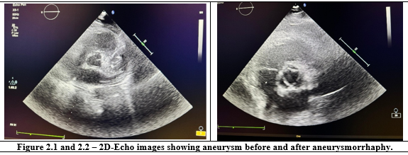
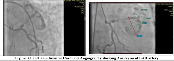
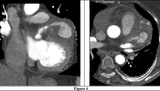
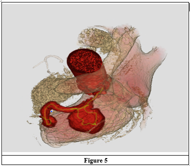
2D-Echo done showed Moderate LV systolic dysfunction with Anterior wall hypokinesia. She underwent CAG which revealed the underlying diagnosis – Lesion in the proximal LAD followed by two well-defined aneurysms measuring 2.5x2.4cm and 1.6x2.2cm proximal to LAD stent with thrombus inside For further evaluation, CT-CAG was done which showed focal outpunching of proximal LAD of size 5.1cm with thrombus followed by large aneurysm in proximal and mid LAD measuring 56x41x54mm with thrombus inside with Hemopericardium of 20mm size without any obvious communication with aneurysm.
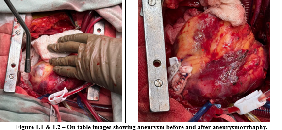
Patient underwent surgical repair for definitive management. During surgery, 2 aneurysms of approximately 4x4x3cm were seen arising from proximal LAD, with fibrotic and small calibre RCA and LAD. LAD aneurysm was opened and clot was evacuated. Aneurysmorrhaphy was done using prolene and FELT. Two venous grafts were put distal to LAD and RCA bypassing atherosclerotic lesions. The patient tolerated surgery and post Op recovery well and was discharged in a stable condition.
REFERENCES:
1. Matta AG, Yaacoub N, Nader V, et al. Coronary artery aneurysm: a review. World Journal of Cardiology 2021;13(9):446.
2. Sheikh AS, Hailan A, Kinnaird T, et al. Coronary artery aneurysm: evaluation, prognosis, and proposed treatment strategies. Heart Views 2019;20(3):101-8.
3. Huizen J. What to know about coronary artery aneurysm. Medical News Today 2022.
4. Kawsara A, Núñez Gil IJ, Alqahtani F, et al. Management of coronary artery aneurysms. JACC: Cardiovascular Interventions 2018;11(13):1211-23.