Prevalence and Determinants of Osteoporosis Among Adults Over 40 Years in Urban Belgaum Using Calcaneal Quantitative Ultrasound
- Abhinandan R Wali , Associate Professor, Department of Community Medicine, KLE Jagadguru Gangadhar Mahaswamigalu Moorusavirmath Medical College and Hospital, Hubli, KLE Academy of Higher Education and Research, Deemed to be University, Belagavi, Karnataka, India.
- S Yogeshkumar , Professor, Department of Community Medicine, Jawaharlal Nehru Medical College, KLE Academy of Higher Education and Research, Deemed to be University, Belagavi, Karnataka, India.
- R. B. Uppin , Professor, Department of Orthopaedics, Jawaharlal Nehru Medical College, KLE Academy of Higher Education and Research, Deemed to be University, Belagavi, Karnataka, India.
- Shruti B Hiremath , Statistician, Department of Community Medicine, KLE Jagadguru Gangadhar Mahaswamigalu Moorusavirmath Medical College and Hospital, Hubli, KLE Academy of Higher Education and Research, Deemed to be University, Belagavi, Karnataka, India.
Article Information:
Abstract:
Background: Osteoporosis is a silent, degenerative bone disease characterized by decreased bone mineral density (BMD), increasing the risk of fractures. In India, underdiagnosis is common, particularly among individuals aged over 40 years. Objective: To determine the prevalence of osteoporosis in adults aged over 40 years in urban Belgaum using Calcaneal Quantitative Ultrasound (QUS) and to identify associated socio-demographic and lifestyle factors. Methods: A community-based cross-sectional study was conducted from January to December 2015 in three urban health centre regions of Belgaum: Rukmini Nagar, Ashok Nagar, and Ram Nagar. A total of 450 adults aged over 40 years were selected through probability proportionate sampling. Data were collected using a pre-tested questionnaire, clinical examination, and calcaneal QUS for BMD classification. Statistical analysis was performed using SPSS version 20, and associations were assessed using the Chi-square test with p<0.05 considered significant. Results: Of 450 participants, 305 (68.8%) were females and 145 (32.2%) males. BMD findings showed 165 (36.7%) had osteoporosis, 168 (37.3%) had osteopenia, and 117 (26%) had normal BMD. Osteoporosis prevalence was higher in females (43.3%) than males (22.1%). Significant associations were observed between BMD and age (p<0.001), sex (p=0.002), BMI (p<0.001), socioeconomic status (p=0.012), lifestyle (p=0.023), and menopausal status (p=0.009). Education and family type showed weaker associations. Conclusion: Over one-third of adults over 40 years in urban Belgaum were osteoporotic, with higher prevalence in older, postmenopausal females with lower BMI and sedentary lifestyles. Early, cost-effective screening and lifestyle interventions are crucial to reduce disease burden.
Keywords:
Article :
INTRODUCTION:
Osteoporosis is a long-term, progressive metabolic bone condition that causes bone tissue to lose mass and micro-architecture, making bones more fragile and more likely to break.1 It is a major public health problem that affects both industrialised and developing countries. Its incidence is rising since people are living longer.2 In India, the condition is often missed and not treated properly. It often doesn't show up until someone breaks a bone.3 The World Health Organisation says that osteoporosis is defined by bone mineral density (BMD) scores: a T-score of ≤ -2.5 means osteoporosis, a score between -1 and -2.5 means osteopenia, and a value of ≥ -1 means normal.8
A mix of hereditary and environmental variables can make someone more likely to become osteoporosis. Up to 80% of peak bone mass is controlled by genetics, but things like smoking, drinking alcohol, getting enough calcium in your diet, and getting enough exercise can have a big impact on bone health.3 In India, postmenopausal women are at greater risk since they go through menopause early, don't get enough calcium in their diets, and can't exercise as much. Men, who are often left out of osteoporosis research, also lose a lot of bone as they become older and because they have other health problems including diabetes and high blood pressure.3
Notwithstanding its increasing prevalence, population-based data on osteoporosis in India are still scarce, particularly among middle-aged and elderly demographics. Dual-energy X-ray absorptiometry (DEXA) is the best way to find out if someone has osteoporosis. But in places where resources are limited, it is hard to get and expensive, therefore calcaneal quantitative ultrasound (QUS) has become a good alternative.4 QUS is a good tool for large community screenings since it is portable, non-invasive, cheap, and does not use ionising radiation.
Prior Indian studies have indicated diverse prevalence rates of osteoporosis, affected by factors including geography, sample demographics, and the diagnostic methods employed. A study in New Delhi indicated a 15% prevalence of osteoporosis in premenopausal women and 53% in postmenopausal women, whereas another study in Tamil Nadu revealed elevated rates among women relative to men.5 Nevertheless, literature from Karnataka, particularly urban Belgaum, is limited.
This study intended to evaluate the chronicity of osteoporosis in people over 40 years old in selected urban areas of Belgaum using calcaneal quantitative ultrasound and to identify the socio-demographic and lifestyle features linked with low bone mineral density. Comprehending these associations is essential for executing community-level screening initiatives and preventive strategies customised for urban Indian populations.
MATERIALS AND METHODS:
Study Strategy
The study was conducted among urban Belgaum residents over a duration of 1 year. Within the operational realm of the Department of Community Medicine, Jawaharlal Nehru Medical College (JNMC), Belgaum, the study included three Urban Health Centers (UHCs): Rukmini Nagar, Ashok Nagar, and Ram Nagar.
Study Cohort
Adult men and women aged over 40 years who had been permanent residents of the selected metropolitan areas for a minimum of one year made up the study population. Participants were solicited via door-to-door outreach.
Inclusion Criteria
• Adults over 40 years of age
• Permanent residents of the study area for more than one year
Exclusion Criteria
• Individuals with significant disabilities or chronic debilitating conditions
• Individuals with paralysis
Sample Size
A total of 450 participants were included, adjusted for non-response. The populations of Rukmini Nagar (43,600), Ashok Nagar (31,933), and Ram Nagar (32,815) were utilised to proportionally allocate the sample size employing the probability proportionate to size (PPS) method.
• Rukmini Nagar: 180 participants
• Ashok Nagar: 135 participants
• Ram Nagar: 135 participants
Participants were selected using simple random sampling within each UHC.
Data Collection
Data were acquired using a pre-tested, structured questionnaire managed through face-to-face meetings conducted in the local languages (Kannada and Marathi). The survey encompassed the subsequent domains:
• Basic information: gender, religion, education level, occupation, age, marital status, family structure, socioeconomic status.10
•Lifestyle factors: dietary habits (vegetarian/mixed), physical activity levels (active/sedentary), tobacco use, and alcohol intake.
• Medical history: existence and duration of diabetes and hypertension.
• Reproductive history (for women): menopausal status and gravida status.
• Anthropometric measurements: height and weight for Body Mass Index (BMI) assessment.
Bone Mineral Density Assessment (BMD)
Bone mineral density (BMD) was evaluated through calcaneal Quantitative Ultrasound (QUS) for all participants. The classification was conducted according to WHO T-score criteria:
• Normal: T-score is -1 or higher
• Osteopenia: T-score between -1 and -2.5
•Osteoporosis: T-score of -2.5 or lower
Participants were classified under the variable BMD CAT.4
Data Analysis
We loaded the data into Microsoft Excel 2007, cleaned it up, and then used SPSS version 20 to look at it. We used the Chi-square test to look at the links between BMD categories and things like age, sex, BMI, lifestyle, education, and socioeconomic status that were not reliant on one other. Descriptive statistics, including frequencies and percentages, were used for variables that could be put into groups. A p-value of less than 0.05 was considered statistically important.
Ethical Considerations
The Institutional Ethics Committee of J.N. Medical College, Belgaum approved the study ethically under reference MDC/DOME/102 dated 14/11/2014. Every participant had written informed permission before data collecting started.
RESULTS:
Sample Characteristics
The investigation encompassed 450 participants, consisting of 305 females (68.8%) and 145 males (32.2%), all of whom were over the age of 40. The average age of the participants was 53.9 ± 10.12 years. The highest percentage of participants was in the 40–49 years age group (42.2%), followed by individuals aged 50–59 years (24.9%) and those in the 60–69 years range (24.2%).
Table 1: BMD Category Distribution
|
BMD Category |
Number of Participants |
Percentage (%) |
|
Normal |
117 |
26.0 |
|
Osteopenia |
168 |
37.3 |
|
Osteoporosis |
165 |
36.7 |
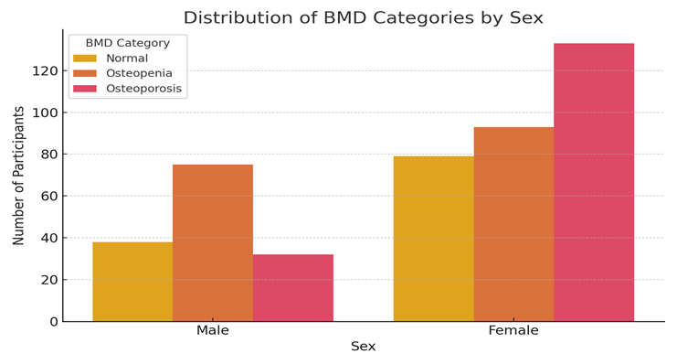
Figure 1: Distribution of BDM categories by sex
Prevalence of Osteoporosis and Osteopenia
Bone Mineral Density (BMD) levels, evaluated using calcaneal Quantitative Ultrasound (QUS), were classified into three categories: osteoporosis, osteopenia, and normal BMD. Of the 450 participants, 165 individuals (36.7%) were diagnosed with osteoporosis, 168 (37.3%) with osteopenia, and 117 (26.0%) had normal bone mineral density (BMD). A pronounced gender gap was noted in osteoporosis prevalence, with females demonstrating a significantly higher rate (43.3%) than males (22.1%). The observed difference was statistically significant (χ² = 14.58, p = 0.002), demonstrating a robust correlation between gender and the prevalence of osteoporosis in the examined group.
Figure 1 and Table 1 shows the distribution of Normal, Osteopenia, and Osteoporosis categories among participants.
Association with Age
A notable association was identified between age groups and BMD categories, indicated by a chi-square value of 52.73 and a p-value below 0.001. The incidence of osteoporosis was seen to rise significantly with increasing age. In the cohort aged 40 to 49 years, 14.2% received a diagnosis of osteoporosis. The proportion jumped to 28.6% in the 50–59 age group, further escalated to 61.5% among persons aged 60 to 69 years, and surpassed 70% in those over 70 years of age. These findings underscore a distinct age-related trend in diminishing bone mineral density.
Association with Socio-Demographic Variables
The correlation between educational status, social class, and family structure and the prevalence of osteoporosis was analysed. Illiterate participants exhibited a greater prevalence of osteoporosis (42.3%) than those with postgraduate education (16.1%); although, this disparity did not achieve statistical significance (p = 0.081). A statistically significant correlation was identified between socioeconomic status and the prevalence of osteoporosis (χ² = 12.91, p = 0.012). Individuals in Class IV and Class V exhibited the highest prevalence of osteoporosis, at 45.5% and 41.2% respectively, suggesting a possible correlation between lower socioeconomic position and diminished bone health (Table 2). The incidence of osteoporosis was marginally greater in persons residing in joint families (39.7%) than in those in nuclear families (35.4%), although this disparity was not statistically significant (p = 0.413).
Table 2: Osteoporosis Prevalence by Socioeconomic Status (SES)
|
Socioeconomic Class |
Osteoporosis Prevalence (%) |
|
Class I |
30.2 |
|
Class II |
33.7 |
|
Class III |
34.2 |
|
Class IV |
45.5 |
|
Class V |
41.2 |
Association with Lifestyle and Behavioural Factors
Lifestyle factors, such as physical activity, food, and body mass index (BMI), shown differing levels of correlation with the occurrence of osteoporosis. A substantial link was identified between physical activity levels and osteoporosis, with sedentary individuals demonstrating a much greater prevalence (44.4%) than those participating in regular physical exercise (27.5%) (χ² = 5.17, p = 0.023). No difference in osteoporosis prevalence was noted between vegetarians and individuals on a mixed diet, with both groups exhibiting a similar prevalence of 36.7% (p = 0.999), indicating that dietary pattern alone may not serve as a distinguishing factor in this population.
BMI exhibited a robust and statistically significant inverse correlation with osteoporosis (χ² = 112.45, p < 0.001). Individuals classified as underweight exhibited the highest prevalence of osteoporosis at 74.1%, whereas those with a normal BMI demonstrated a prevalence of 29.7%. This further diminished among overweight persons (17.1%) and was minimal in the obese cohort (6.4%). These findings emphasise the protective function of elevated BMI in relation to osteoporosis and identify low body weight as a critical risk factor for diminished bone mineral density. Figure 2 displays the inverse relationship between BMI categories and osteoporosis prevalence.
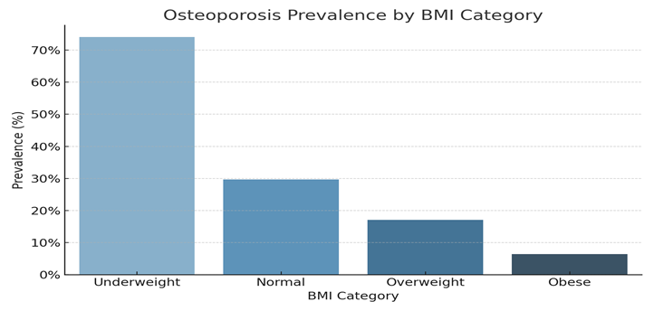
Figure 2: Osteoporosis Prevalence by BMI Category
Female-Specific Risk Factors
The study analysed reproductive characteristics, including menopause and gravida status, among 305 female participants for their correlation with osteoporosis. A notable connection was identified with menopausal status (χ² = 9.41, p = 0.009). Among the total female participants, 229 (75.1%) had attained menopause, and within this cohort, osteoporosis was significantly more common at 49.3%. Conversely, merely 15.3% of premenopausal women and 11.8% of hysterectomised women were identified as having osteoporosis. These findings underscore the increased risk of bone mineral loss post-menopause, presumably attributable to oestrogen shortage.
The gravida status indicated a tendency for increased osteoporosis prevalence in women with two or more pregnancies (44.3%) compared to those with one or no pregnancies. Nonetheless, this correlation did not attain statistical significance (p = 0.124), indicating that although multiparity may influence bone density loss, it is not a conclusive independent risk factor in this population.
DISCUSSION:
This investigation was designed to evaluate the occurrence of osteoporosis and determine its influencing features among adults aged over 40 in urban Belgaum through the use of Calcaneal Quantitative Ultrasound (QUS). The findings revealed that a significant portion of the population (36.7%) was diagnosed with osteoporosis, while another 37.3% showed signs of osteopenia. Merely 26.0% of participants exhibited normal bone mineral density (BMD). These findings underscore the significant burden of impaired bone health in this demographic and indicate an immediate necessity for community-based preventive measures.6
The chronicity of osteoporosis observed in our study aligns with previous Indian studies. A cross-sectional study in Chitwan, India indicated a prevalence of osteoporosis at 26.2% and osteopenia at 39.3% among women utilising peripheral bone assessment methods, statistics that closely correspond with our findings.7 A study conducted in New Delhi utilising quantitative ultrasound (QUS) revealed an osteoporosis prevalence of 15% in premenopausal women and as high as 53% in postmenopausal women, highlighting the increased burden among females.4 Our research confirms this trend, revealing a markedly greater prevalence of osteoporosis in females (43.3%) than in males (22.1%) (p = 0.002), likely attributable to postmenopausal oestrogen deficiency and age-related bone deterioration.
The association between advancing age and diminishing BMD was highly significant (p < 0.001). The highest occurrence of osteoporosis was noted in the 60–69 and ≥70-year age cohorts, corroborating existing literature that designates ageing as a principal non-modifiable risk factor for osteoporosis, attributed to increased bone resorption and diminished bone formation.8
The frequency of osteoporosis was inversely correlated with Body Mass Index (BMI). Individuals classified as underweight exhibited the highest prevalence of osteoporosis at 74.1%, whereas those categorised as obese demonstrated the lowest prevalence at 6.4% (p < 0.001). The protective influence of elevated BMI aligns with both global and Indian literature, suggesting that mechanical loading and increased oestrogen levels linked to adipose tissue may mitigate bone loss.9 Nonetheless, this protective effect must be weighed against the cardiovascular and metabolic risks associated with obesity.
Lifestyle has also emerged as a crucial determinant. Individuals with a sedentary lifestyle exhibited a higher prevalence of osteoporosis (44.4%) compared to their physically active counterparts (27.5%) (p = 0.023). Physical activity is recognised for its ability to enhance bone remodelling via mechanical stimulation of osteoblasts, and our results corroborate the significance of exercise in preserving bone mass.9,3
Socioeconomic status was an additional significant predictor. The prevalence of osteoporosis was greatest among Class IV (45.5%) and Class V (41.2%) participants (p = 0.012), suggesting that lower socioeconomic status may be associated with inadequate nutrition, reduced physical activity, and limited access to healthcare services, all of which adversely affect bone health. 11,12
In female participants, menopause was significantly correlated with reduced bone mineral density (BMD). Postmenopausal women exhibited a greater prevalence of osteoporosis (49.3%) in comparison to premenopausal women (15.3%) and individuals who underwent hysterectomy (11.8%) (p = 0.009). This discovery corresponds with research highlighting estrogen's protective function in bone metabolism and the significant bone loss that occurs post-menopause due to hormonal cessation.5,13
Although variables like educational attainment and family structure exhibited trends of association, they did not achieve statistical significance in our research. This may indicate intricate interactions among education, awareness, and health behaviour that require additional investigation.14
The primary strength of this study lies in its representative sampling from three distinct urban centres, utilising a standardised and validated instrument (Calcaneal QUS) for bone mineral density assessment.4 The substantial sample size and the inclusion of both genders across diverse age groups enhance the validity of the findings. Nonetheless, constraints encompass the utilisation of QUS in lieu of DEXA (the gold standard) and the cross-sectional design, which restricts causal inference. Recall bias may have affected the precision of self-reported data regarding lifestyle and medical history.
Public Health Implications: Our findings underscore the necessity for focused osteoporosis screening initiatives in urban Indian contexts, particularly for women over 50, individuals with low BMI, and inactive older adults. Due to its portability and cost-effectiveness, calcaneal QUS serves as a viable screening instrument for extensive application in primary healthcare environments.8,4.
CONCLUSION:
This study highlights a notable occurrence of osteoporosis and osteopenia among adults aged over 40 in urban Belgaum, with more than 70% of participants showing reduced bone mineral density as assessed through calcaneal quantitative ultrasound. The prevalence was significantly elevated among females, elderly individuals, postmenopausal women, individuals with a low body mass index, sedentary lifestyles, and those of lower socioeconomic status.
The notable correlations identified between bone health and modifiable risk factors, including physical inactivity and undernutrition, highlight the necessity of community-level preventive measures. Public health interventions must prioritise the promotion of consistent physical activity, the assurance of sufficient nutritional intake, and the augmentation of awareness regarding osteoporosis, particularly among vulnerable populations.
Calcaneal QUS is an effective, non-invasive, and cost-efficient screening instrument, appropriate for use in primary care and urban health environments. Timely identification and lifestyle alterations can significantly diminish the likelihood of osteoporotic fractures, enhance quality of life and alleviate the healthcare burden in ageing urban demographics.
Acknowledgments
We extend our heartfelt gratitude to all study participants for their cooperation. We sincerely thank the Department of Community Medicine, J.N. Medical College, Belgaum, for their constant support and guidance. We also acknowledge the efforts of field investigators and staff whose contributions made this research possible.
Conflicts of Interest
The authors declare no affiliations or involvement with any organization or entity with financial or non-financial interests.
REFERENCES:
1. Jiang NS, Newton B, Jiang X. An overview of osteoporosis management. OBM Geriatrics [Internet]. 2021b Jun 28;05(04):1. Available from: https://doi.org/10.21926/obm.geriatr.2104181
2. Rachner TD, Khosla S, Hofbauer LC. Osteoporosis: now and the future. The Lancet [Internet]. 2011 Mar 30;377(9773):1276–87. Available from: https://doi.org/10.1016/s0140-6736(10)62349-5
3. Vilaca T, Eastell R, Schini M. Osteoporosis in men. The Lancet Diabetes & Endocrinology [Internet]. 2022 Mar 3;10(4):273–83. Available from: https://doi.org/10.1016/s2213-8587(22)00012-2
4. Sharma S, Tandon V, Mahajan A, Kour A, Kumar D. Preliminary screening of osteoporosis and osteopenia in urban women from Jammu using calcaneal QUS. Indian Journal of Medical Sciences [Internet]. 2006 Jan 1;60(5):183. Available from: https://doi.org/10.4103/0019-5359.25679
5. Raveendran A, Aggarwal N, Khandelwal N, Sen R, Thakur J, Dhaliwal L, et al. Prevalence and related risk factors of osteoporosis in peri- and postmenopausal Indian women. Journal of Mid-life Health [Internet]. 2011 Jan 1;2(2):81. Available from: https://doi.org/10.4103/0976-7800.92537
6. Kanis JA. Osteoporosis and osteopenia. Journal of Bone and Mineral Research [Internet]. 1990 Mar 1;5(3):209–11. Available from: https://doi.org/10.1002/jbmr.5650050302
7. Aryal DB. Prevalence of Osteoporosis among Middle Aged Women in Chitwan District of Nepal. ijpba [Internet]. 2012 Sep. 23 [cited 2025 Jul. 9];3(4). Available from: https://www.ijpba.info/index.php/ijpba/article/view/734
8. World Health Organization. Assessment of fracture risk and its application to screening for postmenopausal osteoporosis : report of a WHO study group [meeting held in Rome from 22 to 25 June 1992] [Internet]. 1994. Available from: https://iris.who.int/handle/10665/39142
9. Khadilkar A, Mandlik R. Epidemiology and treatment of osteoporosis in women: an Indian perspective. International Journal of Women S Health [Internet]. 2015 Oct 1;841. Available from: https://doi.org/10.2147/ijwh.s54623
10. Sharma N, Aggarwal P. Modified BG Prasad Socio-economic Classification, Update – 2021. Journal of Integrative Medicine and Public Health [Internet]. 2022 Jan 1;1(1):7–9. Available from: https://doi.org/10.4103/jimph.jimph_1_21
11. Paul TV, Thomas N, Seshadri MS, Oommen R, Jose A, Mahendri NV. Prevalence of Osteoporosis in Ambulatory Postmenopausal Women from A Semiurban Region in Southern India: Relationship to Calcium Nutrition and Vitamin D Status. Endocrine Practice [Internet]. 2008 Sep 1;14(6):665–71. Available from: https://doi.org/10.4158/ep.14.6.665
12. Kadam N, Chiplonkar S, Khadilkar A, Divate U, Khadilkar V. Low bone mass in urban Indian women above 40 years of age: prevalence and risk factors. Gynecological Endocrinology [Internet]. 2010 Sep 17;26(12):909–17. Available from: https://doi.org/10.3109/09513590.2010.487604
13. Agrawal T, Verma AK. Cross sectional study of osteoporosis among women. Medical Journal Armed Forces India [Internet]. 2012 Nov 3;69(2):168–71. Available from: https://doi.org/10.1016/j.mjafi.2012.07.024
14. Unni J, Garg R, Pawar R. Bone mineral density in women above 40 years. Journal of Mid-life Health [Internet]. 2010 Jan 1;1(1):19. Available from: https://doi.org/10.4103/0976-7800.66989.