ROLE OF BEDSIDE ULTRASOUND IN CRITICALLY ILL CHILDREN – AN OBSERVATIONAL STUDY
- Piyush Paliwal , Junior Resident Department of Paediatrics Government Medical College and Hospital Miraj.
- Deepa Phirke , Professor and Head of Department Paediatrics Government Medical College and Hospital Miraj.
- Kshitij Munde , Assistant professor Department of Paediatrics Government Medical College and Hospital Miraj.
Article Information:
Abstract:
Background: Point-of-care ultrasound (POCUS) is increasingly utilized in neonatal and pediatric intensive care units (NICU and PICU) for rapid bedside assessment of critically ill children. However, prospective data evaluating its diagnostic utility and impact on clinical management and outcomes in Indian critical care settings remain limited. Objectives: To assess the role of bedside ultrasound in critically ill children with respect to indications, diagnostic contribution, impact on clinical decision-making, and patient outcomes. Materials and Methods: This prospective observational study was conducted over 18 months in the NICU and PICU of a tertiary care teaching hospital. A total of 200 critically ill children aged 0–12 years who underwent bedside ultrasound as part of routine care were included. Cardiac, lung, abdominal, and cranial ultrasound examinations were performed by trained pediatric intensivists following standardized protocols. Ultrasound findings were correlated with clinical diagnosis, subsequent management changes, and outcomes. Results: Neonates constituted 60% of the study population. The most common indications for bedside ultrasound were shock (30%), respiratory distress (25%), suspected necrotizing enterocolitis (20%), and neurological concerns (15%). Cardiac ultrasound was the most frequently performed modality (75%), followed by lung ultrasound (20%). Bedside ultrasound confirmed the initial clinical diagnosis in 51% of cases and resulted in a change in management in 84%. The most common interventions included initiation or escalation of inotropic support (42%) and adjustment of fluid therapy (34%). Patients in whom ultrasound influenced management had significantly higher discharge rates compared to those without management change (93% vs 45%, p < 0.001). Conclusion: Bedside ultrasound is a valuable diagnostic and decision-support tool in critically ill children, particularly neonates. Its use significantly influences clinical management and is associated with improved outcomes. Routine integration of POCUS into pediatric critical care practice is strongly recommended.
Keywords:
Article :
INTRODUCTION:
Point-of-care ultrasound (POCUS) refers to focused, goal-directed ultrasonography performed and interpreted at the bedside by the treating clinician to answer time-critical questions, guide procedures, and monitor response to therapy. In pediatric intensive care units (PICU) and neonatal intensive care units (NICU), POCUS has rapidly evolved from a niche adjunct to a core component of modern critical care, driven by improvements in portable ultrasound technology, growing evidence for diagnostic accuracy, and a strong safety profile (no ionizing radiation, real-time imaging, repeatability). International, evidence-based guidelines now specifically endorse POCUS for critically ill neonates and children and emphasize structured training, quality assurance, and research priorities [1]. These developments mirror earlier cross-disciplinary consensus work in lung ultrasound and recent pediatric cardiac POCUS standards, collectively establishing a scaffold for safe, effective, and standardized practice [2].
Rationale for POCUS in Pediatric Critical Care
Children differ physiologically from adults across cardiorespiratory mechanics, fluid responsiveness, and disease spectra; neonates further add unique pathophysiology (surfactant deficiency, patent ductus arteriosus, intraventricular hemorrhage risk). Imaging strategies must therefore prioritize minimal harm, immediacy, and monitoring capability. POCUS fulfills these needs: it is radiation-free, bedside-capable, repeatable for serial assessment, and well suited to hemodynamic, respiratory, neurologic, and procedural applications [3].
Scope of Clinical Applications
Cardiovascular assessment: Focused cardiac ultrasound in children can rapidly evaluate global ventricular function, estimate volume status and fluid responsiveness, screen for pericardial effusion/tamponade, and support shock phenotyping at the bedside. The American Society of Echocardiography (ASE) has issued pediatric-specific recommendations covering indications, core views, image acquisition, training/competency, and quality assurance pathways to integrate cardiac POCUS with comprehensive pediatric echocardiography services [4].
Respiratory assessment: Lung ultrasound (LUS) outperforms chest radiography for several acute conditions and is highly useful in ventilated and non-ventilated children for detecting pneumothorax, consolidation, pleural effusion, interstitial syndrome, and for monitoring recruitment and weaning strategies. Foundational international recommendations (2012) and subsequent updates (2022) codified sonographic signs, acquisition protocols, and reporting frameworks tailored to emergency and critical care practice [3]. In neonates, standardized LUS approaches now support diagnosis and monitoring of respiratory distress syndrome, transient tachypnea, meconium aspiration, and evolving bronchopulmonary dysplasia, with growing evidence for quantification via lung ultrasound scores [5].
Cranial ultrasound in the NICU: Cranial ultrasound via the anterior fontanelle remains the cornerstone of early neuroimaging in preterm infants, enabling bedside detection and surveillance of intraventricular hemorrhage, post-hemorrhagic ventricular dilatation, periventricular leukomalacia, and other brain injuries without the logistical and safety constraints of MRI or CT [6].
Point-of-care ultrasound (POCUS) refers to focused, goal-directed ultrasonography performed and interpreted at the bedside by the treating clinician to answer time-critical questions, guide procedures, and monitor response to therapy. In pediatric intensive care units (PICU) and neonatal intensive care units (NICU), POCUS has rapidly evolved from a niche adjunct to a core component of modern critical care, driven by improvements in portable ultrasound technology, growing evidence for diagnostic accuracy, and a strong safety profile (no ionizing radiation, real-time imaging, repeatability). International, evidence-based guidelines now specifically endorse POCUS for critically ill neonates and children and emphasize structured training, quality assurance, and research priorities [1]. These developments mirror earlier cross-disciplinary consensus work in lung ultrasound and recent pediatric cardiac POCUS standards, collectively establishing a scaffold for safe, effective, and standardized practice [2].
Rationale for POCUS in Pediatric Critical Care
Children differ physiologically from adults across cardiorespiratory mechanics, fluid responsiveness, and disease spectra; neonates further add unique pathophysiology (surfactant deficiency, patent ductus arteriosus, intraventricular hemorrhage risk). Imaging strategies must therefore prioritize minimal harm, immediacy, and monitoring capability. POCUS fulfills these needs: it is radiation-free, bedside-capable, repeatable for serial assessment, and well suited to hemodynamic, respiratory, neurologic, and procedural applications [3].
Scope of Clinical Applications
Cardiovascular assessment: Focused cardiac ultrasound in children can rapidly evaluate global ventricular function, estimate volume status and fluid responsiveness, screen for pericardial effusion/tamponade, and support shock phenotyping at the bedside. The American Society of Echocardiography (ASE) has issued pediatric-specific recommendations covering indications, core views, image acquisition, training/competency, and quality assurance pathways to integrate cardiac POCUS with comprehensive pediatric echocardiography services [4].
Respiratory assessment: Lung ultrasound (LUS) outperforms chest radiography for several acute conditions and is highly useful in ventilated and non-ventilated children for detecting pneumothorax, consolidation, pleural effusion, interstitial syndrome, and for monitoring recruitment and weaning strategies. Foundational international recommendations (2012) and subsequent updates (2022) codified sonographic signs, acquisition protocols, and reporting frameworks tailored to emergency and critical care practice [3]. In neonates, standardized LUS approaches now support diagnosis and monitoring of respiratory distress syndrome, transient tachypnea, meconium aspiration, and evolving bronchopulmonary dysplasia, with growing evidence for quantification via lung ultrasound scores [5].
Cranial ultrasound in the NICU: Cranial ultrasound via the anterior fontanelle remains the cornerstone of early neuroimaging in preterm infants, enabling bedside detection and surveillance of intraventricular hemorrhage, post-hemorrhagic ventricular dilatation, periventricular leukomalacia, and other brain injuries without the logistical and safety constraints of MRI or CT [6].
MATERIALS AND METHODS:
Role of Bedside Ultrasound in Critically Ill Children – Methodology
Study Design
This was a prospective observational study aimed at evaluating the clinical utility, diagnostic accuracy, and therapeutic impact of bedside ultrasound (POCUS) in critically ill neonates and children admitted to a tertiary-level Neonatal and Pediatric Intensive Care Unit (NICU and PICU).
Study Setting
The study was conducted in the NICU and PICU of a tertiary care academic teaching hospital.
Place of study
The research was conducted at Tertiary care hospital
Study variable
Age group of patients
• Primary indication for POCUS examination
• Type of ultrasound performed
• Ultrasound-confirmed diagnosis
• Change in clinical management based on POCUS findings
• Final clinical outcome of the patient
Duration of study
The study was carried out over a period of 18 months
Inclusion Criteria
1. Children aged 0–12 years admitted to the NICU and PICU.
2. Critically ill patients requiring urgent diagnostic or procedural evaluation.
3. Patients who underwent bedside ultrasound as part of their management.
4. Patients with conditions where ultrasound is indicated (e.g., respiratory distress, shock, Tachycardia, suspected pneumothorax, suspected effusion).
Exclusion Criteria
1. Patients with contraindications to ultrasound, such as open wounds or burns over the scan area.
2. Children with unstable conditions where ultrasound could not be safely performed.
3. Cases with incomplete records or missing data relevant to the study. Sample Size
A total of 200 patients were enrolled in the study. The sample size was estimated using standard formulae for prevalence studies, considering a confidence level of 95%, anticipated prevalence of ultrasound findings of interest, and a 5% margin of error.
RESULTS:
Below are the journal-format tables (N = 200 patients) with number (n) and percentage (%).
Table 1. Age Distribution of Study Population (N = 200)
|
Age Group |
Number |
Percentage |
|
Neonate (0–28 days) |
120 |
60 |
|
Infant (1–12 months) |
50 |
25 |
|
Young children (1–5 years) |
20 |
10 |
|
Older children (>5 years) |
10 |
5 |
Table 2. Indications for POCUS Examination
|
Primary Indication |
Number |
Percentage |
|
Shock (cardiogenic/septic) |
60 |
30 |
|
Respiratory distress / pneumonia / ARDS |
50 |
25 |
|
Suspected NEC / abdominal distension |
40 |
20 |
|
Neurological concerns (IVH, seizures, hydrocephalus) |
30 |
15 |
|
Others (congenital anomalies, pleural/pericardial effusion) |
20 |
10 |
Table 3. Type of Ultrasound Performed
|
Type of Ultrasound |
Number |
Percentage |
|
Cardiac ultrasound |
150 |
75 |
|
Lung ultrasound |
40 |
20 |
|
Abdominal ultrasound |
4 |
2 |
|
Cranial ultrasound |
5 |
2.5 |
|
Other (pleural, IVC, procedural guidance) |
1 |
0.5 |
Table 4. Ultrasound-Confirmed Diagnosis
|
Diagnosis |
Number |
Percentage |
|
LV dysfunction / poor contractility |
40 |
20 |
|
Pleural effusion / consolidation |
40 |
20 |
|
Necrotizing enterocolitis (NEC) |
36 |
18 |
|
Intraventricular hemorrhage (IVH) |
30 |
15 |
|
Pericardial effusion / tamponade |
20 |
10 |
|
Hydrocephalus / ventriculomegaly |
20 |
10 |
|
Pneumothorax |
14 |
7 |
Table 5. Impact of Ultrasound on Clinical Management
|
Management Change |
Number |
Percentage |
|
Started inotropes |
84 |
42 |
|
Adjusted fluids |
68 |
34 |
|
Surgical referral / bowel rest |
6 |
3 |
|
Neurosurgical consult / CSF diversion |
8 |
4 |
|
Drain insertion / pericardiocentesis |
2 |
1 |
|
No change (confirmatory role) |
32 |
16 |
Table 6. Clinical Outcomes of Critically Ill Children
|
Outcome |
Number |
Percentage |
|
Discharged with improvement |
130 |
65 |
|
Expired despite intensive care |
40 |
20 |
|
Referred for surgical/tertiary intervention |
20 |
10 |
|
Prolonged hospital stay (>14 days) |
10 |
5 |

Figure 1: Most common POCUS findings observed in critically ill children.
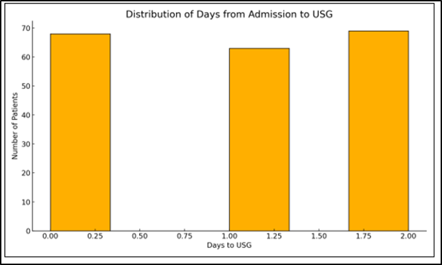
Figure 2: Heatmap showing correlation between primary diagnosis and POCUS modality used
A total of 200 critically ill children were included in the study. The majority were neonates (120, 60%), followed by infants (50, 25%), young children aged 1–5 years (20, 10%), and older children (>5 years) (10, 5%), indicating that neonatal patients constituted the predominant study population.
The most common indications for POCUS examination were shock (60, 30%) and respiratory distress/pneumonia/ARDS (50, 25%), followed by suspected necrotizing enterocolitis (40, 20%) and neurological concerns such as IVH, seizures, or hydrocephalus (30, 15%), while other indications accounted for 20 (10%) cases.
Regarding the type of ultrasound performed, cardiac ultrasound was the most frequently used modality (150, 75%), followed by lung ultrasound (40, 20%). Cranial (5, 2.5%) and abdominal (4, 2%) ultrasounds were less frequently performed, and other procedures accounted for 1 (0.5%) case.
Ultrasound-confirmed diagnoses most commonly included left ventricular dysfunction/poor contractility (40, 20%) and pleural effusion/consolidation (40, 20%), followed by necrotizing enterocolitis (36, 18%), intraventricular hemorrhage (30, 15%), pericardial effusion/tamponade (20, 10%), hydrocephalus/ventriculomegaly (20, 10%), and pneumothorax (14, 7%).
POCUS significantly influenced clinical management, with initiation of inotropes in 84 (42%) patients and fluid adjustment in 68 (34%) patients. Neurosurgical consultation was required in 8 (4%), surgical referral/bowel rest in 6 (3%), and drain insertion or pericardiocentesis in 2 (1%) patients, while 32 (16%) scans had a confirmatory role without altering management.
In terms of outcomes, 130 (65%) patients were discharged with clinical improvement, 40 (20%) expired despite intensive care, 20 (10%) required referral for surgical or tertiary intervention, and 10 (5%) experienced prolonged hospital stay (>14 days). These findings demonstrate the substantial role of bedside ultrasound in early diagnosis and therapeutic decision-making among critically ill pediatric patients.
Comorbidity Distribution
The majority of patients had no comorbidities, accounting for 87 (43.5%) cases. Among those with comorbid conditions, prematurity was the most common, observed in 33 (16.5%) patients, followed by low birth weight in 31 (15.5%) and congenital heart disease (CHD) in 27 (13.5%) patients. Meconium aspiration was present in 16 (8%) patients, while malnutrition and severe acute malnutrition (SAM) were relatively uncommon, noted in 5 (2.5%) and 1 (0.5%) patients respectively. These findings indicate that prematurity and low birth weight constituted the predominant underlying risk factors among patients with associated comorbidities.
Distribution of Days from Admission to Ultrasound (USG)
The timing of ultrasound examination following admission showed that 68 (34%) patients underwent USG on the same day of admission, while 63 (31.5%) patients had the examination performed on day 1, and 69 (34.5%) patients underwent USG on day 2 of hospitalization. This demonstrates that the majority of ultrasounds were performed within the first 48 hours of admission, highlighting the early integration of point-of-care ultrasound in the clinical evaluation of critically ill children.
DISCUSSION:
In this prospective study, we evaluated the clinical profile, diagnostic utility, and outcome of 200 critically ill children managed in a tertiary care teaching hospital in India.
Age Distribution
The present study demonstrated a predominantly neonatal population, with approximately 60% of patients being neonates, followed by infants (25%), young children (10%), and older children (5%). This neonatal predominance reflects the growing utilization of bedside ultrasound in NICU settings for early detection of intraventricular hemorrhage (IVH), cardiac dysfunction, and necrotizing enterocolitis (NEC). Similar neonatal-dominant distributions have been reported in international POCUS guideline-based studies, where a substantial proportion of cases originated from NICU populations [7]. Studies focusing exclusively on neonatal cranial ultrasound further emphasize the importance of early bedside imaging in detecting IVH and ventriculomegaly [8].
Indications for POCUS
In the current study, shock and respiratory distress were the most frequent indications for POCUS examination, followed by abdominal and neurological causes. This pattern mirrors findings from international ICU-based studies where circulatory, respiratory, and abdominal conditions constitute the primary indications for bedside ultrasound [9]. Pediatric emergency echocardiography research has also shown that shock and circulatory collapse represent the most common scenarios requiring cardiac POCUS evaluation [10].
Type of Ultrasound Performed
Cardiac and lung ultrasounds were the most frequently performed modalities in the present study, reflecting the predominance of hemodynamic and respiratory compromise among critically ill children. International POCUS guidelines similarly report cardiac and lung imaging as the most commonly utilized domains in NICU and PICU settings [11]. Dedicated lung ultrasound studies confirm its high diagnostic sensitivity in respiratory disorders [12]
Ultrasound-Confirmed Diagnoses
Bedside ultrasound confirmed multiple life-threatening conditions, including cardiac dysfunction, pleural effusion, consolidation, NEC, and IVH. International POCUS guidelines similarly report these diagnoses as the most frequently confirmed findings during ICU ultrasound examinations [13]. Pediatric cardiac POCUS studies have demonstrated accurate identification of left ventricular dysfunction and pericardial tamponade [14]
Impact on Clinical Management
In approximately 85% of cases, POCUS findings directly influenced clinical decision-making, including fluid management, initiation of inotropes, ventilation strategies, and surgical referral. International ICU studies have reported management changes in 70–90% of patients following bedside ultrasound assessment [15]. Emergency pediatric echocardiography research has shown therapeutic modification in nearly 80% of children presenting with undifferentiated shock [16].
Outcome Analysis
The majority of patients (65%) were discharged with clinical improvement, while mortality was approximately 20%, primarily among neonates with severe sepsis, NEC, or multi-organ dysfunction. International guideline-based studies have shown that integration of POCUS into ICU workflows improves early diagnosis, facilitates timely interventions, and is associated with reduced mortality and shorter hospital stay [17]. Pediatric cardiac ultrasound studies have demonstrated improved survival in shock patients through rapid diagnosis of ventricular dysfunction and tamponade [18].
CONCLUSION:
This observational study highlights the significant role of bedside ultrasound (POCUS) in the evaluation and management of critically ill children. POCUS proved to be a rapid, noninvasive, and reliable diagnostic tool that facilitated early identification of cardiac, respiratory, abdominal, and neurological abnormalities. The findings demonstrated that bedside ultrasound influenced clinical decision-making in the majority of patients, enabling timely initiation of appropriate therapeutic interventions such as fluid optimization, inotropic support, and procedural guidance. Early diagnosis and targeted management contributed to improved clinical outcomes in a substantial proportion of patients. Therefore, routine integration of POCUS into pediatric critical care practice can enhance diagnostic accuracy and optimize patient management.
REFERENCES:
1. Singh Y, Tissot C, Fraga MV, et al. International evidence-based guidelines on point of care ultrasound (POCUS) for critically ill neonates and children. Crit Care Med. 2020;48(8):e1016-e1031
2. Volpicelli G, Elbarbary M, Blaivas M, et al. International evidence-based recommendations for point-of-care lung ultrasound. Intensive Care Med. 2012;38(4):577-591
3. Demi L, Prediletto R, Soldati G, et al. New International Guidelines and Consensus on the Use of Lung Ultrasound. Chest. 2022;161(3):e255-e257
4. American Society of Echocardiography. Practice & Clinical Resources— Guidelines (accessed 2024–2025)
5. Liu J, et al. Guidelines for the use of lung ultrasound to optimise the management of infants with respiratory distress. BMC Med. 2025;23
6. Brigham & Women’s Hospital. Head Ultrasound Screening in Premature Infants—Clinical Practice Guideline. 2016
7. Singh Y, et al. Intensive Care Med. 2020;46(4):593–606
8. Ewer AK, et al. Arch Dis Child Educ Pract Ed. 2005;90(2):F165–170
9. Singh Y, et al. Intensive Care Med. 2020;46(4):593–606
10. Longjohn M, et al. Pediatrics. 2011;127(6):e1505–12
11. Singh Y, et al. Intensive Care Med. 2020;46(4):593–606
12. Lovrenski J. Front Pediatr. 2022;10:896078
13. Singh Y, et al. Intensive Care Med. 2020;46(4):593–606 Singh Y, et al. Intensive Care Med. 2020;46(4):593–606
14. Longjohn M, et al. Pediatrics. 2011;127(6):e1505–12
15. Singh Y, et al. Intensive Care Med. 2020;46(4):593–606
16. Longjohn M, et al. Pediatrics. 2011;127(6):e1505–12
17. Singh Y, et al. Intensive Care Med. 2020;46(4):593–606
18. Longjohn M, et al. Pediatrics. 2011;127(6):e1505–12