Ambiguous Genitalia in Pediatric Patients: Clinical, Radiological, and Cytogenetic Correlation from Bagalkote, Karnataka
- Sujayendra Kulkarni , Department of Genetics and Genomics, S Nijalingappa Medical College and HSK Hospital, Navanagar, Bagalkote-587102.
- Anil Satyaraddi , Department of Endocrinology, Diabetes and Metabolism, S Nijalingappa Medical College and HSK Hospital, Navanagar, Bagalkote-587102.
- Naveen Charantimath , Department of Endocrinology, Diabetes and Metabolism, S Nijalingappa Medical College and HSK Hospital, Navanagar, Bagalkote-587102.
- Suyamindra S.K , Kalyan Karnataka Institute for Human Genome Research, Department of Microbiology, Adikavi shri Maharshi Valmiki University, Raichur-584133.
- Ashwini H , Department of Genetics and Genomics, S Nijalingappa Medical College and HSK Hospital, Navanagar, Bagalkote-587102.
- Kavitha MM. , Department of Biochemistry, S Nijalingappa Medical College and HSK Hospital, Navanagar, Bagalkote-587102.
- Sangappa V Kashinakunti , Department of Biochemistry, S Nijalingappa Medical College and HSK Hospital, Navanagar, Bagalkote-587102.
Article Information:
Abstract:
Background: Ambiguous genitalia, categorized under disorders of sex development (DSD), requires prompt identification and accurate diagnosis to ensure appropriate management and gender assignment. In resource-constrained settings, an integrated diagnostic approach becomes particularly important. Objective: To assess the clinical presentation, endocrine profile, imaging findings, and cytogenetic characteristics of children presenting with ambiguous genitalia at a tertiary care hospital in North Karnataka. Methods: This observational study included 12 pediatric patients. Clinical evaluation with Prader staging, hormonal assays, and ultrasonography were performed. Chromosomal analysis was carried out using conventional karyotyping and further validated by fluorescence in situ hybridization (FISH) using DXZ1, DYZ3, and SRY probes. Results: Out of 12 cases, 8 (66.6%) had a 46,XY karyotype and 4 (33.3%) were 46,XX. All 46,XX individuals exhibited biochemical evidence of androgen excess, consistent with congenital adrenal hyperplasia. The 46,XY group showed varied endocrine abnormalities including reduced androgen levels and possible defects in androgen action. Imaging findings correlated with chromosomal sex in all cases. A high proportion of patients had consanguineous parentage. Karyotyping and FISH findings were fully concordant. Conclusion: A multidisciplinary evaluation combining clinical, biochemical, radiological, and cytogenetic methods is essential for accurate diagnosis of DSD. Conventional karyotyping with FISH confirmation remains a reliable and cost-effective diagnostic strategy.
Keywords:
Article :
INTRODUCTION:
Ambiguous genitalia in children represents a complex clinical entity within the spectrum of disorders of sex development (DSD), characterized by atypical differentiation of chromosomal, gonadal, or anatomical sex [1,2]. These conditions often present at birth or during early infancy and necessitate immediate evaluation to facilitate appropriate gender assignment and clinical management.
The causes of DSD are diverse and include chromosomal abnormalities, defects in gonadal development, and disorders of steroidogenesis or hormone action [2]. Among individuals with a 46,XX karyotype, congenital adrenal hyperplasia (CAH), particularly due to 21-hydroxylase deficiency, is the most frequent cause of virilization [3,16]. In contrast, 46,XY DSD is commonly associated with androgen insensitivity syndrome, defects in testosterone biosynthesis, or gonadal dysgenesis [15].
A systematic diagnostic approach is critical and typically involves clinical examination, hormonal evaluation, imaging studies, and cytogenetic analysis [4,5]. Conventional karyotyping remains a cornerstone investigation, particularly in resource-constrained settings, where access to advanced molecular techniques may be limited [6].
The clinical spectrum and etiology of ambiguous genitalia vary across populations due to genetic, environmental, and sociocultural factors [7,8]. In India, delayed diagnosis and underreporting are common, highlighting the importance of region-specific studies to better understand disease patterns and improve management strategies.
MATERIALS AND METHODS:
Study Design and Study Population
This hospital-based cohort study was conducted in the Cytogenetics Laboratory of S Nijalingappa Medical College and HSK Hospital, Bagalkote a tertiary care teaching hospital in North Karnataka. Children presenting with ambiguous genitalia were referred from the Departments of Pediatric, Endocrinology and Pediatric Endocrinology for chromosomal evaluation. During the study period, 12 children with clinically ambiguous genitalia were identified and included in the study. All cases underwent conventional karyotyping and confirmatory fluorescence in situ hybridization (FISH) for sex chromosome analysis.
Clinical Assisment
Detailed clinical history, including antenatal details, family history, and parental consanguinity, was obtained. Physical examination focused on genital phenotype, and Prader staging was used to classify virilization.
Sample CollectionPeripheral venous blood (2–3 mL) was collected aseptically in sterile sodium heparin vacutainers and in plain vacutainers after obtaining written informed consent from parents or legal guardians. Samples were processed immediately for cytogenetic analysis.
Ethical Considerations
The study was approved by the Institutional Ethics Committee. Written informed consent was obtained from parents or legal guardians prior to sample collection. The study was conducted in accordance with the principles of the Declaration of Helsinki.
Biochemical Evaluation
Hormonal analysis was performed to evaluate adrenal and gonadal function. Parameters included 17-hydroxyprogesterone, cortisol, ACTH, testosterone, LH, FSH, and DHEA-S. Results were interpreted using age-specific reference ranges [11].
Hormonal Assays
The following serum biochemical parameters were analyzed based on clinical presentation and suspected etiology:
• 17-Hydroxyprogesterone (17-OHP): measured to screen for congenital adrenal hyperplasia (CAH), particularly 21-hydroxylase deficiency
• Serum cortisol and adrenocorticotropic hormone (ACTH): assessed to evaluate adrenal function and stress response.
• Testosterone: measured to assess androgen production, especially in suspected 46,XY DSD.
• Luteinizing hormone (LH) and follicle-stimulating hormone (FSH): evaluated to assess hypothalamic–pituitary–gonadal axis function.
• Dehydroepiandrosterone sulfate (DHEA-S): assessed as a marker of adrenal androgen secretion.
Hormonal assays were performed using chemiluminescence immunoassay (CLIA) or enzyme-linked immunosorbent assay (ELISA)( The Autobio AutoLumo A1000- Manufectured 2023, Autobio Diagnostics Co., Ltd.) techniques, according to the manufacturer’s instructions. Age- and sex-specific reference ranges were used for interpretation.
For interpretation of biochemical findings, age- and sex-appropriate reference ranges were used based on established pediatric endocrinology guidelines. Serum 17-hydroxyprogesterone (17-OHP) levels are typically less than 200 ng/dL in newborns, less than 100 ng/dL in infants, and generally below 90 ng/dL in children; markedly elevated levels are suggestive of congenital adrenal hyperplasia, particularly 21-hydroxylase deficiency. Serum cortisol levels normally range from approximately 5–25 µg/dL in the morning (08:00–09:00 h) and 2–14 µg/dL in the evening, reflecting normal diurnal variation of adrenal activity. Adrenocorticotropic hormone (ACTH) concentrations usually range between 10–60 pg/mL, with elevated values indicating possible adrenal insufficiency or increased adrenal stimulation. Serum testosterone levels vary with age and sex: in prepubertal children they are typically less than 20 ng/dL, while higher levels are observed during puberty and in male infants during the period of mini-puberty. Luteinizing hormone (LH) levels in prepubertal children generally range from 0.02–0.3 IU/L and increase during puberty to approximately 1–9 IU/L, while follicle-stimulating hormone (FSH) levels are typically between 0.3–3 IU/L in prepubertal children and may rise to 1–12 IU/L during adolescence. Dehydroepiandrosterone sulfate (DHEA-S) concentrations, reflecting adrenal androgen secretion, generally range from about 5–85 µg/dL in children and increase during adolescence to approximately 40–350 µg/dL. These reference intervals were used to interpret hormonal abnormalities and were correlated with clinical findings, Prader grading, imaging studies, and cytogenetic results to support the diagnosis of disorders of sex development.
Interpretation and Correlation
Biochemical findings were interpreted in conjunction with clinical phenotype, Prader grading, radiological findings, karyotype, and FISH results. Elevated 17-OHP levels in genetically female (46,XX) patients with virilization were suggestive of congenital adrenal hyperplasia, while abnormal androgen or gonadotropin profiles in genetically male (46,XY) patients along with clinical data supported diagnosis such as defects in androgen synthesis or action.
Where available, biochemical results were used to guide further management, including endocrine referral, initiation of hormonal therapy, and genetic counseling.
Conventional Cytogenetic Analysis (Karyotyping)
Peripheral blood lymphocyte cultures were used for chromosomal analysis using GTG banding as described by standard cytogenetic protocols [Seabright, 1971]. A minimum of 20 metaphases were examined, and results were reported according to ISCN guidelines.
Fluorescence In Situ Hybridization (FISH) Analysis
FISH was performed using chromosome-specific probes for X (DXZ1), Y (DYZ3), and SRY regions, following established methodologies [14]. At least 200 nuclei were evaluated per case.
• DXZ1: Alpha-satellite probe specific for the centromeric region of the X chromosome
• DYZ3: Alpha-satellite probe specific for the centromeric region of the Y chromosome
• SRY: Locus-specific probe targeting the sex-determining region on Yp11.3
FISH was carried out on interphase nuclei and metaphase spreads following the manufacturer’s protocol. Hybridized slides were counterstained with DAPI and analyzed using (Olympus BX53 with ASI software, GenASIs v8.4.1) a fluorescence microscope equipped with appropriate filter sets. For each case, a minimum of 200 interphase nuclei were scored for signal enumeration. The presence or absence of DXZ1, DYZ3, and SRY signals was recorded and interpreted according to ISCN 2020/2024 guidelines.
Interpretation and Confirmation of Chromosomal Sex
Results obtained from conventional karyotyping were correlated with FISH findings to accurately determine chromosomal sex. The detection of DXZ1 signals confirmed the presence of X chromosome(s), while DYZ3 and/or SRY signals confirmed Y chromosome material. This integrated cytogenetic approach enabled definitive chromosomal sex assignment in all 12 cases of ambiguous genitalia, supported by clinical and hormonal findings where available.
RESULTS:
A total of 12 children with clinically ambiguous genitalia were referred for cytogenetic evaluation during the study period. All cases underwent conventional karyotyping supplemented by fluorescence in situ hybridization (FISH) using DXZ1, DYZ3, and SRY probes for confirmation of chromosomal sex.
Chromosomal Sex Distribution
Cytogenetic analysis successfully established chromosomal sex in all 12 cases.
- Eight cases (66.6%) were genetically confirmed as male (46,XY or variants) based on the presence of DYZ3 and/or SRY signals.
- Four cases (33.3%) were genetically confirmed as female (46,XX), identified by the presence of DXZ1 signals with absence of DYZ3 and SRY.
Karyotyping revealed normal sex chromosome complements in the majority of cases. FISH provided additional clarity in children with poor-quality metaphases or suspected low-level mosaicism. No structural abnormalities involving X or Y chromosomes were detected in this cohort.
Clinical and Demographic Profile
Among the 12 children,
- 7 cases (58.3%) belonged to urban areas, and
- 5 cases (41.7%) were from rural regions of North Karnataka.
Urban referrals were predominantly from tertiary-care pediatric departments, while rural cases were mainly referred following delayed presentation or diagnostic uncertainty at peripheral health centers.(Table:1)
Table 1. Demographic character of the confirmed cases by GTG-banded karyotyping and confirmed Consanguinity using FISH with DXZ1 (X-centromere), DYZ3 (Y-centromere), and SRY (Yp11.3) probes, interpreted according to ISCN
2020/2024.
|
Category |
Number of cases (n) |
Percentage (%) |
||
|
Chromosomal sex (confirmed by karyotyping + FISH) |
Genetically male (46,XY / variants) |
8 |
66.7 |
||
|
4 |
|
|||
|
Place of residence |
Urban |
7 |
58.3 |
||
|
Rural |
5 |
48.7 |
|||
|
Parental consanguinity |
Present |
10 |
83.3 |
||
|
Absent |
2 |
16.7 |
|||
|
Cytogenetic technique used |
Conventional karyotyping |
12 |
100 |
||
|
FISH (DXZ1, DYZ3, SRY probes) |
12 |
100 |
|||
|
Concordance between karyotype and FISH |
Concordant |
12 |
100 |
A high rate of consanguineous parentage was observed:
- 10 out of 12 cases (83.3%) reported third-degree or closer parental consanguinity.
The frequency of consanguinity was comparable in both chromosomal male and female groups, indicating a strong genetic or autosomal recessive component as a possible underlying etiology in several cases.
Biochemical Evaluation
Biochemical investigations were performed in all 12 children with ambiguous genitalia as part of the diagnostic workup to assess adrenal steroidogenesis, androgen production, and gonadotropin status. Hormonal results were interpreted using age- and sex-appropriate reference ranges and correlated with Prader grading, radiological findings, karyotyping, and FISH results to establish the final diagnosis.
Hormonal Parameters Assessed
The following biochemical parameters were analyzed based on clinical suspicion:
- 17-Hydroxyprogesterone (17-OHP)
- Serum cortisol
- Adrenocorticotropic hormone (ACTH)
- Serum testosterone
- Luteinizing hormone (LH)
- Follicle-stimulating hormone (FSH)
- Dehydroepiandrosterone sulfate (DHEA-S)
Assays were performed using chemiluminescence immunoassay (CLIA) techniques as per manufacturer protocols using fully automated analyser Autobio AutoLumo A1000 by Autobio Diagnostics Co., Ltd
Biochemical Findings
Genetically Female Cases (46XX; n = 4)
All four genetically female children, who demonstrated Prader grades II–V virilization, showed biochemical profiles suggestive of androgen excess:
- Elevated 17-OHP levels were observed in all cases, consistent with impaired adrenal steroidogenesis.
- Serum cortisol levels were low to low-normal, with elevated ACTH in selected cases.
- Raised androstenedione and DHEA-S levels supported an adrenal source of excess androgens.
- LH and FSH levels were appropriate for age, with no evidence of primary gonadal failure.
These findings, in conjunction with preserved Mullerian structures on imaging and 46,XX karyotype, were highly suggestive of virilized 46,XX DSD, most likely congenital adrenal hyperplasia (CAH).
Genetically Male Cases (46XY; n = 8)
The eight genetically male children exhibited variable biochemical patterns, reflecting heterogeneity in etiology:
- Serum testosterone levels were reduced or inappropriately low for age in several cases, correlating with undervirilized genitalia.
- LH and FSH levels showed variable elevation, suggesting possible testicular dysfunction or defects in androgen synthesis/action.
- Normal or mildly elevated 17-OHP levels helped exclude CAH in most 46,XY cases.
- DHEA-S and androstenedione levels were within normal limits in the majority of cases.
Case-wise biochemical evaluation demonstrated markedly elevated 17-hydroxyprogesterone with suppressed cortisol and elevated ACTH in all 46,XX cases, while 46,XY cases showed variable androgen deficiency with elevated gonadotropins. Group mean values highlighted distinct biochemical patterns between 46,XX and 46,XY DSD (Table 2).
Table 2. Case-wise biochemical profile of children with ambiguous genitalia (n = 12)
|
Case No |
Prader grade / phenotype |
Karyotype |
17-OHP (ng/ml) |
Cortisol (µg/dL) |
ACTH (pg/mL) |
Testosterone (ng/dL) |
LH (IU/L) |
FSH (IU/L) |
DHEA-S (µg/dL) |
Biochemical interpretation |
||||||
|
1 |
Grade II virilization
|
46,XX |
150 |
|
|
22 |
|
2.1 |
210 |
Androgen excess – CAH likely |
||||||
|
2 |
Grade III virilization |
46,XX |
250 |
|
|
60 |
|
2.4 |
265 |
Androgen excess – CAH |
||||||
|
3 |
Grade IV virilization |
46,XX |
280 |
|
|
92 |
|
2.6 |
450 |
Classic CAH |
||||||
|
4 |
Grade V virilization |
46,XX |
500 |
|
|
100 |
|
2.0 |
700 |
Severe CAH |
||||||
|
5 |
Male phenotype with hypospadias |
46,XY |
|
|
|
20 |
|
4.6 |
95 |
Defect in androgen synthesis / Low androgen |
||||||
|
6 |
Male phenotype with hypospadias |
46,XY |
|
|
|
18 |
|
5.0 |
102 |
46,XY DSD/ Low androgen |
||||||
|
7 |
Undervirilized male genitalia |
46,XY |
|
|
|
18 |
|
31 |
88 |
|
||||||
|
8 |
Undervirilized male genitalia |
46,XY |
|
|
|
12 |
|
38 |
90 |
Gonadal dysfunction |
||||||
|
9 |
Micropenis with hypospadias |
46,XY |
|
|
|
10 |
|
6.7 |
92 |
Androgen deficiency |
||||||
|
10 |
Ambiguous genitalia (male pattern) |
46,XY |
|
|
|
78 |
|
27 |
96 |
Partial androgen insensitivity |
||||||
|
11 |
Ambiguous genitalia (male pattern) |
46,XY |
|
|
|
46 |
|
20 |
94 |
Androgen action defect |
||||||
|
12 |
Ambiguous genitalia (male pattern) |
46,XY |
|
|
|
44 |
|
19 |
98 |
Androgen action defect |
Results are shown separately for 46XX (n = 4) and 46XY (n = 8) groups. Hormonal assays included 17-hydroxyprogesterone, cortisol, ACTH, testosterone, LH, and FSH. Values were analyzed using age-specific reference intervals and correlated with Prader classification, ultrasonographic findings, and cytogenetic results (karyotyping and FISH). Elevated 17-hydroxyprogesterone with low cortisol and high ACTH in 46XX cases is indicative of adrenal steroidogenic defects, while altered androgen and gonadotropin profiles characterize 46XY DSD.(Table.3)
Table 3. Group mean ± standard deviation (SD) of biochemical parameters in children with ambiguous genitalia
|
Parameter |
46,XX cases (n = 4) |
46,XY cases (n = 8) |
|
17-OHP (ng/mL) |
27.8 ± 8.6 |
3.4 ± 0.4 |
|
Cortisol (µg/dL) |
5.3 ± 0.9 |
11.1 ± 0.6 |
|
ACTH (pg/mL) |
175.5 ± 28.7 |
43.8 ± 3.6 |
|
Testosterone (ng/dL) |
31.3 ± 8.1 |
54.5 ± 12.1 |
|
LH (IU/L) |
1.7 ± 0.3 |
4.7 ± 0.6 |
|
FSH (IU/L) |
2.5 ± 0.3 |
5.9 ± 0.8 |
|
DHEA-S (µg/dL) |
286.3 ± 63.4 |
94.4 ± 4.7 |
Statistical analysis
Biochemical parameters are presented as mean ± SD. Normality of data distribution was assessed prior to analysis. Comparisons between genetically female (46XX) and genetically male (46XY) groups were performed using the independent samples t-test for normally distributed variables and the Mann–Whitney U test for non-parametric data. All statistical tests were two-tailed, and a p-value < 0.05 was considered statistically significant.
Correlation with Prader Grading and Cytogenetics
A strong correlation was observed between biochemical abnormalities and Prader grading:
- Higher Prader grades (II–V) in 46XX cases correlated with greater biochemical evidence of androgen excess.
- Undervirilized phenotypes in 46XY cases correlated with low or inadequate androgen levels or abnormal gonadotropin profiles.
Radiological Findings
Radiological evaluation was performed in all 12 children with ambiguous genitalia as part of the diagnostic workup. Ultrasonography (USG) of the abdomen and pelvis was the primary imaging modality used to assess the presence and morphology of internal reproductive organs, including the uterus, ovaries, testes, and associated structures.
Among the four genetically female cases (46,XX), pelvic ultrasonography demonstrated the presence of Mullerian structures, including a uterus and ovaries, in all cases. The uterus was variably sized for age, and bilateral ovaries were identified in the majority of cases. No testicular tissue was visualized in these patients. These findings were consistent with virilized 46,XX disorders of sex development, likely related to androgen excess.
In the eight genetically male cases (46,XY), ultrasonography revealed absence of Mullerian structures in all cases. Testicular tissue was visualized in the inguinal canal or labioscrotal region in several cases, while a subset showed non-visualization of testes, suggestive of undescended or intra-abdominal testes. No ovarian tissue or uterus was identified in any of the genetically male patients.
Additional imaging findings included varying degrees of urogenital anomalies, such as hypospadias and ambiguous urogenital sinus, which correlated with the clinical phenotype. Imaging findings were concordant with cytogenetic results and contributed significantly to the confirmation of sex of rearing and further clinical management. (Figure.1,2)
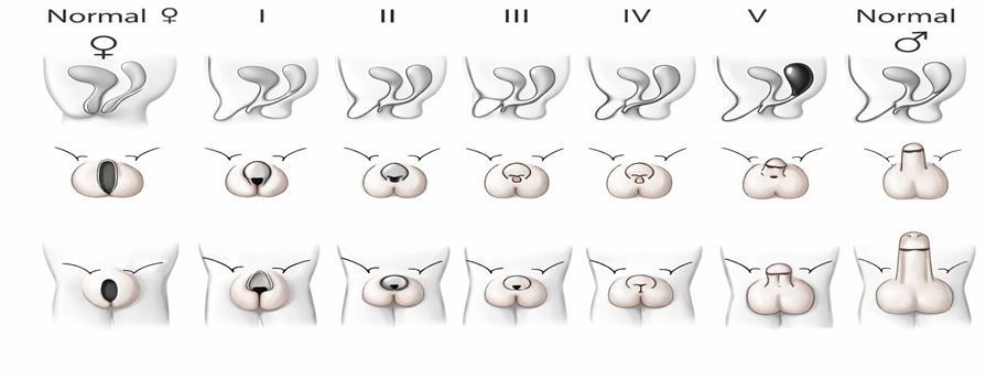
Figure 1. Prader classification of virilization of external genitalia
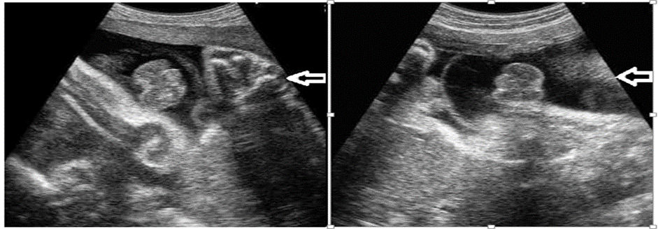
Figure 2. Ultrasonographic appearance of external genitalia
Cytogenetic–Clinical Correlation
In all cases, the cytogenetic diagnosis was concordant with the FISH findings.
- Chromosomal males predominantly presented with micropenis, bifid scrotum, hypospadias, or varying degrees of undervirilization.
- Chromosomal females mainly presented with clitoromegaly or labioscrotal fusion.
The combined karyotype and FISH data were essential for definitive sex assignment and guided appropriate clinical decision-making for each child.
Clinical photograph of a children presenting with ambiguous external genitalia, demonstrating an undervirilized phenotype. The child shows a slender body habitus with absence of clearly differentiated male or female external genital structures, suggestive of a disorder of sex development (DSD). Facial features are unremarkable, and no gross dysmorphic features are evident. The genital ambiguity prompted detailed cytogenetic and molecular evaluation. The eyes are masked to maintain patient confidentiality. Chromosomal sex was subsequently confirmed using GTG-banded karyotyping and fluorescence in situ hybridization (FISH) with sex chromosome–specific probes. (Figure.3,4,5,6a,6b).
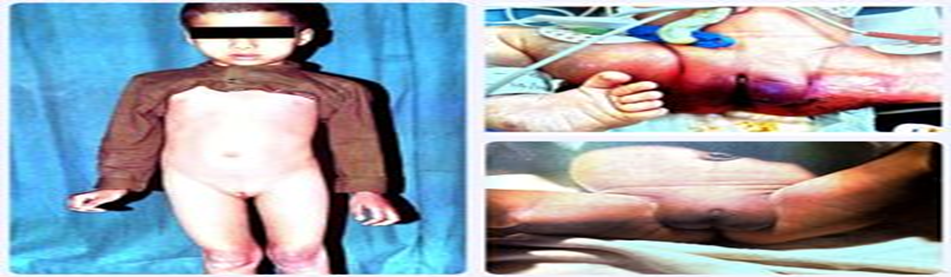
Figure 3. Clinical photograph of a children with ambiguous genitalia
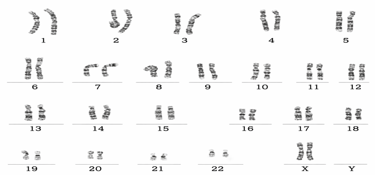
Figure 4 : The sex chromosomes consist of Two X chromosomes, confirming a 46,XX karyotype.
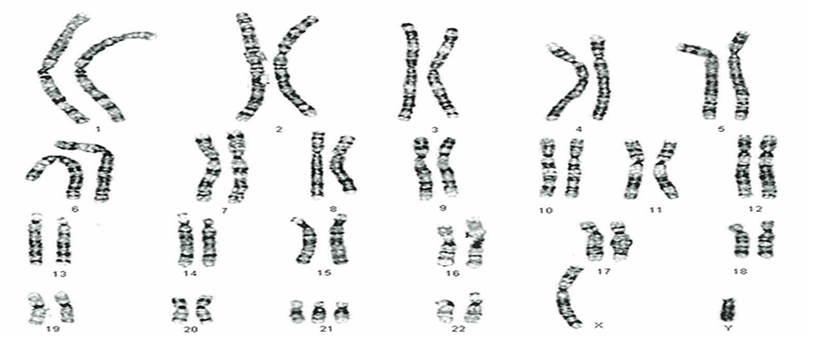
Figure 5: The sex chromosomes consist of one X chromosome and one Y chromosome, confirming a 46,XY karyotype.
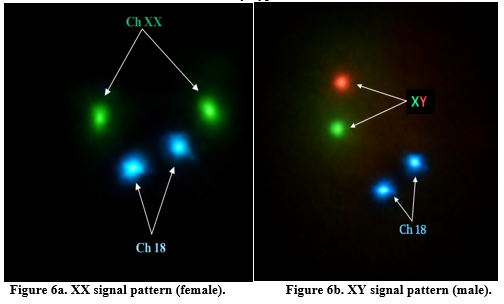
Among the 12 children with ambiguous genitalia, cytogenetic analysis confirmed 4 cases as genetically female (46,XX), all of whom demonstrated varying degrees of virilization corresponding to Prader grades II–V. The remaining 8 cases were genetically male (46,XY) and predominantly presented with hypospadias or undervirilized external genitalia. In all cases, FISH findings were concordant with conventional karyotyping (Table.4).
|
|||||
|
|||||
|
|||||
|
|||||
|
|||||
|
|||||
|
|||||
|
|||||
|
|||||
|
|||||
|
|||||
|
|||||
|
Table. 4 Case-wise correlation of Prader grading with cytogenetic and FISH findings in children with ambiguous genitalia (n = 12)
DISCUSSION:
Ambiguous genitalia represents one of the most challenging presentations within the spectrum of disorders of sex development (DSD) and requires prompt and systematic evaluation to establish chromosomal sex, determine underlying etiology, and guide appropriate sex assignment and management. In the present study conducted at a tertiary care center in North Karnataka, an integrated approach involving clinical examination, Prader grading, biochemical profiling, radiological evaluation, and cytogenetic analysis was utilized to characterize 12 pediatric patients presenting with ambiguous genitalia. The findings emphasize the importance of combining conventional cytogenetic techniques with molecular confirmation methods such as fluorescence in situ hybridization (FISH) for accurate diagnosis.
In the current cohort, 46,XY karyotype was observed in 66.6% of cases, whereas 46,XX karyotype accounted for 33.3% of cases. Similar distributions have been reported in several studies where disorders affecting androgen synthesis, action, or gonadal development contribute significantly to ambiguous genitalia in genetically male individuals. Previous studies have demonstrated that 46,XY DSD often presents with hypospadias, micropenis, or undervirilization, which were also the predominant clinical manifestations observed in our cohort.[15] These findings highlight the heterogeneity of etiologies underlying 46,XY DSD, including androgen insensitivity syndrome, defects in testosterone biosynthesis, and gonadal dysgenesis.
All four 46,XX cases in the present study demonstrated Prader grades II–V virilization, along with elevated 17-hydroxyprogesterone levels and increased adrenal androgen markers, strongly suggestive of congenital adrenal hyperplasia (CAH). CAH due to 21-hydroxylase deficiency is recognized as the most common cause of virilized 46,XX DSD worldwide, accounting for nearly 90–95% of cases.[16] The biochemical profiles observed in these patients, including markedly elevated 17-OHP levels with low cortisol and elevated ACTH, were consistent with classical CAH. Early recognition of this condition is crucial, as affected infants may develop life-threatening adrenal crisis if untreated.
Radiological evaluation using pelvic ultrasonography proved to be a valuable adjunct in the diagnostic workflow, particularly for identification of internal reproductive structures. In the present study, all genetically female patients demonstrated Müllerian structures, including uterus and ovaries, whereas these structures were absent in genetically male patients. These imaging findings correlated well with cytogenetic results and clinical phenotype, reinforcing the importance of ultrasound as a non-invasive and accessible diagnostic tool in resource-limited settings. Similar observations have been reported in previous studies where ultrasonography played a key role in the early identification of internal genital anatomy in children with DSD.[17]
An important observation in this study was the high prevalence of parental consanguinity (83.3%) among affected children. Consanguinity is known to increase the likelihood of autosomal recessive genetic disorders, including enzyme deficiencies involved in steroidogenesis and other metabolic pathways associated with DSD. Several studies conducted in South Asian and Middle Eastern populations have reported a similarly high frequency of consanguinity among families with children affected by DSD.[13] This finding highlights the need for genetic counseling and community awareness programs in regions where consanguineous marriages are common.
Cytogenetic analysis remains a cornerstone in the diagnostic evaluation of ambiguous genitalia. In the present study, conventional GTG-banded karyotyping successfully established chromosomal sex in all cases, and the results were fully concordant with FISH findings using DXZ1, DYZ3, and SRY probes. The use of FISH was particularly valuable in confirming the presence or absence of Y-chromosomal material and in cases where metaphase quality was suboptimal. Previous studies have demonstrated that the integration of cytogenetics with targeted molecular techniques significantly improves diagnostic accuracy in DSD evaluation.[14]
The present study also highlights the utility of Prader classification in correlating clinical phenotype with underlying endocrine abnormalities. Higher Prader grades in genetically female patients corresponded with more pronounced biochemical evidence of androgen excess. Similarly, undervirilized phenotypes in genetically male patients correlated with low testosterone levels or abnormalities in gonadotropin secretion. These correlations underscore the importance of integrating clinical scoring systems with laboratory investigations to improve diagnostic precision.
Despite the small sample size, the findings of this study provide valuable insights into the clinical and cytogenetic spectrum of ambiguous genitalia in North Karnataka. Limited access to advanced molecular diagnostics remains a challenge in many resource-constrained settings. However, the results demonstrate that conventional cytogenetics supplemented with targeted FISH analysis remains a reliable and cost-effective strategy for establishing chromosomal sex and guiding further diagnostic evaluation.
Future studies involving larger cohorts and incorporation of molecular genetic testing such as next-generation sequencing (NGS) may further improve diagnostic yield and help identify novel genetic variants responsible for DSD in the Indian population.
CONCLUSION:
This study demonstrates that a multidisciplinary diagnostic approach integrating clinical, biochemical, radiological, and cytogenetic findings is essential for accurate diagnosis of ambiguous genitalia.
Conventional karyotyping, supported by FISH, remains a dependable and cost-effective diagnostic strategy, particularly in resource-limited settings. Early diagnosis, along with appropriate counseling and management, is crucial for improving long-term outcomes.
REFERENCES:
1. Lee PA, Houk CP, Ahmed SF, Hughes IA. Consensus statement on management of intersex disorders. Pediatrics. 2006;118(2):e488–e500.
2. Hughes IA, Houk C, Ahmed SF, Lee PA. Disorders of sex development: an update. Lancet. 2006;366(9480):69–78.
3. Speiser PW, White PC. Congenital adrenal hyperplasia. N Engl J Med. 2003;349(8):776–788.
4. Ahmed SF, Achermann JC, Arlt W, Balen A, Conway G, Edwards ZL, et al. UK guidance on the initial evaluation of an infant or an adolescent with a suspected disorder of sex development. Arch Dis Child. 2016;101(7):638–645.
5. Witchel SF. Disorders of sex development. Best Pract Res Clin Obstet Gynaecol. 2018;48:90–102.
6. Thangaraj K, Chaubey G, Kivisild T. Genetic landscape of the people of India: a review. Curr Sci. 2010;98(3):349–356.
7. Warne GL, Raza J. Disorders of sex development (DSDs), their presentation and management in different cultures. Endocr Dev. 2008;13:198–208.
8. Ammini AC, Gupta R. Ambiguous genitalia in India: diagnostic and management issues. Indian J Pediatr. 2006;73(8):675–681.
9. Sax L. How common is intersex? A response to Anne Fausto-Sterling. J Sex Res. 2002;39(3):174–178.
10. Joshi RR, Deshpande SS. Ambiguous genitalia—An overview. Indian J Pediatr. 2010;77(2):163–170.
11. White PC. Neonatal screening for congenital adrenal hyperplasia. Nat Rev Endocrinol. 2009;5(9):490–498.
12. Achermann JC, Hughes IA. Disorders of sex development. In: Melmed S, Polonsky KS, Larsen PR, Kronenberg HM, editors. Williams Textbook of Endocrinology. 14th ed. Philadelphia: Elsevier; 2020.
13. Raza J, Mazen I. Disorders of sex development in the Middle East and South Asia. Semin Reprod Med. 2012;30(5):389–395.
14. Wolff DJ, Van Dyke DL, Powell CM. Laboratory guideline for constitutional cytogenomic analysis using fluorescence in situ hybridization. Genet Med. 2018;20(6):655–663.
15. Ahmed SF, Bashamboo A, Lucas-Herald A, McElreavey K. Understanding the genetic aetiology in disorders of sex development. Nat Rev Endocrinol. 2016;12(7):433–444.
16. Speiser PW, Arlt W, Auchus RJ, Baskin LS, Conway GS, Merke DP, et al. Congenital adrenal hyperplasia due to steroid 21-hydroxylase deficiency: an Endocrine Society clinical practice guideline. J Clin Endocrinol Metab. 2018;103(11):4043–4088.
17. Ogilvy-Stuart AL, Brain CE. Early assessment of ambiguous genitalia. Arch Dis Child. 2004;89(5):401–407.
18. Bashamboo A, McElreavey K. Mechanisms of sex determination in humans: insights from disorders of sex development. Sex Dev. 2015;9(1):3–7.