GASTRIC DUPLICATION CYST WITH PANCREATIC HETEROTOPIA PRESENTING AS CHEST WALL SWELLING WITH FISTULOUS COMMUNICATION: A RARE CASE REPORT
- Mayank Mohit , Senior Resident, Department of General Surgery, B. J. Medical College, Ahmedabad, Gujarat, India
- Nina M. Shah , Professor (Additional) & Head of Unit Department, Department of General Surgery, B. J. Medical College, Ahmedabad, Gujarat, India.
- Hardik B. Bhatt , Associate Professor, Department of General Surgery, B. J. Medical College, Ahmedabad, Gujarat, India.
- Gauravkumar Govindbhai Patel , Senior Resident, Department of General Surgery, B. J. Medical College, Ahmedabad, Gujarat, India.
Article Information:
Abstract:
Background: Gastrointestinal duplication cysts are uncommon congenital anomalies, of which gastric duplication cysts constitute only a small proportion. These lesions are typically identified in early childhood but may occasionally present later in life. We describe the case of a 16-year-old female who presented with a progressively enlarging cystic swelling over the right chest wall. Radiological evaluation suggested a gastric duplication cyst having ectopic pancreatic tissue and a fistulous communication with a subcutaneous collection. Surgical exploration confirmed the diagnosis. Complete excision of the cyst along with the fistulous tract along with splenectomy was performed.. The postoperative recovery was uneventful. This case underscores an unusual clinical presentation and highlights the diagnostic challenges associated with gastric duplication cysts.
Keywords:
Article :
INTRODUCTION:
Gastrointestinal duplication cysts are rare congenital anomalies that may arise anywhere along the gastrointestinal tract. Gastric duplication cysts account for approximately 4% of all duplication cysts and are most often diagnosed within the first two years of life [1,2]. The clinical presentation depends on factors such as size, location, and the presence of heterotopic mucosa, which often makes diagnosis challenging [2, 3]. Patients may present with abdominal pain, vomiting or a palpable mass while some lesions are detected incidentally [3,4]. In addition, these cysts can resemble other intra-abdominal conditions, including pancreatic pseudocysts, biliary or hepatic cysts, splenic cystic lesions. and gastrointestinal stromal tumors, thereby further complicating diagnosis [4, 5]. Given their rarity and variable presentation, a high index of clinical suspicion, supported by appropriate imaging, is essential for accurate diagnosis and management [5, 6].
CASE PRESENTATION:
A 16-year-old female presented with a progressively enlarging swelling over the right chest wall for duration of three months. The swelling measured approximately 20 × 10 cm and extended from the clavicle to the epigastric region. On examination, it was cystic in consistency and fluctuant (Figure 1).
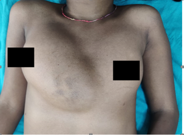
Figure 1: Clinical photograph showing a large cystic swelling over the right chest wall extending from the clavicle to the epigastric region.
Course of pastevents
● Age 6 years (Dec 2013):
o Presented with abdominal pain (2 months) → managed conservatively
o USG suggested aortic aneurysm → conservative treatment
● Jan 2014:
o CECT abdomen:
▪ Multiple necrotic masses in hepato-gastric region
▪ Mesenteric lymphadenopathy
▪ Terminal ileum thickening
o Impression: Koch’s abdomen (tuberculosis)
o Patient received AKT for 2 months, then stopped after symptom relief
● May 2014:
o CECT: Organized epigastric collection + sternum, suggestive of osteomyelitis
o Diagnostic laparoscopy:
▪ Drainage + biopsy
▪ Findings: Old organized hematoma, high amylase (4038 IU)
o Histopathology: Mild inflammation, no malignancy
● Nov 2021:
o Recurrence of swelling
o CECT: Multiloculated collection with hepatosplenomegaly with cardiomegaly
o Swelling resolved spontaneously in 10 days → asymptomatic for 3 years
● May 2024:
o Large right chest wall swelling (18×8 cm) extending to epigastrium
o Fluid analysis: very high amylase (59,714 IU/L)
Clinical Examination
Per abdominal examination did not reveal any palpable epigastric mass or ascites; however, hepato-splenomegaly was noted.
Laboratory and Radiological Findings
Laboratory investigations revealed hemoglobin of 5.4 g/dL, WBC count of 3090 cells/mm³, platelet count of 42,000 cells/mm³, INR of 1.5, creatinine of 0.5 mg/dL, SGPT of 6 IU/L, total bilirubin of 0.8 mg/dL, serum amylase of 178 IU/L, and serum lipase of 425 IU/L. Aspiration of the swelling yielded brownish turbid fluid with markedly elevated amylase (5258 IU/L) and lipase (1448 IU/L) levels.
Ultrasonography demonstrated a large collection over the right chest wall suggestive of a pseudocyst, along with a cystic lesion anterior to the stomach consistent with a duplication cyst. Contrast-enhanced computed tomography (CECT) of the abdomen and pelvis suggested a benign cystic lesion, likely representing an enteric (gastric) duplication cyst with ectopic pancreatic tissue, communicating with a large subcutaneous collection (Figure 2). Features suggestive of portal hypertension with moderate hepato-splenomegaly were also noted. MRCP was done to rule out communication with pancreatic duct.
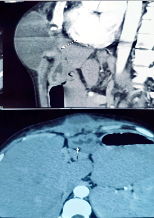
Figure 2: Contrast-enhanced computed tomography of the abdomen showing a cystic lesion suggestive of a gastric duplication cyst with communication to a subcutaneous collection.
Intraoperative Findings
Surgical exploration through an upper midline incision revealed a 5 × 3 cm cyst located near the lesser curvature of the stomach, extending up to the gastroesophageal junction. The cyst was found to communicate with the chest wall through a fistulous tract and had no communication with the gastric lumen (Figure 3). Complete excision of the cyst and fistulous tract was performed. Curettage of the sequestrum was carried out, and the raw area healed over a period of three months. A splenectomy was also performed for hypersplenism secondary to portal hypertension. The postoperative course was uneventful (Figure 4).
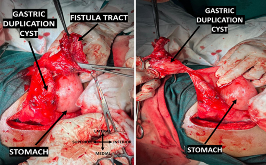
Figure 3: Intraoperative image showing a gastric duplication cyst near the lesser curvature of the stomach with a fistulous tract communicating with the chest wall.
Histopathology Findings(Post surgery)
● Biopsy suggestive of gastric duplication cyst with including gut-musculature with heterotopic pancreatic tissue with fistulous tract showing fibrocollagenous tissue, inflammatory infiltrate, hemorrhage and vascular proliferation.
● Changes of congestive splenomegaly were noted in specimen labelled ‘spleen’.
Final Diagnosis: Gastric duplication cyst with ectopic pancreatic tissue and fistulous communication to chest wall.
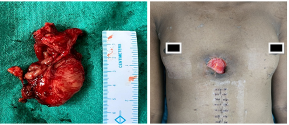
Figure 4: Post-operative images
DISCUSSION:
Gastric duplication cysts are uncommon congenital lesions that can present with a wide range of clinical manifestations. Although most cases are identified during early childhood, some may remain undetected until adolescence or adulthood [7]. The presence of heterotopic pancreatic tissue within these cysts may lead to complications such as pseudocyst formation and fistulous communication [8]. In the present case, the patient presented with an unusual chest wall swelling, which initially raised the possibility of conditions such as a pancreatic pseudocyst or other cystic lesions. Radiological findings, followed by surgical exploration, established the diagnosis [9]. Because of their rarity and diverse presentation, gastric duplication cysts can mimic several other intra-abdominal cystic conditions, making diagnosis challenging [10]. Surgical excision remains the definitive treatment and is generally associated with favorable outcomes [7].
CONCLUSION:
Gastric duplication cyst is a rare congenital condition that may present in an atypical manner, particularly in adolescence. Its resemblance to other cystic lesions can lead to diagnostic uncertainty. Early recognition with appropriate imaging and timely surgical intervention is essential for optimal outcomes.
Conflict of Interest: None declared
Funding: None
Declaration of patient consent: The authors certify that they have obtained appropriate patient consent.
Author Contributions: All authors contributed equally and approved the final manuscript
REFERENCES:
1. Liu R, Adler DG. Duplication cysts: Diagnosis, management, and the role of endoscopic ultrasound. Endosc Ultrasound. 2014; 3(3):152–160.
2. Gjeorgjievski M, Manickam P, Ghaith G, Cappell MS. Safety and efficacy of endoscopic ultrasound in duplication cysts. World J Gastroenterol. 2016; 22(35):8031–8041.
3. Yang DM, Kim HC, Choi SI, Lim SJ. Sonographic diagnosis of gastric duplication cyst communicating with the gastric lumen. J Clin Ultrasound. 2011 Nov-Dec; 39(9):550-2. doi: 10.1002/jcu.20828. Epub 2011 May 2. PMID: 21538375.
4. Singh JP, Rajdeo H, Bhuta K, Savino JA. Gastric duplication cyst: Two case reports and review of the literature. Case Rep Surg. 2013; 2013:605059.
5. Doepker MP, Ahmad SA. Gastric duplication cyst: a rare entity. J Surg Case Rep. 2016 May 5; 2016(5):rjw073. doi: 10.1093/jscr/rjw073. PMID: 27150283.
6. Kayastha K, Sheikh A. Gastric duplication cyst presenting as acute abdomen: a case report. APSP J Case Rep. 2010 Jan; 1(1):6. Epub 2010 Aug 14. PMID: 22953249
7. Li X, Zhang J, et al. Diagnosis and management of gastric duplication cysts in adults. Medicine (Baltimore). 2018; 97(51):e13603.
8. Jongmee Lee, Cheol Min Park, Kyeong Ah Kim, Chang Hee Lee, Jae Woong Choi, Bong Kyung Shin, Soon Jin Lee, Dongil Choi, Kee-Taek Jang, Cystic Lesions of the Gastrointestinal Tract: Multimodality Imaging with Pathologic Correlations, Korean Journal of Radiology 2010; 11(4): 457-468.
9. Seng Pei Khaw, Antonio T S Tan, Martin T W Kueh, Alexander Z Y Lim, Pei Pei Lee, Aizat Aziz, Wei Keat Ooi, Gastric duplication cyst in an adult: a rare case presentation and literature review, Journal of Surgical Case Reports, Volume 2025, Issue 8, August 2025, rjaf651, https://doi.org/10.1093/jscr/rjaf651
10. Li Y, Li C, Wu H, Wang Q, Gao ZD, Yang XD, Jiang KW, Ye YJ. Clinical features of gastric duplications: evidence from primary case reports and published data. Orphanet J Rare Dis. 2021 Aug 19; 16(1):368. doi: 10.1186/s13023-021-01992-1. PMID: 34412674; PMCID: PMC8377950.