Investigation of Klebsiella pneumoniae outbreak in a neonatal intensive care unit in a tertiary care hospital in North Delhi.
- Shipra Gupta , Senior Resident, Department of Microbiology, NDMC Medical College and Hindu Rao Hospital Delhi, India.
- Manoj Kumar , Professor, Department of Microbiology, Institution Maharaja Agrasen kedarnath gupta medical college nuna Majra Bahadurgarh Haryana India.
- Sanjay Jain. , Sanjay Jain, CMO, SAG, Department of Microbiology, NDMC Medical College and Hindu Rao Hospital Delhi, India.
Article Information:
Abstract:
Background: Objective: Klebsiella pneumoniae commonly causes neonatal sepsis and can quickly disseminate within Neonatal Intensive Care Units (NICUs) via contaminated surfaces or through healthcare workers’ (HCWs) hands. The objective was to investigate an outbreak of Klebsiella pneumoniae among neonates in the NICU of a tertiary care hospital in North Delhi, identify potential sources, and implement infection control measures to reduce associated mortality. Material and Methods: This retrospective study was conducted from July to September 2022. Active surveillance and comprehensive environmental sampling were undertaken in the NICU and Labour Room. Clinical isolates of Klebsiella pneumoniae from neonates were analyzed using standard microbiological methods and antimicrobial susceptibility testing (AST). Random Amplified Polymorphic Deoxyribonucleic Acid Polymerase Chain Reaction (RAPD−PCR) analysis was performed to determine genetic relatedness among the isolates. Breaches in infection control were identified using structured audits. Results: A total of 24 neonates with Klebsiella pneumoniae bloodstream infections (BSIs) were documented. Among the neonates, prematurity (66.7%) and low birth weight (70.8%) were frequently observed. Additionally, 79.2% had intravenous catheters placed, and 58.3% required mechanical ventilation. Environmental sampling yielded Klebsiella pneumoniae, coagulase-negative staphylococci (CoNS), Acinetobacter, and Methicillin Resistant Staphylococcus aureus (MRSA). RAPD-PCR revealed distinct Klebsiella pneumoniae strain patterns, including two environmental isolates indistinguishable from clinical strains. Infection control lapses were addressed. Conclusions: Strengthening infection control measures—particularly meticulous hand hygiene, thorough disinfection, and prudent antibiotic usage, proved crucial in controlling the outbreak and reducing neonatal morbidity and mortality. Ongoing surveillance and staff training remain vital for preventing future Klebsiella. pneumoniae outbreaks in resource-limited NICU settings.
Keywords:
Article :
INTRODUCTION:
Neonatal intensive care units (NICUs) are at elevated risk for healthcare-associated infections (HAIs), which can severely impact newborns. Klebsiella pneumoniae, a Gram-negative bacillus, is well known for causing nosocomial infections, such as pneumonia and bloodstream infections (BSIs) in this vulnerable population (1). Spread may occur during delivery or through contact with nursery personnel, equipment, or contaminated surfaces."
Klebsiella pneumoniae is frequently implicated in NICU outbreaks, partly because it can maintain antibiotic resistance with minimal impact on its viability (2). In low- and lower-middle-income settings, where resources are constrained, lapses in care protocols or infection prevention often precipitate neonatal sepsis (3).
Because Klebsiella pneumoniae survives within hospital environments, it can colonize skin and multiple organ systems. Preterm and low-birth-weight neonates are particularly at risk due to their underdeveloped immunity and the frequent need for invasive interventions (4).
Between July and September 2022, multiple isolates of Klebsiella pneumoniae were recovered from the blood cultures of neonates in our hospital’s NICU, raising concern for a potential outbreak. Therefore, a targeted investigation was initiated to identify the sources and routes of transmission, and surveillance samples were collected from various NICU sites.
The aim of this study was to investigate and characterise NICU outbreak of Klebsiella pneumoniae, identify potential sources and transmission routes, and implement infection control measures to minimize neonatal sepsis–related illness and death.
MATERIALS AND METHODS:
Study design, settings and participants:
This hospital-based retrospective observational study was conducted at the Departments of Microbiology and Paediatrics (Neonatal Intensive Care Unit, NICU) at NDMC Medical College and Hindu Rao Hospital, Delhi. The NICU is a 24-bedded unit (eight cots on the right wing, seven cots on the left wing, and five cots in the step-down area). It is staffed by 17 paediatricians, 2 nurses, 3 (multi-task staff) MTS workers, and 3 aaya (ward helper) workers. The nurse-to-doctor ratio is 2:17, and the nurse-to-patient ratio is 2:24.
Since July 2022, multiple cases of Klebsiella pneumoniae bloodstream infections were observed among neonates admitted to this NICU. Active surveillance of the NICU and Labour room was conducted from July to September 2022. A probable case was defined as Klebsiella species isolated from the blood of an infant (<28 days of age) admitted to the NICU between July 1 and September 30, 2022. A confirmed case was an isolate identified specifically as Klebsiella pneumoniae through biochemical analysis.
A standardised proforma was used to collect neonates’ clinical and demographic data, including gestational age, birth weight, Appearance Pulse Grimace Activity Respiration (APGAR) scores, mode of delivery, and use of invasive devices (e.g., indwelling catheters), and patient outcomes. Ethical approval for this research was obtained from the Institutional Ethics Committee (IEC/NDM/2023/203).
For statistical analysis data were compiled in Microsoft Excel and analysed using SPSS version (V.28). Categorical variables (e.g. sex, gestation type, delivery mode) were compared between outcome groups (survived vs. expired) using chi-square or Fisher’s exact tests, as appropriate. Continuous variables (e.g., birth weight) were reported as mean ± SD. A p-value of < 0.05 was considered statistically significant.
Environmental sampling and microbiological investigations
To identify potential oversights in infection control, the Hospital Infection Control Team performed daily inspections of the NICU. Healthcare workers (HCWs) were interviewed regarding any procedural changes that might have contributed to the outbreak.
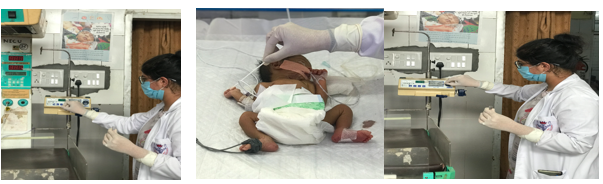
Extensive environmental sampling was conducted using sterile cotton swabs pre-moistened with thioglycolate broth (HiMedia Laboratories Pvt. Ltd., Mumbai, India). These swabs were rolled over targeted surfaces (neonates’ intravenous line insertion sites, umbilical venous catheters (UVCs), hands and feet of neonates, cots, incubators, ventilators, Ambu bags, weighing machines, refrigerators, phototherapy units, laryngoscopes, continuous positive airway pressure (CPAP) machines, pulse oximeters, warmers, feeding chairs, side trolleys, suction catheters, tables, medicine trays, baby mattresses, infusion pumps, log registers, etc.). Further samples were taken from the hands and mobile phones of HCWs, stethoscopes, and intravenous fluids (5–10 per cent dextrose or 0.9 per cent sodium chloride).
All specimens were inoculated onto Blood agar and MacConkey agar and incubated aerobically at 37°C for 18-24 hours. Culture plates were observed for microbial growth and any isolated colonies were subjected to standard biochemical tests. Antimicrobial susceptibility testing (AST) was performed using the Kirby-Bauer disk-diffusion method in accordance with Clinical and Laboratory Standards Institute (CLSI) guidelines (2022).
Random Amplified Polymorphic Deoxyribonucleic Acid–Polymerase Chain Reaction (RAPD-PCR) for molecular typing
Genomic DNA was extracted from Klebsiella pneumoniae isolates using Qiagen DNA extraction kits (Qiagen GmbH, Hilden, Germany) as per the manufacturer’s protocol. RAPD-PCR was performed to assess the genetic relatedness among isolates.
• Reaction mixture (25 µL): 18.95 µL distilled water, 1 µL primer, 0.15 µL Taq DNA polymerase, 0.4 µL deoxynucleotide triphosphates (dNTPs), 2.5 µL buffer, 2 µL DNA template.
• Thermal cycling program: Initial denaturation at 94°C for 2 minutes, followed by 40 cycles of 94°C for 1 minute, 36°C for 2 minutes, and 72°C for 2 minutes, then a final extension at 72°C for 7 minutes.
• Analysis: Amplified PCR products were electrophoresed on a 1.2% agarose gel in Tris-borate-EDTA (TBE) buffer and visualized by ethidium bromide staining. Dendrograms were constructed using a similarity threshold of ≥85% to define identical patterns.
RESULTS:
During the three-month period (July–September 2022), a total of 24 neonates presented with Klebsiella pneumoniae bloodstream infection. The baseline characteristics of these neonates are summarized in Table 1.
Table 1: Outbreak overview & patient characteristics
|
Characteristic
|
Measure |
Result |
|
Sex |
Male Female |
17 (70.8%) 7 (29.2%) |
|
Delivery mode |
Normal vaginal delivery (NVD) Lower segment Caesarean section (LSCS) |
13 (54.2%)
11 (45.8%) |
|
Gestation |
Preterm |
16(66.7%) |
|
Birth Weight (kg)
|
Mean ± standard deviation (range) |
2.14 ± 0.74 (0.74 – 3.75) |
|
Low birth weight (≤2.5 kg) |
Proportion of neonates |
17 (70.8%) |
|
Very low birth weight (≤1.5 kg) |
Proportion of neonates |
6 (25.0%) |
|
APGAR Score (at 1, 5, 10 min) |
Range |
3 – 9 |
|
Invasive Procedures |
UVC IV Catheter CPAP Mechanical Ventilation Endotracheal Tube Urinary Catheter |
3 (12.5%) 20 (83.3%) 2 (8.3% 14 (58.3%) 6 (25%) 1 (4.2%) |
|
Outcome
|
Expired Survived |
7 (29.2%) 17 (70.8%) |
The index case had remained in the NICU for several days before additional cases emerged. Low birth weight (70.8 per cent) and prematurity (66.7 per cent) were the most common risk factors, with birth weight ranging from 0.74 to 3.75 kg and gestational age from 29 to 40 weeks. APGAR scores varied from as low as 3-5-7 to as high as 8-9-9.
No statistically significant association was found between outcome (survival vs. expired) and factors such as sex, gestational age, delivery mode, or birth weight category (p ≥ 0.05, by chi-square or Fisher’s exact test).
Common clinical features included respiratory distress, hypoglycaemia, thrombocytopenia, aspiration pneumonia, and other signs of sepsis (e.g., poor feeding, hypothermia). Seven neonates (29.2%) died due to severe infection and its complications; the others survived.
Environmental sampling and microbiological findings
Extensive swab sampling of NICU and labour room surfaces detected multiple organisms, including Klebsiella pneumoniae, coagulase-negative staphylococci, Acinetobacter spp., and methicillin-resistant Staphylococcus aureus. A summary of the environmental isolates is shown in Table 2, Figure 1,2.
Table 2. Selected environmental/healthcare worker samples and organisms isolated
|
Sample source |
Organism isolated |
|
Hand swabs of HCWs |
Coagulase-negative staphylococci |
|
Mobile phone of an HCW |
Acinetobacter spp. |
|
Neonate’s skin swab |
Acinetobacter spp. |
|
Ambu bag |
Methicillin-resistant Staphylococcus aureus |
|
Baby mattress |
Klebsiella pneumoniae |
|
Infusion pump |
Acinetobacter spp. |
|
Another Neonate’s skin swab |
Klebsiella pneumoniae |
A subset (approximately 8.3 per cent) of these Klebsiella pneumoniae environmental isolates displayed phenotypic similarity (antibiogram) with the clinical strains.
Antimicrobial susceptibility
All Klebsiella pneumoniae isolates underwent antimicrobial susceptibility testing using the disk-diffusion (Kirby-Bauer) method according to CLSI guidelines (2022). Although most isolates were susceptible to amikacin (AK), gentamicin (GEN), and ciprofloxacin (CIP), sporadic resistance was observed against cephalosporins (e.g., cefotaxime (CTX), ceftriaxone (CTR)). Two environmental isolates mirrored the resistance patterns found in clinical isolates cefotaxime/ceftriaxone resistance profiles of clinical isolates.
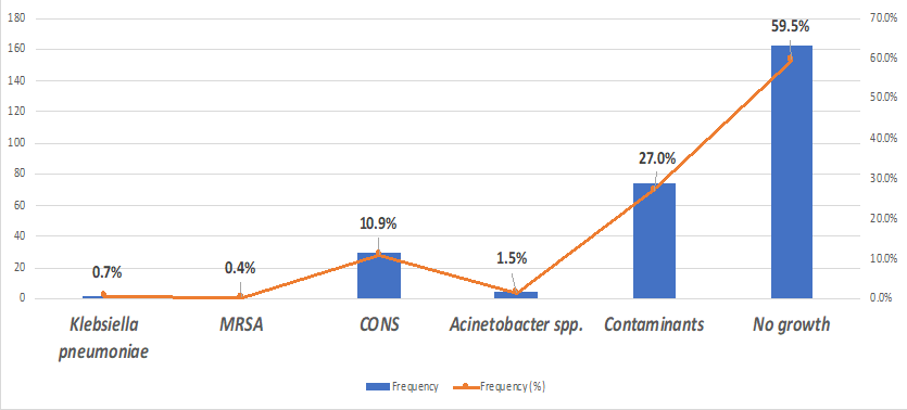
Figure 1: Distribution of organisms isolated from environmental/healthcare workers samples
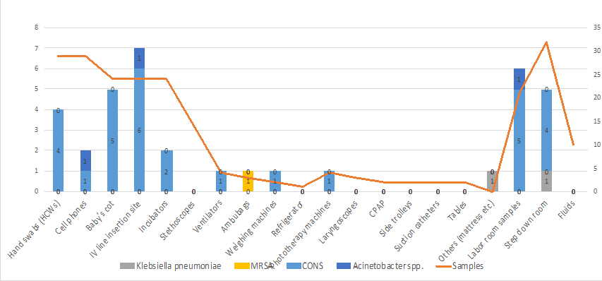
Figure 2: Distribution of isolates based on source of sample collection
Molecular typing: RAPD-PCR
RAPD-PCR analysis of a subset of 14 Klebsiella pneumoniae isolates (12 clinical and 2 environmental) revealed nine distinct patterns (D1–D9), suggesting polyclonal spread. However, two environmental isolates (one from a baby mattress, one from a neonate’s skin) were genetically indistinguishable from specific clinical isolates.
Similarity threshold: ≥85% to define isolates as the same clone/ pattern and highest homology (>96.3%) was observed between isolate 5 and 11.
A representative dendrogram illustrated in Figure 3, 4 shows clustering of certain clinical isolates with environmental ones, confirming a possible transmission link within the NICU environment.
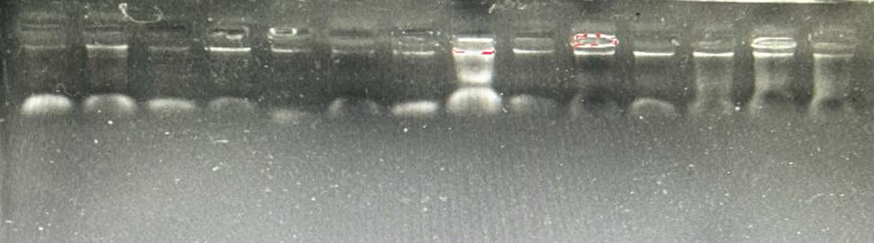
Figure 2 - RAPD profiles of Klebsiella pneumoniae isolates amplified by primer AP4. From left to right, respectively K. pneumoniae isolates number 537, 538, 541, 543, 544, 546, 548, 549, 552, 553, 555, 557, 558, 561,563 and 573. M: DNA molecular weight marker (1Kb DNA ladder;Fermentase, Germany).
RAPD profiles of Klebsiella pneumoniae isolates amplified by primer AP4. From left to right, respectively K. pneumoniae isolates number 537, 538, 541, 543, 544, 546, 548, 549, 552, 553, 555, 557, 558, 561, RAPD profiles of Klebsiella pneumoniae isolates amplified by primer AP4. From left to right, respectively K. pneumoniae isolates number 537, 538, 541, 543, 544, 546, 548, 549, 552, 553, 555, 557, 558, 561.
Figure 3: RAPD-PCR agarose gel. Representative electrophoresis gel image of RAPD-PCR products for selected Klebsiella pneumoniae isolates (lanes 1–12). M: 1 kb DNA ladder. Distinct band patterns highlight the polyclonal nature of the outbreak.

Figure 2 - RAPD profiles of Klebsiella pneumoniae isolates amplified by primer AP4. From left to right, respectively K. pneumoniae isolates number 537, 538, 541, 543, 544, 546, 548, 549, 552, 553, 555, 557, 558, 561, 563 and 573. M: DNA molecular weight marker (1Kb DNA ladder; Fermentase, Germany).According to the dendrograms, Random Amplified Polymorphic DNA (RAPD) analyses revealed and distinct patterns of K. pneumoniae isolates with similarity >80%, respectively
Figure 4: A dendrogram showing nine distinct RAPD patterns (D1–D9), (Figure 1). The isolates were considered as the same pattern if the level of similarity was ≥85%. Between isolate 5 and 11, the level of similarity was >96.3%.
DISCUSSION:
This outbreak investigation demonstrated that 24 neonates in our tertiary care NICU developed Klebsiella pneumoniae bloodstream infections (BSIs) over a three-month period (July–September 2022). Similar to the previous studies, Klebsiella pneumoniae is among the most frequently reported pathogens in neonatal units and is particularly threatening to preterm neonates, who constituted nearly two-thirds (66.7%) of our cases. These neonates tend to have underdeveloped immune systems and often require invasive supportive measures (e.g., ventilators, intravenous catheters), both of which heighten infection risk.
Environmental sampling revealed Klebsiella pneumoniae, Acinetobacter spp., and MRSA on multiple high-touch surfaces such as baby mattresses, Ambu bags, and infusers. Our RAPD-PCR analysis confirmed that two environmental isolates were genetically indistinguishable from clinical isolates, suggesting cross-transmission via contaminated equipment or healthcare workers’ (HCWs) hands. Similar outbreaks have been reported, underscoring that surfaces and devices can serve as key reservoirs of infection (5,6). Molecular approaches—such as RAPD-PCR and pulsed-field gel electrophoresis (PFGE)—are widely used to confirm genetic relatedness in hospital outbreaks (5,6).
Although most isolates in our setting were susceptible to aminoglycosides (amikacin, gentamicin) and fluoroquinolones (ciprofloxacin), sporadic cephalosporin resistance raises concern about evolving multidrug resistance. Such resistance underscores the necessity of prudent antibiotic use and routine surveillance, consistent with World Health Organization and Centres for Disease Control and Prevention (CDC) guidelines (7,8).
Our investigation identified breaches in infection control (e.g., inadequate bed spacing, inconsistent use of PPE) including suboptimal hand hygiene, inadequate bed spacing, and inconsistent personal protective equipment (PPE) usage—which were rectified through staff education, reorganisation of cot placement, and improved aseptic protocols. Similar infection prevention bundles have effectively reduced NICU sepsis rates in other settings (9). Furthermore, environmental disinfection and co-horting infected neonates proved pivotal in interrupting transmission cycles—an approach also supported by Li et al. (10) and Crispim et al. (11), who emphasize that consistent decontamination of high-touch surfaces limits the spread of multidrug-resistant organisms.
The corrective steps taken—reconfiguring bed spacing, reinforcing PPE usage, and ensuring stricter asepsis protocols—helped reduce new cases. Despite these successes, sustainability of these measures can be challenging. Staff turnover, resource limitations, and infrastructure constraints (e.g., no dedicated airborne isolation room) complicate efforts to maintain long-term compliance. A 2022 multicenter study found that continuous staff training and internal audits correlated with a sustained reduction in neonatal HAIs. Notably, empowering front-line HCWs to monitor adherence to aseptic techniques can foster a culture of safety that withstands changes in personnel (12,13).
Additionally, recent studies continue to highlight gaps in preventing neonatal sepsis. For example, prophylactic measures might not fully avert early-onset sepsis (14), and robust infection control strategies remain crucial in North Indian NICUs (15).
CONCLUSION:
In summary, our investigation of a Klebsiella pneumoniae NICU outbreak revealed that environmental sources and breaches in infection control facilitated pathogen spread among highly susceptible neonates. The measures implemented—reinforced hand hygiene, equipment decontamination, PPE adherence, and antibiotic stewardship—were effective in curtailing the outbreak. Nevertheless, sustaining these gains requires vigilant surveillance, ongoing staff education, and institutional commitment to infection prevention. With the increasing prevalence of MDR strains, proactive strategies to limit transmission and minimize antibiotic misuse are indispensable for reducing neonatal morbidity and mortality worldwide.
Limitations and Future Perspectives
Our study’s constraints include single-center design, limited sample size, and the absence of advanced molecular assays like whole genome sequencing (WGS). Future research could expand sampling to multicenter cohorts, integrate resistance gene profiling, and assess longitudinal post-discharge outcomes—since post-NICU sequelae are significant for survivors of severe sepsis. Furthermore, cost-effective interventions that consistently reduce NICU infection rates in resource-limited settings must be explored to address the global burden of neonatal mortality.
Declarations
Conflicts of Interest:
The authors declare no conflict of interest.
Funding Statement:
This research received no external funding.
REFERENCES:
1. Gastmeier P, Loui A, Stamm-Balderjahn S, Hansen S, Zuschneid I, Sohr D, et al. Outbreaks in neonatal intensive care units—They are not like others. Am J Infect Control. 2007;35(3):172–176.
2. Viswanathan R, Singh AK, Mukherjee S, Mukherjee R, Das P, Basu S. An outbreak of neonatal sepsis presenting with exanthematous rash caused by Klebsiella pneumoniae. Epidemiol Infect. 2011;139(2):226–228.
3. Mukherjee S, Mitra S, Dutta S, Basu S. Neonatal sepsis: The impact of carbapenem-resistant and hypervirulent Klebsiella pneumoniae. Front Med (Lausanne). 2021;8:634349.
4. Fabbri G, Panico M, Dallolio L, Suzzi R, Ciccia M, Sandri F, et al. Outbreak of ampicillin/piperacillin-resistant Klebsiella pneumoniae in a neonatal intensive care unit (NICU): Investigation and control measures. Int J Environ Res Public Health. 2013;10(3):808–815.
5. Rastogi V, Nirwan PS, Jain S, Kapil A. Nosocomial outbreak of septicaemia in neonatal intensive care unit due to extended-spectrum β-lactamase producing Klebsiella pneumoniae showing multiple mechanisms of drug resistance. Indian J Med Microbiol. 2010;28(4):380–384.
6. Ruiz E, Rojo-Bezares B, Sáenz Y, Olarte I, Esteban I, Rocha-Gracia R, et al. Outbreak caused by a multi-resistant Klebsiella pneumoniae strain of new sequence type ST341 carrying new genetic environments of aac(6ʹ)-Ib-cr and qnrS1 genes in a neonatal intensive care unit in Spain. Int J Med Microbiol. 2010;300(7):464–469.
7. World Health Organization. Implementation manual to prevent and control the spread of carbapenem-resistant organisms at the national and health care facility level. Geneva: WHO; 2020.
8. Centers for Disease Control and Prevention (CDC). Core Infection Prevention and Control Practices for Safe Healthcare Delivery in All Settings—Recommendations of the Healthcare Infection Control Practices Advisory Committee (HICPAC). 2022.
9. Auriti C, Ronchetti MP, Pezzotti P, Marrocco G, Quondamcarlo A, Seganti G, et al. Determinants of nosocomial infection in 6 neonatal intensive care units: An Italian multicentre prospective cohort study. Infect Control Hosp Epidemiol. 2010;31(9):926–933.
10. Li J, Zhang Y, Peng X, Chen G, et al. Environmental contamination as a factor in neonatal intensive care unit-associated infections: A multicentre cross-sectional study. J Hosp Infect. 2021;110:34–41.
11. Crispim JA, Ribeiro VB, Oliveira ACP, et al. Emergence of extended-spectrum beta-lactamase (ESBL)-producing Klebsiella pneumoniae in neonatal intensive care: A systematic review of epidemiology, risk factors, and outcomes. BMC Infect Dis. 2022;22(1):512.
12. Parente V, Colnaghi MR, Bonucci S, et al. A multifaceted infection control programme reduces invasive neonatal candidiasis and systemic fungal infections in NICU: A multicentre study. Pediatr Infect Dis J. 2022;41(9):747–753.
13. Sax H, Allegranzi B, Chraïti MN, et al. The World Health Organization hand hygiene observation method and outcomes measures. Infect Control Hosp Epidemiol. 2017;38(5):542–548.
14. Darlow BA, Voss LM, Lennon DR, Grimwood K. Early-onset neonatal sepsis following maternal group B Streptococcus screening and intrapartum antibiotic prophylaxis: still room for improvement. J Paediatr Child Health. 2023;59(2):301–308.
15. Kakkar N, Gupta P, Kanodia P. Neonatal septicaemia: A scenario in a tertiary care hospital in North India. Indian J Pathol Microbiol. 2023;66(1):72–78.