Myocardial Infarction below Fifth Decade – A Retrospective Analysis
- Geetha J. , Professor, Department of Anaesthesia, Dhanalakshmi Srinivasan Medical College and Hospital, Siruvachur, Perambalur, Tamil Nadu, India.
- Reddy Shetty Swetha , Postgraduate in Anesthesia, Department of Anaesthesia, Dhanalakshmi Srinivasan Medical College and Hospital, Siruvachur, Perambalur, Tamil Nadu, India.
- Pavithra J. , Postgraduate in Anesthesia, Department of Anaesthesia, Dhanalakshmi Srinivasan Medical College and Hospital, Siruvachur, Perambalur, Tamil Nadu, India.
- Mangala Roobini V. , CRMI, Department of Anaesthesia, Dhanalakshmi Srinivasan Medical College and Hospital, Siruvachur, Perambalur, Tamil Nadu, India.
- Mirudulaa S. , CRMI, Department of Anaesthesia, Dhanalakshmi Srinivasan Medical College and Hospital, Siruvachur, Perambalur, Tamil Nadu, India.,
- Madhan Bharath S. , CRMI, Department of Anaesthesia, Dhanalakshmi Srinivasan Medical College and Hospital, Siruvachur, Perambalur, Tamil Nadu, India.
Article Information:
Abstract:
INTRODUCTION: The present era scientific advancements focus on early detection and diagnosis of various illnesses. This demand is due to the rising risk of premature illnesses such as coronary artery disease occurring in patients below 50 years of age. Within a span of six months, 26 out of 86 admissions to our coronary care unit were acute coronary syndromes below the age of fifty. These patients did not have the effects of end organ damage due to long-standing diabetes mellitus nor were very obese with BMI more than 35. This led to the probing of causes and precipitating factors for coronary artery disease below fifth decade. The study was done to throw some light upon occurrence and prevention of coronary artery disease even in patients below 50 years of age. MATERIALS AND METHODS: This Retrospective study was conducted from April 2025 – Jan 2026 in CCU, DSMCH, Siruvachur. The Study Population involved all patients admitted with chest pain to the Coronary Care Unit (CCU). Patients presenting with chest pain diagnosed due to non ‑coronary causes were excluded. Study Sample: Convenient study sample including admissions to the CCU satisfying the study criteria. The total sample size was 86 patients. OBSERVATIONS AND RESULTS: 1. The study included 86 MI patients with a mean age of 55.36 years. 2. Hypertension (58.8%) and diabetes (48.2%) were the most prevalent risk factors. 3. No statistically significant associations were observed between major risk factors and MI type. 4. BMI showed no significant difference across MI or treatment groups. 5. Mutual information analysis suggested stress and hypertension may have minimal predictive value, though the effect was weak. 6. Cluster analysis identified three possible patient subgroups. 7. Smoking demonstrated no statistically significant relationship with STEMI, with a relative risk close to 1. CONCLUSION: About 30% of cases admitted with acute coronary syndrome were below the age of 50. Our cluster of patients did not include any patient with BMI more than 35. Neglected hypertension or indigenous treatment in these patients was the prominent contributory risk factor. ST elevation myocardial infarction occurred in the youngest patient (age of 19), and non-STEMI was common between 30–50 years. Rupture of atherosclerotic plaque and occlusive coronary thrombus were the causes beyond 5th decade presenting as STEMI. Smoking and alcohol contributed to anatomic and physiologic differences that made the cardiovascular system more susceptible to coronary artery disease. Aetiological factors in the coronary patients below 5th decade were usually modifiable. Recognition of STEMI and non-STEMI by ECG was contributory for initiation and management outcome. In-depth elaboration of details of risk factors and small study group are our limitations.
Keywords:
Article :
INTRODUCTION:
Coronary artery disease (CAD) has been recognized as one of the leading causes of mortality worldwide, second only to trauma in certain populations. It contributes to a significant socioeconomic burden. Age and systemic comorbidities such as diabetes mellitus are major factors that enhance atherosclerosis and contribute to the development of coronary artery disease. Traditionally, coronary artery disease is more commonly observed beyond the fifth decade of life due to progressive accumulation of atherosclerotic plaque. However, recent trends indicate an increasing number of cases occurring in younger individuals. During the last six months of admissions to coronary care unit, we noted significant numbers of patient below fifth decade who presented with acute coronary syndrome. This triggered this study to examine risk factors, type of infarction and management.
Aim
To analyse the risk factors, type of infarction, and interventions required for patients admitted to our coronary care unit during the study period in the age group below the fifth decade.
Objectives
1. To evaluate age, body mass index (BMI), smoking habits, alcohol consumption, stress factors, and interventions performed in all admissions to the coronary care unit among patients below the fifth decade.
2. To examine the presence or absence of contributing comorbidities such as diabetes mellitus and hypertension in the study population.
MATERIALS AND METHODS:
Study Design
Retrospective study.
Study Population
All patients admitted with chest pain to the Coronary Care Unit (CCU).
Study Period
April 2025 – Jan 2026.
Exclusion Criteria
Patients presenting with chest pain diagnosed due to non ‑coronary causes were excluded.
Study Place
CCU, DSMCH, Siruvachur.
Study Sample
Convenient study sample including admissions to the CCU satisfying the study criteria. The total sample size was 86 patients.
Methods
Case sheets, nominal registers, and treatment registers were reviewed to obtain the required study parameters including:
• Age
• Gender
• Body Mass Index (BMI)
• Type of myocardial infarction (STEMI / NSTEMI)
• Presence of comorbidities such as diabetes mellitus and hypertension
• Habits such as smoking and alcohol consumption
• Presence of stress (family or occupational)
• Need for medical or surgical/procedural intervention
RESULTS:
Study Population Characteristics
The dataset consisted of 86 patients diagnosed with myocardial infarction (MI). The dataset included demographic factors, cardiovascular risk factors, and treatment details.
|
Variable |
Mean ± SD |
Median |
Min |
Max |
|
Age(years) |
55.36±11.31 |
56.5 |
25 |
76 |
|
BMI(kg/m²) |
25.67±4.08 |
26.3 |
18 |
31 |
|
Table 1. Baseline Characteristics of Study Population (N = 86) |
||||
|
Risk |
Prevalence (%) |
|
Diabetes Mellitus |
48.20 |
|
Hypertension |
58.80 |
|
Smoking |
24.40 |
|
Alcohol consumption |
30.20 |
|
Psycological stress |
22.40 |
|
STEMI among MI cases |
55.80 |
|
Table 2. Prevalence of Cardiovascular Risk Factors |
|
Hypertension was the most common risk factor, present in 58.8% of patients, followed by diabetes mellitus (48.2%).
Association between Risk Factors and STEMI
|
Risk Factor |
Relative Risk |
|
Diabetes Mellitus |
0.99 |
|
Hypertension |
0.95 |
|
Smoking |
1.03 |
|
Alcohol |
1.05 |
|
Stress |
0.71 |
|
Table 3. Relative Risk of STEMI for Major Risk Factors |
|
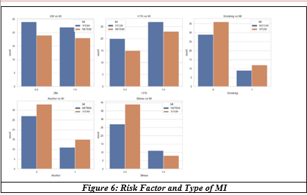
Relative risk values were close to 1.0, suggesting no meaningful increase in STEMI risk associated with these variables in this cohort.
|
Risk Factor |
Odd's Ratio |
|
Diabetes Mellitus |
0.97 |
|
Hypertension |
0.88 |
|
Smoking |
1.07 |
|
Alcohol |
1.12 |
|
Stress |
0.5 |
|
Table 4. Odds Ratios for STEMI |
|
Smoking and alcohol showed slightly elevated odds ratios, though the magnitude was small.
Statistical Association Tests
|
Variables Tested |
P-value |
|
Smoking vs MI type |
1.00 |
|
Hypertension vs MI type |
0.948 |
|
Diabetes vs MI type |
1.000 |
|
Gender vs MI type |
0.996 |
|
Smoking vs treatment |
0.302 |
|
Table 5. Chi-Square Test Results |
|
No statistically significant associations were identified (p>0.05 for all tests).
BMI and Myocardial Infarction Type
|
Test |
Statistic |
P-value |
|
Independent t-test |
----- |
0.867 |
|
Mann-Whitney U |
----- |
0.841 |
|
ANOVA(BMI vs Treatment |
F=1.33 |
0.251 |
|
Kruskal-Wallis(BMI vs MI type) |
------ |
0.837 |
|
Effect Size |
||
|
Metric |
Value |
|
|
Cohen's d |
0.037 |
|
|
Table 6. BMI comparison across groups |
||
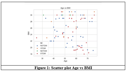
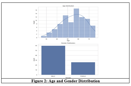
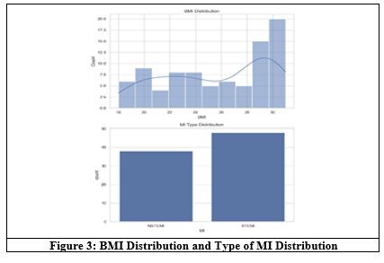
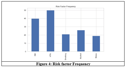
The effect size indicates a negligible difference in BMI between groups
Exact Test for smoking and STEMI
|
Comparison |
P-value |
|
Smoking vs STEMI |
1.000 |
|
Table 7. Fisher Exact Test |
|
No statistically significant association was observed.
Machine Learning Feature Importance
|
Variable |
Mutual Information |
|
Stress |
0.020 |
|
Hypertension |
0.019 |
|
Smoking |
0.005 |
|
Age |
0.000 |
|
BMI |
0.000 |
|
Diabetes |
0.000 |
|
Alcohol |
0.000 |
|
Table 8. Mutual Information Scores for Predicting STEMI |
|
Stress and Hypertension showed the highest predictive signals, although the values remained very small.
Interaction Analysis
|
Interaction |
Count |
|
Smoking+Hypertension |
11 |
|
Diabetes+Hypertension |
35 |
|
Smoking+Alcohol |
15 |
|
Table 9. Interaction risk factors counts |
|
The most common interaction observed was diabetes with hypertension.
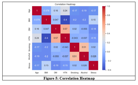
Cluster Analysis
|
Cluster |
Number of Patients |
|
Cluster 0 |
35 |
|
Cluster 1 |
34 |
|
Cluster 2 |
17 |
|
Table 10. Patient Clusters identified |
|
Cluster analysis revealed three patient groups, suggesting possible sub-populations with differing risk profiles.
Smoking Risk Estimation
|
Metric |
Value |
|
Relative Risk |
1.02 |
|
Smoking Lift |
1.02 |
|
Bootstrap 95% CI (RR) |
0.63-1.55 |
|
Table 11. Smoking Risk Metrics |
|
The confidence interval includes 1.0, indicating no statistically significant association between smoking and STEMI in this dataset.
Summary of Key Findings
1. The study included 86 MI patients with a mean age of 55.36 years.
2. Hypertension (58.8%) and diabetes (48.2%) were the most prevalent risk factors.
3. No statistically significant associations were observed between major risk factors and MI type.
4. BMI showed no significant difference across MI or treatment groups.
5. Mutual information analysis suggested stress and hypertension may have minimal predictive value, though the effect was weak.
6. Cluster analysis identified three possible patient subgroups.
7. Smoking demonstrated no statistically significant relationship with STEMI, with a relative risk close to 1.
DISCUSSION:
Derek Rowlands et al have extensively dealt on various topics in cardiology regarding the trend of disorders, presentation and complication apart from examining factors of ethnicity. South Asians have greater predisposition to cardiometabolic dysfunction[1] and socioeconomic drivers existing within the South Asian population contribute to adverse risk factors precipitating coronary diseases. The American Medical Association released articles in 2007 designating smoking and early onset of diabetes mellitus in the South Asian population as the risk factors dominating the onset and severity of coronary artery involvement.[2] Elevated blood pressure, smoking, physical inactivity, stress, alcohol consumption, increased weight and male gender shifted the occurrence of CAD to the younger population below fifth decade.[3] Cigarette smoking is the central and most common risk factor among the younger individuals while hypertension, diabetes and dyslipidemia are common in older population.
Smoking is the most common risk factor among younger individuals. Smoking tobacco speeds up the onset of atherosclerosis by decreasing tissue oxygenation, causing vascular endothelial dysfunction and eventual increase in platelet activity namely aggregation which aids in developing intravascular clots.[4]
Tobacco smoke has mixed chemical composition. The main ingredients are 1] nicotine that increases myocardial oxygen demand. 2] Carbonmonoxide that interferes with oxygen carrying capacity reducing tissue oxygenation and 3] tar [total aerosol] which coats the airways and alveoli increasing airway resistance and interfering with gas exchange. Comparing smokers and non-smokers, smokers have twice increased risk for coronary artery disease, twice increased risk for cerebrovascular accident and more than five times risk for peripheral arterial disease. Passive smoking causes 30% increased risk for heart disease and sudden infant death.
Cigarrette use activates platelets, increases circulating fibrinogen, increases heart rate and elevates blood pressure. Smoking appears to promote plaque disruption. Duration of smoking and the daily amount markedly influence the risk of CAD. The number of cigarettes smoked per day is directly proportional to the risk of MI.
The benefits of quitting smoking includes
20 minutes quit: heart rate and blood pressure drop.
12 hours: Carbonmonoxide levels in blood normalise.
1 year: Quitting smoking for 1 year reduces risk of heart disease by 50%.
15 years of quitting smoking brings down the risk of cardiovascular disease comparable to non-smokers.
Though exact mechanisms of cardiovascular damages are not well known, endothelial dysfunction is well recognised. Smoking elicits oxidative processes, negatively affects platelet function, fibrinolysis, inflammation and vasomotor function; all these are proatherogenic and double the 10 years risk of fatal events in smokers compared to non-smokers.
Cigarrette smoke is a mixture of chemical compounds that are bound to aerosol particle are free in the gaseous phase. It has been estimated that cigarette smoke has over 7000 chemical compounds from many different classes, including at least 72 carcinogens.[6] Fowles et al in 2003 associated 1, 3 butadiene to cancer risk and cyanide arsenic and cresols to cardiovascular risk. Ammonia, arsenic, lead, mercury and radioactive elements are constituents of cigarettes which may also cause impotence.
Smoking generates free radicals such as reactive oxygen species (ROS) which overwork the antioxidant system leading to oxidative stress towards cellular macromolecules, including lipids, proteins and DNA.[7] This stress also promotes inflammation with increased levels of inflammatory cytokines and influx of activated immune cells into the damaged area. Nitric oxide is produced by endothelial cells that is essential for vascular dilatation and relaxation. Smoking causes deficiency of nitric oxide leading to marked vascular constriction, impaired vasodilation and rise in blood pressure.[8]
Metabolic Syndrome
The metabolic syndrome is the terminology coined to represent complex pathophysiology constructed from clustering various interrelated processes.[9] Specific metabolic factors namely obesity, hypertension, insulin resistance and dyslipidemia commonly occur together and influence risk for cardiovascular diseases. Energy homeostasis is coordinated by hormone signal pathways that integrate metabolic activities of multiple tissues and organ systems. Insulin resistance disrupts metabolic efficiency thus driving chronic metabolic diseases such as type 2 diabetes, atherosclerosis, hypertriglyceridemia, fatty liver, polycystic ovarian disease and obesity. The metabolism in adipose tissue, skeletal muscle, vascular endothelium, bone, liver, kidneys, pancreatic islets of Langerhans, hypothalamus and immune system are all affected by insulin resistance. About 20 to 25% of worldwide population have metabolic syndrome increasing the risk of atherosclerotic disease.[10]
Though body mass index above 30 kg/m² is generally considered as obese, cut off points as low as 25–27 kg/m² in various Asian regions may indicate obesity while 23–26.9 kg/m² ranges indicate overweight.[11] About 10% of obese population do not demonstrate signs of insulin resistance and hence termed as metabolically healthy people. Cardiovascular risk is nearly doubled in people with metabolic syndrome.
Pathogenesis of Metabolic Syndrome
Molecular basis for metabolic syndrome as a true emergent syndrome is described by an interconnected cascade of dysregulated energy metabolism. The accumulation of fat in adipose and non-adipose tissue is the common starting pathway, which increases systemic inflammation and insulin resistance. Fat deposits in extremities are classed as peripheral adipose tissue, those in the abdomen as central adipose tissue, those in pericardial and perinephric regions as ectopic adipose tissue and fat deposits in visceral tissues, skeletal muscle and liver are classified as non-adipose tissue with fat deposits.
Insulin resistance and enhanced fat accumulation are precipitated by the following biologic and metabolic pathways:
1. Degeneration and inactivation of pancreatic β cells
2. Reduced release and activity of glucagon-like peptide by L cells of distal ileum
3. Impaired nutrient sensing within paraventricular nucleus of hypothalamus
4. Reduced adiponectin production within adipose tissue
5. Reduced endothelial and adipose lipoprotein lipase activity yielding elevated triglycerides
6. Highly atherogenic dense LDL particles enter circulation due to impaired hepatic lipoprotein synthesis
7. Impaired nitric oxide synthase activity leads to vasoconstriction and essential hypertension
8. Release of inflammatory cytokines such as tumor necrosis factor α, interleukins 1 and 6 promote degradation of insulin receptor substrate-1
Lifestyle and environmental factors involved in pathogenesis of metabolic syndrome include sedentary behaviour, modern agriculture, exposure to manmade industrial chemicals such as pesticides, plasticizers and preservatives, artificial sweeteners which act as endocrine disruptors, sleep hygiene and drug use of antidepressant and antipsychotic medications.
Alcoholic Heart Disease
Habitual heavy alcohol consumption is associated with increased risk of left ventricular dilatation and dysfunction namely alcoholic cardiomyopathy,[12] arrhythmias, systemic hypertension, ischemic heart disease, stroke and skeletal muscle abnormalities. Standard sized portions of wine, liquor or beer contain the same amount of alcohol. Hence the daily amount of ethanol is usually measured per number of standard drinks.
BMI Classification
· >30 kg/m²: Obese
· 25–27 kg/m² (Asian): Obese
· 23–24.9 kg/m²: Overweight
About 10% obese population do not demonstrate signs of insulin resistance and hence termed metabolically healthy.
|
|
|
Table 12: Metabolic Syndrome |
Pathogenesis of Metabolic Syndrome
Molecular basis for metabolic syndrome as a true emergent syndrome is described by an interconnected cascade of dysregulated energy metabolism. The accumulation of fat in adipose and non-adipose tissue is the common starting pathway which increases systemic inflammation and insulin resistance. Fat deposits in extremities are classed as peripheral adipose tissue, those in the abdomen as central adipose tissue, those in pericardial and perinephric regions as ectopic adipose tissue and fat deposits in visceral tissues, skeletal muscle and liver are classified as non-adipose tissue with fat deposits.
Insulin resistance and enhanced fat accumulation are precipitated by the following biologic and metabolic pathways:
· Degeneration and inactivation of pancreatic β cells.
· Reduced release and activity of glucagon-like peptide by L cells of distal ileum.
· Impaired nutrient sensing within paraventricular nucleus of hypothalamus.
· Reduced adiponectin production within adipose tissue.
· Reduced endothelial and adipose lipoprotein lipase activity yielding elevated triglycerides.
· Highly atherogenic dense LDL particles enter circulation due to impaired hepatic lipoprotein synthesis.
Alcoholic Cardiomyopathy
Consumption corresponds to less than three standard sized drinks or less than 80 grams per day. Alcoholic cardiomyopathy is an acquired form of dilated cardiomyopathy with patients often presenting with heart failure as a result of reduced cardiac output and atrial or ventricular arrhythmias, with a history of heavy alcohol intake.
The toxic damage of ethanol is attributed to a non-oxidative metabolic pathway for alcohol related to fatty acid metabolism in the heart, skeletal muscle, pancreas and brain.[13] Apoptosis, abnormal excitation-contraction coupling, functional and structural mitochondrial damage, loss of contractile filaments, deregulation of protein synthesis and activation of renin-angiotensin and sympathetic nervous systems are all involved in the pathogenic mechanisms of alcoholic cardiomyopathy. Alcohol induced mitochondrial toxicity results in mitochondrial dysfunction, loss of mitochondrial membrane potential and increase in mitochondrial oxidative stress eventually culminating in cell death.[14] These explain the multi-organ damage in alcohol usage disorders.
The next risk factor for coronary artery disease in less than 50 years is drug abuse or recreational drug usage, which is not noted in our case analysis and general population does not disclose drug usage.
Atherosclerosis and atheromatous plaque rupture are common from fifth decade of life. Hence dyslipidemias are not elaborated in this article. Stress factor, which is highly variable between individuals and this was noted down after a simple questionnaire whether the patient had anything worrying in the family or work place. This retrospective analysis has only case sheet derived data as it is a short observational study to derive differing risk factors in ACS affecting age groups less than fifty.
STEMI and NSTEMI
Acute coronary syndrome is a dynamic process that involves cyclical transitioning among complete vessel occlusion, partial vessel occlusion and reperfusion.[15] Occlusive thrombus in the absence of significant, collateral vessels most often results in acute ST segment elevation infarction (STEMI).
The pathophysiology of STEMI and non-ST segment elevation myocardial infarction is similar and explains the overlap in acute coronary syndromes with regard to ultimate outcome, extent of necrosis and mortality rates. If the ECG demonstrates acute ST segment elevation or new LBBB, emergent reperfusion treatment with primary PCI or fibrinolysis is indicated.
Coronary plaque rupture is the initiating event in acute MI. Rupture of the fibrous cap of coronary atheroma exposes the underlying subendothelial matrix to formed elements of circulating blood, leading to activation of platelets, thrombin generation and thrombus formation.
|
|
|
Table 13: Clinical classification of types of Myocardial Infarction |
Clinical Diagnosis
Signs and symptoms crushing substernal chest pain radiating to left arm is the classical presentation. The chest discomfort may radiate to jaw, neck, right arm, shoulder and back, epigastrium even without chest pain especially in the elderly and diabetes mellitus. Sudden tearing pain indicates aortic dissection.
|
|
|
Table 14: Differential Diagnosis |
Unstable angina and non-ST elevation myocardial infarction are a part of continuum of acute coronary syndrome ranging from NSTEMI to STEMI. Clinical presentation is variable ranging from progressive exertional angina to post infarction angina. Elevated serum levels of cardiac biomarkers namely Troponins and creatine kinase are the differentiating features between unstable angina and NSTEMI. Troponin T and I assays are rapid and highly sensitive, accessible and available bedside indicating their usefulness in NSTEMI detection. Creatine kinase is more useful to detect ST segment elevation MI and may help in gauging the size and timing of acute MI rather than diagnosis.
|
Gusto Risk Score |
|
|
Risk Score |
30 Day Mortality (%) |
|
0-5 |
0.4 |
|
6-10 |
2.8 |
|
11-15 |
8.7 |
|
16-19 |
25.0 |
|
20-22 |
41.7 |
|
Gusto Scoring System |
|
|
Criteria |
Points |
|
Age 50-59 |
2 |
|
Age 60-69 |
4 |
|
Age 70-79 |
6 |
|
Age ⪰80 |
8 |
|
Prior Heart Failure |
2 |
|
Prior Stroke/TIA |
2 |
|
Prior MI/Revascularization/Angina |
1 |
|
HR ⪰90 bpm |
3 |
|
Elevated Troponin/CK-MB |
3 |
|
Creatinine >1.4 mg/dL |
2 |
|
CRP >20 µg/L |
2 |
|
CRP 10-20 µg/L |
1 |
|
Anemia |
1 |
|
Table 15: Gusto Risk Score |
|
|
|
|
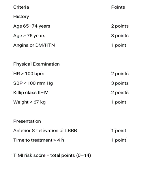
Table 16: TIMI Risk Score For STEMI
Therapy
Patients with ACS, classical symptoms and ST elevation in ECG need immediate initiation of therapy.
A. Prior to reperfusion: Aspirin, Oxygen, nitroglycerine, Platelet receptor antagonists namely clopidogrel and prasugrel, parenteral anticoagulants namely unfractionated heparin.
B. Reperfusion Therapy: Fibrinolysis and PCI.
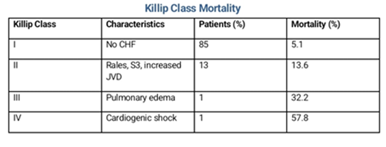
Table 17: Killip Class Mortality
Treatment Statistics
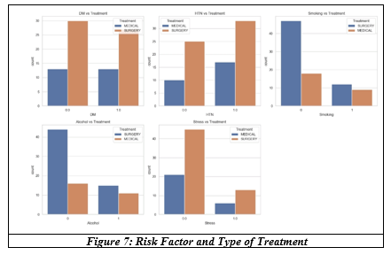
NSTEMI Management Strategy
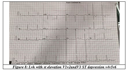
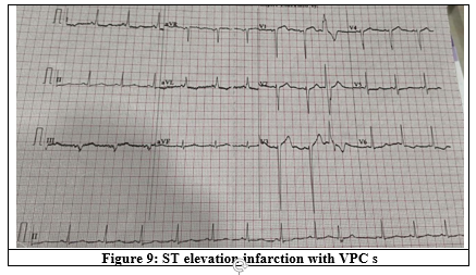
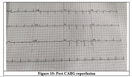
Electrocardiogram
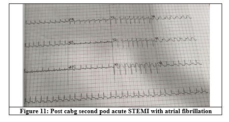
Differentiating STEMI and NSTEMI patterns can significantly impact initiation of treatment and patient outcomes.
ST Elevation Patterns[16]
V2-V3 leads: >/ 2.5mm for men <40 years,>/ 2mm in men more than 40 years and 1.5mm for women
In other leads: more than 1mm elevation in two or more contiguous leads.
NSTEMI Pattern[17]
ST depression and T wave changes.
· Horizontal or downsloping ST depression >0.5mm in at least two anatomically contiguous leads.
· T wave inversion of >1mm in leads with prominent R waves or R/S ratio >1.
· New horizontal or downsloping ST segment depressions in V4, V5, V6.[17]
CONCLUSION:
Acute coronary syndrome can present with or without classical symptoms of chest pain and its typical distribution.
The youngest patient in our study had STEMI, suggesting occlusive coronary artery disease. Social habits such as smoking, alcohol, and drug abuse (not included in our study due to unavailable or undisclosed data) may contribute to anatomical and physiological differences, making cardiovascular tissues more susceptible to harmful effects apart from the impacts of lifestyle factors such as sedentary habits and dietary patterns.
Electrocardiographic recognition of STEMI and NSTEMI plays a crucial role in initiating effective treatment and achieving successful outcomes
REFERENCES:
[1] Zaman MJ. Impact of South Asian ethnicity on incidence and prognosis of CAD. In: Rowlands D, Clarke B, eds. Recent advances in cardiology. Jaypee Medical 2015.
[2] Joshi P, Islam S, Pais P, et al. Risk factors for early myocardial infarction in South Asians compared with individuals in other countries. JAMA 2007;297(3):286-94.
[3] Vaccarino V, Sullivan S, Hammadah M, et al. Mental stress–induced-myocardial ischemia in young patients with recent myocardial infarction: sex differences and mechanisms. Circulation 2018;137(8):794-805.
[4] Matsis K, Holliday, Alsman A, et al. Differences in clinical characteristics between young and older patients presenting with myocardial infarction. Heart Lung Circ 2014;26:566-91.
[5] Smoking and heart disease / better health. NCBI.gov.
[6] Cowther CA, Grasso AW. Non lipid cardiovascular risk factors. Chap- 44. In: Griffin BP, ed et al. Manual of cardiovascular medicine. 4th edn. Wolters Kluwer 2017:722-4.
[7] Caliri AW, Tommasi S, Besaratinia A. Relationships among smoking, oxidative stress, inflammation, macromolecular damage, and cancer. Mutation Research/Reviews in Mutation Research 2021;787:108365.
[8] Bloom SI, Islam MT, Lesniewski LA, et al. Mechanisms and consequences of endothelial cell senescence. Nature Reviews Cardiology 2023;20(1):38-51.
[9] Via MA, Mechanick JJ. The metabolic syndrome. Chap- 26. In: Fuster V, Narula J, Vaishnava P, et al, eds. Fuster & Hurst's the Heart. McGraw Hill Education 2017:767-71.
[10] Miller JM, Kaylor MB, Johannsson M, et al. Prevalence of metabolic syndrome and individual criterion in US adolescents: 2001–2010 National Health and Nutrition Examination Survey. Metabolic Syndrome and Related Disorders 2014;12(10):527-32.
[11] Zheng W, McLerran DF, Rolland B, et al. Association between body-mass index and risk of death in more than 1 million Asians. N Engl J Med 2011;364(8):719-29.
[12] Klatsky AL. Alcohol and cardiovascular diseases. J Int Med 2005;278:238-50.
[13] Guzzo-Merello G, Cobo-Marcos M, Gallego-Delgado M, et al. Alcoholic cardiomyopathy. World J Cardiol 2014;6(8):771-81.
[14] Varga ZV, Ferdinandy P, Liaudet L, et al. Drug-induced mitochondrial dysfunction and cardiotoxicity. American Journal of Physiology-Heart and Circulatory Physiology 2015;309(9):H1453-67.
[15] Huff CM Acute Myocardial Infarction. Chap- 1. In: Griffin BP, Callahan TD, Menon V, et al, eds. Manual of cardiovascular medicine. 4th edn. New Delhi: Wolters Kluwer (India) Pvt. Ltd 2013.
[16] Cardiology /learn the heart/ eg series https://ecg.ecgwares.com
https://ecgwares.com/non-st elevation myocardial infarction. Understanding STEMI vs. NSTEMI information. TACCog /accelerating cardiac case published Dec 31/ 2024 updated Apr 6 2025.