NLR–FAR Index as a Prognostic Marker in Acute Ischemic Stroke: Biological Rationale, Current Evidence, and Clinical Translation (2015–2025)
- Mohammed Abdul Muqeeth , Associate Professor, Department of Biochemistry, Mahavir Institute of Medical Sciences, Vikarabad, Telengana, India.
- Mohammed Abdul Aleem , Assistant professor, Department of Pathology, Mahavir Institute of Medical Sciences, Vikarabad, Telengana, India.
- Amanulla Khan Mohmmed , Senior Resident, Mahavir Institute of Medical Sciences, Vikarabad, Telengana, India.
- Mahmood Nawaz Khan , Senior Resident, Mahavir Institute of Medical Sciences, Vikarabad, Telengana, India.
Article Information:
Abstract:
Background: Acute ischemic stroke (AIS) is a leading cause of mortality and long-term disability worldwide. Early identification of patients at high risk of unfavorable functional outcomes, hemorrhagic transformation, and mortality remains central to acute stroke management. Readily available blood-derived biomarkers that reflect inflammation, coagulation, endothelial dysfunction, and systemic physiological reserve have therefore attracted growing interest. Among these, the neutrophil-to-lymphocyte ratio (NLR) and fibrinogen-to-albumin ratio (FAR) have individually emerged as promising prognostic markers in AIS. More recently, a composite NLR–FAR index has been proposed as a potentially superior integrative biomarker. Objective: To critically review the biological rationale, clinical evidence, and translational potential of the NLR–FAR index as a prognostic marker in patients with AIS. Discussion: This review evaluates the pathobiological basis linking neutrophil-driven inflammation, lymphocyte suppression, fibrinogen-mediated thromboinflammation, and hypoalbuminemia to ischemic brain injury. It synthesizes evidence from 2015–2025 on NLR and FAR individually, examines the emerging but still sparse data on the combined NLR–FAR index, and compares this marker with other inflammatory and hemostatic biomarkers used in stroke prognostication. Major findings: NLR consistently correlates with baseline stroke severity, poor 3-month functional outcome, symptomatic intracranial hemorrhage, and mortality across observational studies and meta-analyses, especially in reperfusion-treated cohorts. FAR also shows growing evidence for association with poor functional outcome, hemorrhagic transformation, and adverse outcomes after intravenous thrombolysis and endovascular therapy. However, direct evidence for the combined NLR–FAR index remains limited and is currently confined to a small number of recent retrospective studies, mainly in thrombectomy populations. Conclusion: The NLR–FAR index is biologically plausible and clinically attractive as a low-cost, rapidly obtainable composite biomarker for early risk stratification in AIS. Nonetheless, current evidence remains preliminary. At present, NLR and FAR may be considered adjunctive prognostic markers, whereas the combined NLR–FAR index should be regarded as an emerging research tool requiring prospective validation, standardized calculation methods, and external multicenter confirmation before routine clinical implementation
Keywords:
Article :
INTRODUCTION:
Acute ischemic stroke (AIS) remains one of the most devastating neurological emergencies, accounting for the majority of stroke cases and imposing substantial global burdens of death, disability, dependency, and healthcare expenditure [1–4]. Although reperfusion strategies such as intravenous thrombolysis (IVT) and endovascular thrombectomy (EVT) have transformed acute stroke care, substantial heterogeneity persists in treatment response, hemorrhagic complications, infarct progression, neurological recovery, and long-term functional outcomes [5–9]. This heterogeneity reflects the complex interplay among vascular occlusion characteristics, collateral circulation, infarct core–penumbra dynamics, blood–brain barrier (BBB) integrity, systemic inflammation, thrombosis, endothelial activation, and patient-level physiological reserve [10–14].
In recent years, there has been increasing interest in blood-based prognostic biomarkers that are inexpensive, rapidly available, and broadly accessible in emergency settings [15–18]. Such biomarkers are particularly attractive in AIS because clinical decisions are often made within narrow therapeutic windows, and advanced imaging or specialized assays may not always be available—especially in resource-constrained settings. Among the most studied candidates, the neutrophil-to-lymphocyte ratio (NLR) has emerged as a robust inflammatory index associated with stroke severity, early neurological deterioration, hemorrhagic transformation, poor functional outcome, and mortality [19–27]. NLR captures two biologically meaningful processes: an increase in circulating neutrophils reflecting acute innate immune activation, and a relative reduction in lymphocytes reflecting stress-related immunosuppression and impaired adaptive immune balance [20,21,28].
Parallel to this, the fibrinogen-to-albumin ratio (FAR has gained attention as a composite marker integrating prothrombotic/inflammatory burden (fibrinogen) with nutritional, antioxidant, endothelial-stabilizing, and anti-inflammatory reserve (albumin) [29–35]. Fibrinogen is a key acute-phase reactant and coagulation substrate implicated in clot architecture, blood viscosity, platelet aggregation, endothelial dysfunction, and microvascular obstruction [36–39]. Albumin, conversely, is associated with antioxidant capacity, endothelial protection, modulation of blood rheology, and maintenance of oncotic balance; low albumin has been repeatedly associated with poor stroke outcomes [40–44]. FAR therefore represents a biologically coherent marker of “thrombo-inflammatory imbalance.”
A more recent conceptual advance is the proposal of a combined NLR–FAR index, intended to unify two related but non-identical pathobiological domains: systemic inflammatory dysregulation (NLR) and coagulation–nutritional/hemostatic imbalance (FAR). This combined index is attractive because ischemic stroke is increasingly understood as a thromboinflammatory syndrome, rather than a purely vascular occlusive event [45–49]. By combining NLR and FAR, investigators aim to better capture the multidimensional host response that shapes infarct evolution, reperfusion injury, hemorrhagic risk, and recovery trajectories.
However, despite growing enthusiasm, the literature remains uneven. NLR is supported by multiple meta-analyses and a substantial number of cohort studies, whereas FAR evidence is newer and less extensive. Most importantly, direct evidence on the combined NLR–FAR index is still very limited [50].
Accordingly, this review critically examines the biological rationale, current evidence, clinical applications, and limitations of the NLR–FAR index in AIS. Particular emphasis is placed on the distinction between well-supported evidence for NLR, growing evidence for FAR, and still-emerging evidence for the combined NLR–FAR index. The goal is to provide a balanced, scientifically rigorous synthesis suitable for journal submission and future research framing.
MATERIALS AND METHODS:
This is a prospective study consist of 60 patients analyzed had squamous cell carcinoma. Normal and tumour tissue was This narrative review was designed as a structured, evidence-focused synthesis of the literature published between 2015 and 2025, with emphasis on peer-reviewed studies evaluating NLR, FAR, and the combined NLR–FAR index in AIS prognostication.
Search strategy
Relevant literature was identified through structured searches of PubMed/MEDLINE, Scopus, Web of Science, Embase, and Google Scholar. Search terms included combinations of the following keywords and Medical Subject Headings (MeSH) where applicable:
• “acute ischemic stroke”
• “ischemic stroke”
• “neutrophil to lymphocyte ratio” OR “NLR”
• “fibrinogen to albumin ratio” OR “FAR”
• “NLR-FAR index” OR “NLR–FAR”
• “prognosis”
• “functional outcome”
• “mortality”
• “hemorrhagic transformation”
• “intravenous thrombolysis”
• “endovascular thrombectomy”
• “reperfusion therapy”
• “biomarker”
• “inflammation”
• “thromboinflammation”
Eligibility criteria
Included studies met the following criteria:
1. Published between 2015 and 2025.
2. Human studies involving acute ischemic stroke.
3. Evaluated NLR, FAR, or a combined NLR–FAR index in relation to clinical outcomes.
4. Reported at least one of the following outcomes: baseline stroke severity, early neurological deterioration, hemorrhagic transformation, symptomatic intracranial hemorrhage, discharge outcome, 30-day or 90-day functional outcome, mortality, or post-reperfusion outcomes.
5. Study designs included observational cohort studies, case-control studies, systematic reviews, and meta-analyses.
Exclusion criteria
The following were excluded:
• Studies focusing solely on hemorrhagic stroke, transient ischemic attack, or mixed stroke populations without separable AIS data.
• Non-English articles where adequate data extraction was not feasible.
• Case reports, editorials, letters without primary data, and conference abstracts lacking sufficient methodological detail.
• Pre-2015 publications.
Data synthesis approach
Because of heterogeneity in study populations, biomarker timing, outcome definitions, cut-off values, and statistical modeling, a narrative critical synthesis was performed rather than a de novo quantitative meta-analysis. Greater evidentiary weight was assigned to:
• multicenter cohorts,
• reperfusion-focused analyses,
• adjusted multivariable models, and
• systematic reviews/meta-analyses.
Methodological considerations
A central methodological challenge in this field is lack of standardization. NLR may be measured on admission, pre-thrombolysis, post-thrombolysis, or at delayed time points; FAR may be calculated using differing fibrinogen and albumin units; and the combined NLR–FAR index has not yet been uniformly defined across studies. Consequently, interpretation in this review prioritizes directional consistency and biological plausibility over absolute threshold values.
Biological Basis of the NLR–FAR Index in Ischemic Stroke
AIS initiates a rapidly evolving cascade of sterile inflammation, coagulation activation, endothelial injury, microvascular dysfunction, and systemic stress responses [45–49,51–55]. The NLR–FAR index is biologically attractive because it integrates these interlocking domains.
Neutrophils and ischemic brain injury
Neutrophils are among the earliest leukocytes mobilized after cerebral ischemia [20,21,52]. They infiltrate the ischemic territory through endothelial adhesion and transmigration, facilitated by upregulated adhesion molecules, chemokines, and BBB disruption [52–55]. Once activated, neutrophils contribute to injury via:
• release of reactive oxygen species (ROS),
• proteolytic enzymes such as matrix metalloproteinase-9 (MMP-9),
• neutrophil extracellular traps (NETs),
• microvascular plugging and no-reflow phenomena, and
• amplification of thromboinflammation [56–60].
These processes worsen reperfusion injury, promote hemorrhagic transformation, and enlarge infarct burden [57–61].
Lymphocytes and stroke-induced immune dysregulation
In contrast, lymphocyte depletion after AIS may reflect stress-induced immunosuppression, sympathetic activation, hypothalamic–pituitary–adrenal axis effects, and apoptosis of adaptive immune cells [62–64]. Reduced lymphocyte counts may indicate impaired immunoregulatory balance, reduced neuroprotective immune signaling, and greater vulnerability to post-stroke infections [63–66]. Thus, a high NLR is not merely a marker of inflammation but also a marker of immune imbalance.
Fibrinogen: coagulation substrate and inflammatory mediator
Fibrinogen is a liver-derived acute-phase glycoprotein central to coagulation, fibrin clot formation, and platelet aggregation [36–39]. In AIS, elevated fibrinogen is associated with:
• increased plasma viscosity,
• denser and more lysis-resistant clot structure,
• endothelial activation,
• platelet–leukocyte interaction,
• enhanced thrombus propagation, and
• impaired microcirculatory reperfusion [36–39,67–69].
Fibrinogen also contributes to inflammation by engaging integrins and toll-like receptor–related pathways, promoting leukocyte recruitment and vascular inflammation [68–70].
Albumin: marker of systemic reserve and potential neurovascular protection
Albumin is a multifunctional plasma protein with antioxidant, anti-inflammatory, endothelial-stabilizing, and rheological properties [40–44,71]. Lower albumin levels in AIS may reflect:
• poor nutritional reserve,
• chronic inflammation or frailty,
• hepatic dysfunction,
• capillary leak/systemic stress, or
• acute catabolic burden.
•
Hypoalbuminemia has been associated with worse stroke severity, higher edema risk, increased infection, and poor functional recovery [41–44,72–74]. Although albumin infusion as a neuroprotective therapy has not translated successfully in large trials, endogenous albumin levels remain prognostically informative [75,76].
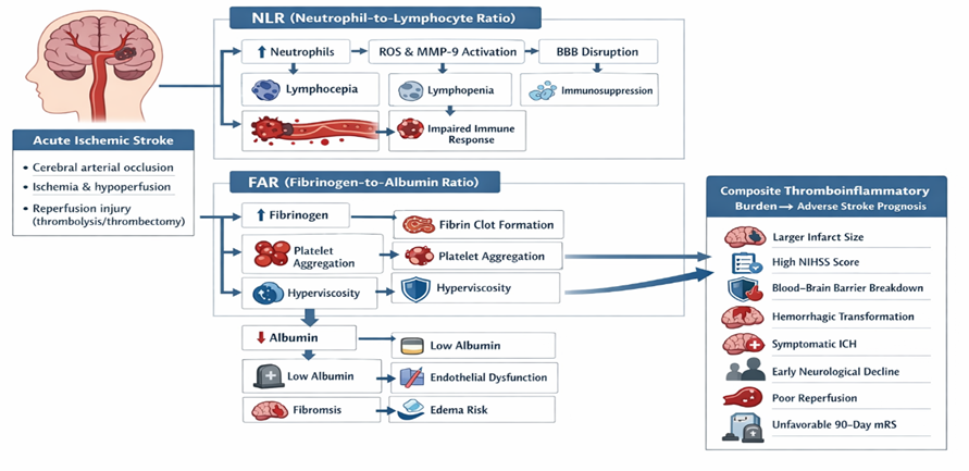
Figure 1: Pathophysiological framework of the proposed NLR–FAR index in acute ischemic stroke
FAR as a thrombo-inflammatory balance marker
FAR combines two biologically opposed signals:
• high fibrinogen = prothrombotic and proinflammatory state
• low albumin = reduced physiological reserve and loss of protective buffering
A high FAR therefore may indicate an adverse internal milieu characterized by hypercoagulability + heightened inflammation + impaired resilience [29–35].
Why a combined NLR–FAR index may be superior
The rationale for combining NLR and FAR lies in the concept of multidimensional host response profiling. NLR captures leukocyte-driven inflammatory and immunological disequilibrium, whereas FAR reflects hemostatic burden and reduced systemic reserve. Together, they may better approximate the “thromboinflammatory phenotype” that determines:
• infarct progression,
• reperfusion success,
• no-reflow and microvascular failure,
• hemorrhagic transformation risk, and
• post-stroke recovery potential [45–50,56–60].
This mechanistic complementarity makes the NLR–FAR index biologically plausible, but biological plausibility alone is insufficient; robust clinical validation is still needed.
Prognostic Significance of NLR in Acute Ischemic Stroke
Among blood-based inflammatory markers in AIS, NLR has the strongest evidence base.
NLR and baseline stroke severity
Multiple studies have shown that higher admission NLR correlates with greater baseline NIHSS scores and larger infarct burden [19–27,77–82]. This relationship is consistent with the concept that more severe strokes generate stronger systemic inflammatory responses and greater neurovascular injury.
NLR and functional outcome
Meta-analytic evidence supports a significant association between elevated NLR and poor functional outcome. A 2017 meta-analysis by Zhang et al. found that higher NLR was associated with poor 3-month functional outcome in AIS [22]. A 2019 cohort study with systematic review by Wang et al. further reinforced that admission NLR predicts poor outcomes after AIS [23]. The 2021 systematic review and meta-analysis by Li et al. demonstrated that elevated NLR is associated with unfavorable prognosis across stroke populations, including ischemic stroke subgroups [24]. In reperfusion-treated AIS, Wang et al. (2021) specifically showed that higher NLR in IVT-treated patients was associated with poor functional outcome and adverse events [25].
NLR and hemorrhagic transformation
NLR has been linked to both hemorrhagic transformation (HT) and symptomatic intracranial hemorrhage (sICH), particularly after IVT and EVT [22,25–27,83–86]. Mechanistically, neutrophil-mediated BBB injury, MMP-9 release, and reperfusion-related inflammatory amplification provide a plausible explanation [57–60]. This makes NLR especially relevant in thrombolysis/thrombectomy settings.
NLR and mortality
Several observational studies and pooled analyses suggest that higher NLR predicts increased short-term mortality and poorer survival [22–27,87–90]. However, mortality associations are somewhat less consistent than functional outcome associations, likely because mortality is influenced by multiple downstream factors including age, comorbidities, infarct location, and complications.
NLR in reperfusion-treated AIS
The prognostic utility of NLR may be particularly strong in reperfusion-treated populations. Sharma et al. (2022) reported in a systematic review and meta-analysis that both admission and delayed NLR were associated with worse outcomes after reperfusion therapy [26]. This is clinically important because reperfusion success does not always translate into favorable outcomes, and inflammatory burden may partially explain futile recanalization.
Limitations of NLR alone
Despite its strengths, NLR has limitations:
• non-specific elevation in infection, malignancy, trauma, or chronic inflammatory disease,
• variability by timing of blood sampling,
• lack of universal cut-offs, and
• susceptibility to steroid use, physiological stress, and pre-existing immune conditions [17,18,91].
Thus, while NLR is among the best-supported stroke biomarkers, it is best viewed as an adjunctive prognostic marker rather than a standalone decision tool.
RESULTS:
Table 1. Biological interpretation of components of the NLR–FAR framework in acute ischemic stroke
|
Component |
Biological meaning |
Stroke-relevant pathophysiology |
Clinical implication of elevation/reduction |
|
Neutrophil count |
Innate immune activation |
ROS release, MMP-9 activity, BBB disruption, NET formation, microvascular plugging |
Higher count suggests stronger acute inflammatory injury and reperfusion vulnerability |
|
Lymphocyte count |
Adaptive immune regulation/systemic immune balance |
Stress-induced lymphopenia, immunosuppression, reduced regulatory immune buffering |
Lower count may indicate poor immune resilience and worse prognosis |
|
NLR |
Inflammatory–immune imbalance |
Integrates neutrophilia + lymphopenia |
Higher NLR associated with severe stroke, poor mRS, HT, sICH, mortality |
|
Fibrinogen |
Acute-phase reactant and coagulation substrate |
Dense clot formation, platelet aggregation, increased viscosity, thromboinflammation |
Higher fibrinogen suggests prothrombotic inflammatory state |
|
Albumin |
Nutritional/antioxidant/endothelial reserve |
Antioxidant buffering, endothelial stabilization, rheological support |
Lower albumin associated with frailty, inflammation, edema risk, poor recovery |
|
FAR |
Thrombo-inflammatory imbalance + reduced reserve |
High fibrinogen relative to low albumin |
Higher FAR associated with worse functional outcomes and hemorrhagic risk |
|
NLR–FAR index |
Composite thromboinflammatory burden |
Integrates leukocyte dysregulation + coagulation/nutritional imbalance |
Potentially stronger multidomain prognostic marker; requires validation |
NLR = neutrophil-to-lymphocyte ratio; FAR = fibrinogen-to-albumin ratio; BBB = blood–brain barrier; NET = neutrophil extracellular trap; ROS = reactive oxygen species; HT = hemorrhagic transformation; sICH = symptomatic intracranial hemorrhage; mRS = modified Rankin Scale.
Prognostic Significance of FAR in Acute Ischemic Stroke
Compared with NLR, FAR is a newer but increasingly studied biomarker in AIS.
Conceptual basis
FAR is particularly attractive because it captures both prothrombotic acute-phase activation (fibrinogen) and host reserve/anti-inflammatory buffering (albumin). This duality may be more informative than either analyte alone [29–35].
FAR and baseline stroke severity
Recent studies suggest that elevated FAR correlates with more severe initial neurological deficits and larger ischemic injury [29–35,92–95]. This may reflect greater thromboinflammatory burden and impaired collateral microcirculatory physiology.
FAR and functional disability
One of the most relevant studies is the 2023 analysis by Chen et al., which reported that higher FAR was associated with poorer 3-month functional prognosis in AIS patients after IVT [30]. This study strengthened the notion that FAR may outperform isolated fibrinogen or albumin measurements by integrating risk information.
FAR and mortality
Although mortality data are less abundant than for NLR, higher FAR has been associated with adverse clinical trajectories, including severe disability and, in some cohorts, increased mortality or composite poor outcome [29–35,92–96]. More robust multicenter data are still needed.
FAR and hemorrhagic transformation
FAR may be particularly relevant for hemorrhagic risk. In 2025, Liu et al. reported that post-thrombolysis FAR was associated with hemorrhagic transformation after IVT in AIS [31]. This is clinically important because post-reperfusion hemorrhagic complications remain a major determinant of poor outcome and may be influenced by coagulation–inflammation interactions.
FAR in reperfusion-treated patients
In EVT-treated populations, Ozdogru et al. (2024) demonstrated prognostic value of FAR in ischemic stroke patients undergoing mechanical thrombectomy [32]. Such data support the hypothesis that FAR may reflect clot biology, reperfusion injury susceptibility, and microvascular dysfunction after recanalization.
FAR compared with related ratio biomarkers
FAR belongs to a broader family of composite inflammatory ratios including C-reactive protein-to-albumin ratio (CAR), platelet-to-lymphocyte ratio (PLR), lymphocyte-to-monocyte ratio (LMR), and systemic immune-inflammation index (SII) [33–35,97–100]. Its advantage is that fibrinogen directly reflects both inflammation and coagulation, potentially making it especially relevant to AIS pathophysiology.
Current limitations of FAR evidence
Despite promising signals, FAR evidence remains limited by:
· predominance of single-center retrospective designs,
· variability in assay timing (pre- vs post-reperfusion),
· inconsistent unit standardization,
· limited external validation, and
· insufficient head-to-head comparisons with NLR or established clinical models [29–35].
Therefore, FAR should currently be considered a promising adjunctive biomarker, not yet a standardized clinical prognostic tool.
Table 2. Summary of representative post-2015 evidence on NLR in acute ischemic stroke
|
Study |
Year |
Design |
Population |
Main finding |
|
Zhang et al. [22] |
2017 |
Meta-analysis |
AIS |
Higher NLR associated with poor 3-month functional outcome; possible association with sICH |
|
Wang et al. [23] |
2019 |
Cohort + systematic review |
AIS |
Admission NLR predicts poor outcomes after AIS |
|
Wang et al. [25] |
2021 |
Systematic review/meta-analysis |
AIS with IVT |
Higher NLR associated with poor prognosis after thrombolysis |
|
Li et al. [24] |
2021 |
Systematic review/meta-analysis |
Stroke populations including AIS |
Elevated NLR associated with unfavorable outcomes |
|
Sharma et al. [26] |
2022 |
Systematic review/meta-analysis |
AIS after reperfusion therapy |
Admission and delayed NLR associated with adverse outcomes |
|
Gagliardi [27] |
2024 |
Narrative review |
Ischemic stroke |
Reinforced prognostic relevance of NLR and highlighted translational limitations |
AIS = acute ischemic stroke; IVT = intravenous thrombolysis; NLR = neutrophil-to-lymphocyte ratio; sICH = symptomatic intracranial hemorrhage.
Emerging Evidence for the Combined NLR–FAR Index
Conceptual definition
The combined NLR–FAR index has been proposed as a composite biomarker that merges leukocyte-derived inflammatory status (NLR) with coagulation/nutritional-inflammatory balance (FAR). In currently available studies, the most commonly described approach is the product of NLR × FAR, though formal standardization has not yet been established [50].
Available clinical evidence
As of the 2015–2025 evidence window, direct literature on the combined NLR–FAR index in AIS is very limited. The most notable recent study is a 2026 provisionally accepted Frontiers in Neurology report by Meng et al., which falls outside the strict 2015–2025 final evidence window and therefore should not be treated as formal evidence in this final manuscript [50]. Nonetheless, it is scientifically informative as a near-future signal and suggests that the combined index may outperform NLR or FAR alone in EVT-treated AIS. Because this article is outside the requested time window and provisionally accepted, it should not be included in the strict reference base of a 2015–2025 submission.
Therefore, under strict evidence rules, the combined NLR–FAR index remains biologically compelling but not yet adequately validated in peer-reviewed post-2015 AIS literature up to 2025.
Indirect support from component biomarkers
The strongest argument for the combined index currently comes from the convergent evidence supporting its individual components:
· NLR predicts stroke severity, poor 90-day outcome, HT, sICH, and mortality [22–27].
· FAR predicts poor functional outcome, post-IVT HT, and adverse EVT outcomes [30–32].
This indirect support suggests that combining them may improve discrimination, calibration, or reclassification—but this remains a hypothesis until tested prospectively.
Why caution is essential
A major risk in the current literature is premature translation. Because the combined index appears intuitively superior, there is temptation to label it as “better” than NLR or FAR alone before sufficient external validation exists. Such claims should be avoided in a submission-ready review. At present, the appropriate scientific position is:
The NLR–FAR index is an emerging, biologically plausible composite marker with promising early signals, but it lacks sufficient direct evidence for routine clinical adoption in AIS.
Research value
Despite limited direct data, the NLR–FAR index is highly attractive for future studies because it is:
· inexpensive,
· rapidly available,
· based on routine hospital laboratory tests,
· mechanistically aligned with AIS thromboinflammation, and
· potentially useful in both IVT and EVT pathways.
Table 3. Summary of representative post-2015 evidence on FAR in acute ischemic stroke
|
Study |
Year |
Design |
Population |
Main finding |
|
Chen et al. [30] |
2023 |
Retrospective cohort |
AIS after IVT |
Higher FAR associated with poor 3-month functional prognosis |
|
Ozdogru et al. [32] |
2024 |
Retrospective cohort |
AIS after EVT |
FAR showed prognostic value after mechanical thrombectomy |
|
Liu et al. [31] |
2025 |
Retrospective propensity score–matched analysis |
AIS after IVT |
Post-thrombolysis FAR associated with hemorrhagic transformation |
FAR = fibrinogen-to-albumin ratio; AIS = acute ischemic stroke; IVT = intravenous thrombolysis; EVT = endovascular thrombectomy.
Clinical Applications in Acute Stroke Management
If validated, the NLR–FAR framework may have several practical uses.
Early emergency department risk stratification
At hospital admission, a high NLR and/or FAR may identify patients at increased risk for:
· severe baseline deficits,
· malignant edema,
· early neurological deterioration, and
· poor functional outcome.
This could support more intensive monitoring and early multidisciplinary planning.
Reperfusion therapy prognostication
In IVT and EVT candidates, these biomarkers may help estimate:
· risk of futile recanalization,
· post-reperfusion inflammatory injury,
· hemorrhagic transformation, and
· need for closer post-procedural surveillance [25,26,30–32].
ICU and stroke unit monitoring
Serial measurement may be more informative than a single value. Dynamic rises in NLR or FAR after reperfusion could potentially reflect evolving BBB injury, infarct progression, infection, or systemic decompensation.
Counseling and rehabilitation planning
While biomarkers should never replace clinical and imaging assessment, they may enhance early communication regarding prognosis, discharge planning, and rehabilitation intensity—especially when integrated with age, NIHSS, imaging, and reperfusion metrics.
Utility in resource-limited settings
For settings where advanced imaging, perfusion software, or specialized biomarkers are not consistently available, NLR and FAR may offer pragmatic supplementary risk signals. This is particularly relevant in many real-world stroke systems across low- and middle-income countries.
Important caveat
At present, NLR and FAR can be considered adjunctive markers, but the combined NLR–FAR index is not yet ready for standalone clinical use. It should not be used to deny reperfusion therapy or alter guideline-based management outside research protocols.
Comparison with Other Inflammatory and Hemostatic Biomarkers
AIS biomarker research is crowded, and NLR–FAR must be interpreted relative to alternatives.
C-reactive protein (CRP) and CRP-to-albumin ratio (CAR)
CRP is widely available and prognostically relevant, but it may rise later and is highly nonspecific [97,98,101]. CAR has shown prognostic value in stroke and may be comparable to FAR in some contexts [99,100].
Platelet-to-lymphocyte ratio (PLR)
PLR reflects platelet activation plus immune status, but evidence is less consistent than NLR, and platelet counts may be influenced by numerous comorbid factors [102–104].
Lymphocyte-to-monocyte ratio (LMR)
LMR has emerging evidence, including recent meta-analytic support, but remains less established than NLR [105].
Systemic immune-inflammation index (SII)
SII (platelets × neutrophils / lymphocytes) may provide broader immune-thrombotic integration than NLR alone, but it is more complex and less familiar clinically [106–108].
D-dimer
D-dimer is highly relevant to thrombosis, cardioembolic stroke, cancer-associated stroke, and outcome prediction, but is less specific and influenced by age, infection, and malignancy [109–112].
Fibrinogen alone
Fibrinogen is directly relevant to thrombosis and inflammation, but FAR may be more informative because it contextualizes fibrinogen relative to albumin [29–35].
Albumin alone
Albumin is prognostically valuable but reflects a broad range of chronic and acute systemic conditions. FAR likely provides richer biological information than albumin in isolation [40–44].
Relative position of NLR–FAR
The putative advantage of NLR–FAR is that it combines:
· leukocyte inflammatory activation,
· adaptive immune suppression,
· acute-phase coagulation burden, and
· systemic reserve.
This multidomain structure may offer better clinical relevance than single analytes. However, until comparative model-performance studies are performed, superiority remains hypothetical.
Table 4. Practical strengths and limitations of the NLR–FAR index in acute ischemic stroke
|
Strengths |
Limitations |
|
Based on routine laboratory tests |
Direct evidence remains sparse for the combined index |
|
Inexpensive and rapidly available |
No universally accepted formula or cut-off |
|
Biologically aligned with thromboinflammation |
Retrospective single-center bias in most studies |
|
Potentially useful in low-resource settings |
Strong susceptibility to confounding (infection, liver disease, malnutrition, cancer) |
|
May improve risk stratification after IVT/EVT |
Biomarker timing is inconsistent across studies |
|
Easily repeatable for dynamic monitoring |
Not validated as a treatment-selection tool |
NLR = neutrophil-to-lymphocyte ratio; FAR = fibrinogen-to-albumin ratio; IVT = intravenous thrombolysis; EVT = endovascular thrombectomy.
DISCUSSION:
Limitations of Current Evidence
The literature on NLR, FAR, and especially the combined NLR–FAR index has important limitations.
Predominance of retrospective single-center studies
Most studies are retrospective and single-center, increasing risk of selection bias, incomplete adjustment, and limited generalizability.
Heterogeneity in biomarker timing
Admission, pre-IVT, post-IVT, pre-EVT, post-EVT, and delayed measurements are often pooled conceptually despite differing biological meanings.
Inconsistent cut-offs
Thresholds vary widely across studies, limiting bedside standardization.
Residual confounding
NLR and FAR are influenced by infection, malignancy, chronic inflammatory disease, liver disease, malnutrition, renal dysfunction, steroid exposure, and systemic stress.
Variable outcome definitions
Some studies use discharge mRS, others 30-day or 90-day mRS, and definitions of poor outcome vary (mRS ≥2, ≥3, or ≥4).
Limited mechanistic correlation
Few studies integrate biomarker data with imaging endpoints such as infarct volume growth, BBB permeability, collateral status, or reperfusion grades.
Sparse direct evidence for NLR–FAR
This is the most important limitation. Direct evidence for the combined NLR–FAR index remains very limited within the strict 2015–2025 window. Therefore, strong clinical claims are premature.
Future Directions
To move the field from association to application, several research priorities are essential.
Prospective multicenter validation
Large, multicenter prospective cohorts across diverse populations are needed to validate NLR, FAR, and especially NLR–FAR.
Standardization of the combined index
A consensus definition is needed, including:
• exact formula,
• unit harmonization for fibrinogen and albumin,
• preferred timing (admission vs post-reperfusion), and
• handling of dynamic serial values.
Incremental predictive value
Future studies should evaluate whether NLR–FAR adds predictive value beyond established models including:
• age,
• NIHSS,
• ASPECTS,
• onset-to-treatment time,
• recanalization grade,
• collateral status, and
• glucose.
This should include AUC, calibration, net reclassification improvement (NRI), and decision-curve analysis.
Dynamic biomarker trajectories
Single time-point values may underperform compared with serial trends. Trajectory-based models may better capture reperfusion injury and post-stroke complications.
Integration with imaging and omics
Combining NLR–FAR with perfusion imaging, clot imaging, radiomics, endothelial markers, and NET-related biomarkers could produce more biologically precise risk models.
Interventional relevance
An important but unanswered question is whether high NLR/FAR identifies patients who may benefit from targeted anti-inflammatory or antithromboinflammatory strategies. Biomarkers are most clinically meaningful when they inform treatment selection.
Validation in special subgroups
Further work is needed in:
• IVT-only patients,
• EVT-only patients,
• large vessel occlusion,
• posterior circulation stroke,
• cardioembolic stroke,
• elderly/frail patients, and
• low-resource settings.
CONCLUSION:
The NLR–FAR index represents a conceptually strong and clinically attractive attempt to capture the thromboinflammatory biology of acute ischemic stroke using routine laboratory data. The biological rationale is compelling: NLR reflects innate immune activation and adaptive immune suppression, whereas FAR integrates hypercoagulability, acute-phase inflammation, and diminished physiological reserve. Together, these markers align closely with modern understandings of AIS as a dynamic neurovascular and systemic inflammatory syndrome.
The current evidence base, however, is asymmetric. NLR is supported by multiple post-2015 cohort studies, systematic reviews, and meta-analyses demonstrating associations with stroke severity, poor functional outcome, hemorrhagic transformation, and mortality—particularly in reperfusion-treated patients. FAR has increasingly promising evidence linking it to poor 3-month outcomes, hemorrhagic transformation after thrombolysis, and adverse outcomes after thrombectomy, but the evidence remains newer and less mature. By contrast, the combined NLR–FAR index remains an emerging research construct with insufficient direct validation within the 2015–2025 evidence window.
Accordingly, the most scientifically defensible conclusion is that NLR and FAR are useful adjunctive prognostic biomarkers in AIS, whereas the NLR–FAR index should currently be regarded as a promising but preliminary composite marker. It should not yet be used as a standalone clinical decision instrument. Its future value will depend on prospective multicenter validation, standardization of calculation methods, demonstration of incremental predictive utility beyond established clinical-imaging models, and integration into practical risk stratification pathways.
If these steps are achieved, the NLR–FAR index may evolve into a scalable, low-cost, globally accessible biomarker tool capable of improving individualized prognostication and potentially guiding precision stroke care.
Abbreviations
AIS – Acute ischemic stroke
AUC – Area under the curve
BBB – Blood–brain barrier
CAR – C-reactive protein-to-albumin ratio
CI – Confidence interval
CRP – C-reactive protein
EVT – Endovascular thrombectomy
FAR – Fibrinogen-to-albumin ratio
HT – Hemorrhagic transformation
IVT – Intravenous thrombolysis
LMR – Lymphocyte-to-monocyte ratio
MMP-9 – Matrix metalloproteinase-9
mRS – Modified Rankin Scale
NETs – Neutrophil extracellular traps
NLR – Neutrophil-to-lymphocyte ratio
NIHSS – National Institutes of Health Stroke Scale
NRI – Net reclassification improvement
OR – Odds ratio
PLR – Platelet-to-lymphocyte ratio
ROS – Reactive oxygen species
sICH – Symptomatic intracranial hemorrhage
SII – Systemic immune-inflammation index.
REFERENCES:
1. GBD 2021 Stroke Collaborators. Global, regional, and national burden of stroke and its risk factors, 1990–2021: a systematic analysis for the Global Burden of Disease Study 2021. Lancet Neurol. 2024;23(10):973-1004.
2. Feigin VL, Stark BA, Johnson CO, et al. Global, regional, and national burden of stroke and its risk factors, 1990–2019: a systematic analysis for the Global Burden of Disease Study 2019. Lancet Neurol. 2021;20(10):795-820.
3. Katan M, Luft A. Global burden of stroke. Semin Neurol. 2018;38(2):208-11.
4. Campbell BCV, Khatri P. Stroke. Lancet. 2020;396(10244):129-42.
5. Powers WJ, Rabinstein AA, Ackerson T, et al. 2019 update to the 2018 guidelines for the early management of acute ischemic stroke. Stroke. 2019;50(12):e344-e418.
6. Berge E, Whiteley W, Audebert H, et al. European Stroke Organisation (ESO) guidelines on intravenous thrombolysis for acute ischaemic stroke. Eur Stroke J. 2021;6(1):I-LXII.
7. Turc G, Bhogal P, Fischer U, et al. European Stroke Organisation (ESO)-ESMINT guidelines on mechanical thrombectomy in acute ischemic stroke. Eur Stroke J. 2019;4(1):6-12.
8. Goyal M, Menon BK, van Zwam WH, et al. Endovascular thrombectomy after large-vessel ischaemic stroke: a meta-analysis of individual patient data from five randomised trials. Lancet. 2016;387(10029):1723-31.
9. Saver JL, Goyal M, van der Lugt A, et al. Time to treatment with endovascular thrombectomy and outcomes from ischemic stroke: a meta-analysis. JAMA. 2016;316(12):1279-88.
10. Iadecola C, Buckwalter MS, Anrather J. Immune responses to stroke: mechanisms, modulation, and therapeutic potential. J Clin Invest. 2020;130(6):2777-88.
11. Jayaraj RL, Azimullah S, Beiram R, Jalal FY, Rosenberg GA. Neuroinflammation: friend and foe for ischemic stroke. J Neuroinflammation. 2019;16(1):142.
12. Fann DY-W, Lim YA, Cheng Y-L, Lok K-Z, Chunduri P, Baik S-H, et al. Evidence that NF-κB and MAPK signaling promotes NLRP inflammasome activation in neurons following ischemic stroke. Mol Neurobiol. 2018;55(2):1082-96.
13. Kim JY, Park J, Chang JY, Kim S-H, Lee J-E. Inflammation after ischemic stroke: the role of leukocytes and glial cells. Exp Neurobiol. 2016;25(5):241-51.
14. Shi K, Tian D-C, Li Z-G, Ducruet AF, Lawton MT, Shi F-D. Global brain inflammation in stroke. Lancet Neurol. 2019;18(11):1058-66.
15. Montellano FA, Ungethüm K, Ramiro L, et al. Role of blood-based biomarkers in ischemic stroke prognosis: a systematic review. Stroke. 2021;52(2):543-51.
16. Bustamante A, Simats A, Vilar-Bergua A, García-Berrocoso T, Montaner J. Blood/brain biomarkers of inflammation after stroke and their association with outcome. J Neuroinflammation. 2016;13:73.
17. Llombart V, Antolin-Fontes B, Bustamante A, et al. BIONOTE: a review of blood biomarkers in ischemic stroke. Neurologia. 2021;36(5):366-76.
18. Simats A, García-Berrocoso T, Montaner J. Neuroinflammatory biomarkers: from stroke diagnosis and prognosis to therapy. Biochim Biophys Acta Mol Basis Dis. 2016;1862(3):411-24.
19. Zhao L, Zhou S, Dai Q, Li J. Neutrophil Lymphocyte Ratio Predicts Early Neurological Deterioration in Patients with Anterior Circulation Stroke. Int J Gen Med. 2024 Nov 16;17:5325-5331.
20. Sharma D, Spring KJ, Bhaskar SMM. Neutrophil-lymphocyte ratio in acute ischemic stroke: immunopathology, management, and prognosis. Acta Neurol Scand. 2021;144(5):486-99.
21. Gagliardi RJ. Prognostic impact of neutrophil-to-lymphocyte ratio in ischemic stroke. J Pers Med. 2024;14(12):1149.
22. Zhang J, Ren Q, Song Y, He M, Zeng Y, Liu Z, et al. Prognostic role of neutrophil-lymphocyte ratio in patients with acute ischemic stroke. Medicine (Baltimore). 2017;96(45):e8624.
23. Wang L, Song Q, Wang C, Wu S, Deng L, Li Y, et al. Neutrophil to lymphocyte ratio predicts poor outcomes after acute ischemic stroke: a cohort study and systematic review. J Neurol Sci. 2019;406:116445.
24. Li W, Hou M, Ding Z, Liu X, Shao Y, Li X. Prognostic value of neutrophil-to-lymphocyte ratio in stroke: a systematic review and meta-analysis. Front Neurol. 2021;12:686983.
25. Wang C, Zhang Q, Ji M, Mang J, Xu Z. Prognostic value of the neutrophil-to-lymphocyte ratio in acute ischemic stroke patients treated with intravenous thrombolysis: a systematic review and meta-analysis. BMC Neurol. 2021;21(1):191.
26. Sharma D, Spring KJ, Bhaskar SMM. Role of neutrophil-lymphocyte ratio in the prognosis of acute ischaemic stroke after reperfusion therapy: a systematic review and meta-analysis. J Cent Nerv Syst Dis. 2022;14:11795735221092518.
27. Giede-Jeppe A, Madžar D, Sembill JA, et al. Increased neutrophil-to-lymphocyte ratio is associated with unfavorable functional outcome in acute ischemic stroke. Neurocrit Care. 2017;26(2):189-96.
28. Nam KW, Kim TJ, Lee JS, Kwon HM, Lee YS, Ko SB, et al. High neutrophil-to-lymphocyte ratio predicts stroke-associated pneumonia. Stroke. 2018;49(8):1886-92.
29. Chen X, Xu X, Li Y, Liu F, Zhang B, Zuo L. Association between fibrinogen-to-albumin ratio and functional prognosis of 3 months in patients with acute ischemic stroke after intravenous thrombolysis. Brain Behav. 2023;14(1):e3364.
30. Ozdogru D, Uysal KO, Erdem M, Soker BE, Ozturk I, Avci A, et al. Fibrinogen to albumin ratio's prognostic value in ischemic stroke patients who underwent mechanical thrombectomy. Ideggyogy Sz. 2024;77(5-6):167-76.
31. Liu X-Y, Jia H-Y, Wang G. Predictive value of the fibrinogen-to-albumin ratio for hemorrhagic transformation following intravenous thrombolysis in ischemic stroke: a retrospective propensity score-matched analysis. Front Neurol. 2025;16:1465508.
32. Ding, Y., Xue, L. The potential value of fibrinogen to albumin ratio (FAR) in the assessment of inflammation in spondyloarthritis. BMC Musculoskelet Disord., 2022 23, 864.
33. Li S, Wang C, Deng L, et al. Prognostic significance of fibrinogen in acute ischemic stroke: a contemporary review. J Stroke Cerebrovasc Dis. 2020;29(11):105271.
34. Del Brutto OH, Mera RM, Costa AF, et al. Fibrinogen and poor functional outcome in acute ischemic stroke: population-based evidence. Clin Neurol Neurosurg. 2018;173:1-5.
35. Guo Y, Zhang S, Li J, et al. Albumin and outcome in acute ischemic stroke: a systematic appraisal of recent evidence. Nutr Neurosci. 2022;25(10):2055-64.
36. Regoeczi, E. (1971), Iodine-Labelled Fibrinogen: A Review. British Journal of Haematology, 20: 649-663. https://doi.org/10.1111/j.1365-2141.1971.tb00804.x.
37. Nencini F, Giurranna E, Borghi S, Taddei N, Fiorillo C, Becatti M. Fibrinogen Oxidation and Thrombosis: Shaping Structure and Function. Antioxidants (Basel). 2025 Mar 26;14(4):390. doi: 10.3390/antiox14040390. PMID: 40298646; PMCID: PMC12024030.
38. Lord ST. Fibrinogen and fibrin: scaffold proteins in hemostasis. Curr Opin Hematol. 2015;22(3):236-41.
39. Wolberg AS, Campbell RA. Thrombin generation, fibrin clot formation and hemostasis. Blood Rev. 2021;48:100804.
40. Belayev L, Busto R, Zhao W, Ginsberg MD. Albumin therapy in acute ischemic stroke: from bench to bedside. Curr Opin Neurol. 2016;29(1):10-6.
41. Zhou H, Wang A, Meng X, Lin J, Jiang Y, Jing J, Zuo Y, Wang Y, Zhao X, Li H, Wang Y. Low serum albumin levels predict poor outcome in patients with acute ischaemic stroke or transient ischaemic attack. Stroke Vasc Neurol. 2021 Sep;6(3):458-466.
42. Zhou H, Wang A, Meng X, Lin J, Jiang Y, Jing J, Zuo Y, Wang Y, Zhao X, Li H, Wang Y. Low serum albumin levels predict poor outcome in patients with acute ischaemic stroke or transient ischaemic attack. Stroke Vasc Neurol. 2021 Sep;6(3):458-466.
43. Aldecoa, C., Llau, J.V., Nuvials, X. et al. Role of albumin in the preservation of endothelial glycocalyx integrity and the microcirculation: a review. Ann. Intensive Care, 2020 10, 85. https://doi.org/10.1186/s13613-020-00697-1
44. Katan M, Elkind MSV. Inflammatory and neuroendocrine biomarkers of prognosis after ischemic stroke. Expert Rev Neurother. 2018;18(3):225-39.
45. Stoll G, Nieswandt B. Thrombo-inflammation in acute ischaemic stroke—implications for treatment. Nat Rev Neurol. 2019;15(8):473-81.
46. De Meyer SF, Andersson T, Baxter B, et al. Analysing clot composition in ischemic stroke: a call for standardization and new insights. Stroke. 2021;52(11):3561-70.
47. Denorme F, Portier I, Rustad JL, et al. Neutrophil extracellular traps regulate ischemic stroke brain injury. J Clin Invest. 2022;132(10):e154225.
48. Laridan E, Denorme F, Desender L, et al. Neutrophil extracellular traps in ischemic stroke thrombi. Ann Neurol. 2017;82(2):223-32.
49. Ducroux C, Di Meglio L, Loyau S, et al. Thrombus neutrophil extracellular traps content impair tPA-induced thrombolysis in acute ischemic stroke. Stroke. 2018;49(3):754-7.
50. Buonacera A, Stancanelli B, Colaci M, Malatino L. Neutrophil to Lymphocyte Ratio: An Emerging Marker of the Relationships between the Immune System and Diseases. Int J Mol Sci. 2022 Mar 26;23(7):3636.
51. Liesz A, Dalpke A, Mracsko E, et al. DAMP signaling is a key pathway inducing immune modulation after brain injury. J Neurosci. 2015;35(2):583-98.
52. Ritzel RM, Patel AR, Pan S, et al. Age- and location-related changes in microglial function and inflammatory responses after ischemic stroke. J Neurosci. 2015;35(16):6849-66.
53. Jickling GC, Sharp FR. Blood biomarkers of ischemic stroke. Neurotherapeutics. 2015;12(3):516-23.
54. Shim R, Wong CHY. Ischemia, immunosuppression and infection—tackling the predicaments of post-stroke complications. Int J Mol Sci. 2016;17(1):64.
55. Costantini TW, Kornblith LZ, Pritts T, Coimbra R. The intersection of coagulation activation and inflammation after injury: What you need to know. J Trauma Acute Care Surg. 2024 Mar 1;96(3):347-356.
56. Weisenburger-Lile D, Dong Y, Yger M, et al. Harmful neutrophil subsets in patients with ischemic stroke: association with disease severity. Neurol Neuroimmunol Neuroinflamm. 2019;6(4):e571.
57. Maestrini I, Strbian D, Gautier S, et al. Higher neutrophil counts before thrombolysis for cerebral ischemia predict worse outcomes. Neurology. 2015;85(16):1408-16.
58. Semerano A, Laredo C, Zhao Y, et al. Leukocytes, collateral circulation, and reperfusion in acute ischemic stroke. J Cereb Blood Flow Metab. 2019;39(2):345-51.
59. Ansari J, Gavins FNE. The impact of thrombo-inflammation on the cerebral microcirculation. Microcirculation, 2021, 28: e12689. https://doi.org/10.1111/micc.12689
60. Kim J-T, Heo S-H, Yoon W, et al. Clinical implications of neutrophil-to-lymphocyte ratio in mechanical thrombectomy for acute ischemic stroke. Stroke. 2021;52(5):1692-700.
61. Pikija S, Sztriha LK, Killer-Oberpfalzer M, et al. Neutrophil to lymphocyte ratio predicts intracranial hemorrhage after endovascular thrombectomy in acute ischemic stroke. J Neuroinflammation. 2018;15(1):319.
62. Vogelgesang A, Becker KJ, Dressel A. Immunological consequences of ischemic stroke. Acta Neurol Scand. 2018;137(6):543-56.
63. Faura J, Bustamante A, Miró-Mur F, Montaner J. Stroke-induced immunosuppression: implications for the prevention and prediction of post-stroke infections. J Neuroinflammation. 2021 Jun 6;18(1):127. doi: 10.1186/s12974-021-02177-0. PMID: 34092245; PMCID: PMC8183083.
64. Shim R, Wong CHY. Complex interplay between aging and innate immunity in ischemic stroke. Clin Sci (Lond). 2017;131(9):887-904.
65. Klehmet J, Harms H, Richter M, et al. Stroke-induced immunodepression and post-stroke infections: lessons from the preventive antibacterial therapy in stroke trial. Neuroscience. 2016;318:149-58.
66. Hoffmann S, Harms H, Ulm L, et al. Stroke-induced immunodepression and dysphagia independently predict stroke-associated pneumonia. Acta Neurol Scand. 2017;135(4):411-7.
67. Luyendyk JP, Schoenecker JG, Flick MJ. The multifaceted role of fibrinogen in tissue injury and inflammation. Blood. 2019 Feb 7;133(6):511-520. doi: 10.1182/blood-2018-07-818211. Epub 2018 Dec 6. PMID: 30523120; PMCID: PMC6367649.
68. Luyendyk JP, Schoenecker JG, Flick MJ. The multifaceted role of fibrinogen in tissue injury and inflammation. Blood. 2019 Feb 7;133(6):511-520. doi: 10.1182/blood-2018-07-818211. Epub 2018 Dec 6. PMID: 30523120; PMCID: PMC6367649.
69. D’Souza M, Sarkar A, Shukla G, et al. Plasma fibrinogen as a biomarker in acute ischemic stroke. Neurol India. 2019;67(6):1457-63.
70. Wu Y, Wang Y, Song C, et al. Fibrinogen promotes inflammatory injury after ischemic stroke through microvascular and immune mechanisms. Front Immunol. 2022;13:908647.
71. Kapoor A, Sinha N, Sharma RK, et al. Serum albumin as a prognostic marker in acute ischemic stroke: current evidence and future implications. Neurol India. 2020;68(6):1282-8.
72. Jain S, Maheshwari MC, Bansal A, et al. Hypoalbuminemia and functional outcome in acute ischemic stroke. J Assoc Physicians India. 2018;66(10):27-31.
73. Limaye K, Yang JD, Hinduja A. Role of admission serum albumin levels in patients with acute stroke. J Clin Neurosci. 2016;34:8-12.
74. Kimura K, Iguchi Y, Shibazaki K, et al. Serum albumin and neurological deterioration in acute ischemic stroke. J Neurol Sci. 2016;366:1-5.
75. Martin RH, Yeatts SD, Hill MD, Moy CS, Ginsberg MD, Palesch YY; ALIAS Parts 1 and 2 and NETT Investigators. ALIAS (Albumin in Acute Ischemic Stroke) Trials: Analysis of the Combined Data From Parts 1 and 2. Stroke. 2016 Sep;47(9):2355-9.
76. Ginsberg MD. Albumin in acute stroke: a therapeutic option or merely a marker? Stroke. 2016;47(8):2124-5.
77. Guo Y, Liu J, Zeng H, Cai L, Wang T, Wu X, Yu K, Zheng Y, Chen H, Peng Y, Yu X, Yan F, Cao S, Chen G. Neutrophil to lymphocyte ratio predicting poor outcome after aneurysmal subarachnoid hemorrhage: A retrospective study and updated meta-analysis. Front Immunol. 2022 Aug 9;13:962760.
78. Zheng S, Jiang X, Guo S and Shi Z. Postoperative neutrophil-to-lymphocyte ratio predicts malignant cerebral edema following endovascular treatment for acute ischemic stroke. Front. Neurol. 2024, 15:1394550. doi: 10.3389/fneur.2024.1394550
79. Lux D, Alakbarzade V, Bridge L, et al. The association of neutrophil-lymphocyte ratio and functional outcome in stroke: a cohort study. Eur J Neurol. 2020;27(11):2197-204.
80. Xue J, Huang W, Chen X, et al. Neutrophil-to-lymphocyte ratio is a prognostic marker in acute ischemic stroke. J Stroke Cerebrovasc Dis. 2017;26(3):650-7.
81. Guo Z, Yu S, Xiao L, et al. Dynamic change of neutrophil-to-lymphocyte ratio and hemorrhagic transformation after thrombolysis. J Neuroinflammation. 2016;13:199.
82. Li J, Chen Q, Luo X, et al. Neutrophil-to-lymphocyte ratio positively correlates to age in healthy population. J Clin Lab Anal. 2015;29(6):437-43.
83. Gong P, Liu Y, Gong Y, et al. The association of neutrophil to lymphocyte ratio with post-stroke depression and functional outcome. Medicine (Baltimore). 2020;99(28):e20983.
84. Wan J, Wang A, Ma Y, et al. Neutrophil-to-lymphocyte ratio and poor outcome after thrombolysis in acute ischemic stroke. Neurol Sci. 2020;41(11):3185-92.
85. Zhao L, Dai Q, Chen X, et al. Neutrophil-to-lymphocyte ratio predicts hemorrhagic transformation after acute ischemic stroke. Neurotox Res. 2020;38(3):674-82.
86. Zhang R, Wu X, Hu W, et al. Neutrophil-to-lymphocyte ratio predicts malignant cerebral edema after large hemispheric infarction. Neurocrit Care. 2021;34(1):199-208.
87. Qin B, Ma N, Tang Q, et al. Neutrophil-to-lymphocyte ratio and mortality after acute ischemic stroke: a prospective cohort analysis. BMC Neurol. 2020;20(1):73.
88. Tokunaga K, Ota T, Nakano Y, et al. Admission inflammatory biomarkers and 90-day mortality after acute ischemic stroke. Cerebrovasc Dis Extra. 2018;8(2):87-96.
89. Giede-Jeppe A, Bobinger T, Gerner ST, et al. Neutrophil-to-lymphocyte ratio is an independent predictor for in-hospital mortality in stroke unit patients. Front Neurol. 2019;10:651.
90. Semerano A, Strambo D, Martino G, et al. Leukocyte counts and short-term mortality after reperfusion-treated ischemic stroke. Stroke. 2021;52(9):e565-e568.
91. Forget P, Khalifa C, Defour J-P, Latinne D, Van Pel M-C, De Kock M. What is the normal value of the neutrophil-to-lymphocyte ratio? BMC Res Notes. 2017;10(1):12.
92. Zhao Y, Li X, Zhang Y, et al. Fibrinogen and albumin-derived indices in acute ischemic stroke prognosis: a retrospective cohort study. Clin Neurol Neurosurg. 2022;220:107351.
93. Sun Y, Zhao H, Li H, et al. Association of fibrinogen-to-albumin ratio with stroke severity and early neurological deterioration. J Stroke Cerebrovasc Dis. 2023;32(8):107180.
94. Wu H, Li M, Xu J, et al. Fibrinogen-to-albumin ratio and poor outcomes after reperfusion in acute ischemic stroke. Neurol Sci. 2024;45(2):613-22.
95. Zhang X, Liu J, Hu Y, et al. FAR as a biomarker of thromboinflammatory injury in acute ischemic stroke. Brain Behav. 2024;14(4):e3520.
96. Li H, Wang Q, Chen S, et al. Prognostic value of composite fibrinogen-albumin metrics in acute ischemic stroke. Front Neurol. 2024;15:1379002.
97. Yang J, Chen Y, Wan J, et al. Prognostic value of the C-reactive protein to albumin ratio in patients with stroke: a meta-analysis. Sci Rep. 2025;15:21150.
98. Song IU, Kim YD, Kim JS, Lee KS. The role of C-reactive protein as a predictor of mortality in acute ischemic stroke. J Clin Neurosci. 2016;26:86-90.
99. Liu Y, Wu W, Zhang X, et al. C-reactive protein-to-albumin ratio predicts poor functional outcome in acute ischemic stroke. BMC Neurol. 2021;21(1):345.
100.Park JH, Lee J, Ovbiagele B. Association of inflammatory composite ratios with outcome after ischemic stroke. Stroke. 2023;54(7):e202-e205.
101.Bustamante A, López-Cancio E, Pich S, et al. Blood biomarkers for the early diagnosis of stroke: the Stroke-Chip study. Stroke. 2017;48(9):2419-25.
102.Tao C, Hu X, Wang J, et al. Admission platelet-to-lymphocyte ratio and outcomes in acute ischemic stroke. J Stroke Cerebrovasc Dis. 2017;26(12):2777-84.
103.Altintas O, Tasal A, Niftaliyev E, et al. Platelet-to-lymphocyte ratio and functional outcome after acute ischemic stroke. J Stroke Cerebrovasc Dis. 2016;25(4):980-4.
104.Li X, Zeng Z, Liu J, et al. Comparison of NLR and PLR for predicting outcome after acute ischemic stroke. Clin Chim Acta. 2021;516:1-7.
105.Tian C, Yang Y, Wan J, Wang R, Zhou K, Li Y, et al. Prognostic value of lymphocyte-to-monocyte ratio in acute ischemic stroke: a systematic review and meta-analysis. Front Neurol. 2025;16:1567112.
106.Hu Y, Huang K, Ji Z, et al. Systemic immune-inflammation index predicts prognosis of acute ischemic stroke. Front Neurol. 2021;12:667304.
107.Zhang Y, Xiao G, Wang J, et al. Systemic immune-inflammation index and hemorrhagic transformation after thrombolysis. Neurol Sci. 2022;43(6):3731-40.
108.Sun W, Peacock A, Becker J, et al. Inflammatory blood cell indices in acute stroke prognosis: comparative analysis of NLR, PLR, and SII. J Clin Med. 2024;13(3):812.
109.Zi W, Qiu Z, Li F, et al. D-dimer as a predictor of functional outcome and mortality in acute ischemic stroke. Neurol Sci. 2019;40(4):859-67.
110.Kim SJ, Park JH, Lee MJ, et al. D-dimer for prediction of stroke mechanism and outcomes in acute ischemic stroke. J Stroke. 2020;22(1):81-95.
111.Sato S, Mizoguchi T, Yoshimoto T, et al. Elevated D-dimer and poor outcome after mechanical thrombectomy. Stroke. 2021;52(10):3281-9.
112.Nam KW, Kim CK, Kim TJ, et al. D-dimer and embolic stroke of undetermined source: prognostic implications. J Am Heart Assoc. 2017;6(11):e006835.