To Study Memorial of Sloan Kettering Cancer Centre [MSKCC] Nomogram to Predict Probability of Spread to Sentinel Lymph Nodes in Patients with Early Breast Cancer
- Mrunal Parab , Assistant Lecturer, Department of General Surgery, Terna Medical College, Nerul Navi Mumbai, Consultant Surgical Oncologist, Kokilaben Dhirubhai Ambani, Hospital, India.
- Anjana Ekka , Associate Professor, Department of General Surgery, Terna Medical College, Nerul Navi Mumbai, India
Article Information:
Abstract:
Background: OBJECTIVE: To evaluate feasibility of using MSKCC Nomogram in Indian scenario in clinically node negative early breast cancer patients to predict positive sentinel lymph node METHODOLOGY: This prospective and retrospective study was conducted from February 2013 to February 2016. All early breast cancer patients with clinically node negative status were screened preoperatively sono - mammography to know exact size, site, multi focality and multi centricity, axillary LN status. On true cut biopsy tumor type, grade, estrogen receptor status[ER status], progesterone receptor status [PR status], Her 2 neu status, LVI were checked preoperatively. Probability of SLN metastasis was calculated for every patient by using MSKCC nomogram [which was in percentages] and was recorded. All patients underwent SLNB by dual tracer technique. Intraoperative frozen section analysis of sentinel lymph nodes was done for every patient. Predicted SLNB status by MSKCC nomogram was compared with actual SLNB status intraoperatively by SPSS verion 23. RESULTS: The mean age of study group was 54.81 yrs and range 24 to 78 yrs. The mean tumor size of population was 2.12 cm and range of tumor size in study group was 0.7 to 4.6 cm. Total 16 [ 14.3%] patients had multifocal breast tumor in study group than validation group [n = 390, 25.2%]. Total 107 [95.5%] patients reported IDC out of which grade 3 tumor was seen in 70 [62.5%], grade 2 in 35 [31.3%] and grade 1 in 7 patients [6.3%]. Out of 112 patients 80 [71.4%] were Er positive, 75 patients [67%] were Pr positive, 15 cases tested Her positive. There were 23 [20.5%] triple negative cases. There were only 10 patients who were only Her positive. On analysing it is seen that there were 34 cases of positive SLNB and 78 cases of negative SLNB. The mean of predicted probability for positive cases is 43.59 %, while mean of predicted probability Fro negative cases is 33.92%. The predicted probability range for negative cases is 2% to 62%. The predicted probability range for positive cases is 22% to 81%. The area under the curve is 0.7 [p= 0.001] with 95% CI 0.599 to 0.802. This test showed that for positive SLN cases the its predictive capability is more accurate than for negative cases. The cut off point for maximum sensitivity and specificity for ROC curve of this analysis is 30.5%. The coordinates of ROC have shown that below and above this cut off point either the sensitivity or specificity decreases. CONCLUSION: The study population is younger. The study population had larger tumors at presentation than western population; tumors seen were higher grade. MSKCC nomogram predicts fairly about positive SLNB cases in present study than negative SLNB cases. In view of this nomogram can be applied for Indian scenario but with caution and at cut off of 30.5%, considering younger age, higher tumor size and grade of presentation in Indian population.
Keywords:
Article :
INTRODUCTION:
Every year 1.64 million females or more are diagnosed with breast cancer all over the world. The incidence is showing increasing trend every year especially in developed and developing countries. The trend is increasing due to changing life style which has become more sedentary, reproductive patterns like increasing late first pregnancy rate, decreasing age of menarche and use of hormone replacement therapy to prolong menopause.
The treatment of breast cancer has evolved through years from radical mastectomy in 1890s as described by William Halstead to present year’s breast conservation surgery.[1,2,3] Similarly treatment of axilla has evolved from complete axillary lymph node dissection [ALND], axillary radiation to the recent Sentinel lymph node biopsy[SLNB].[4-9].Sentinel Lymph node biopsy is been considered as standard of care in breast cancer patients. Multiple studies have shown that SLNB identification rate is 95%-98% successful if done by dual tracer technique and accurate up to 8-9% FNR. The norm is that in all clinically negative axilla SLNB is performed, then if even a single SLN becomes positive complete axillary dissection is done.
The recent ACOSOG Z0011[10] trial shows that in low burden patients who are undergoing breast conserving surgery along with tangential field radiation therapy and systemic therapy complete axillary lymph node dissection [ALND] is not warranted even if sentinels lymph node biopsy is positive in less than or equal to 3 nodes. In these patients it is thought that radiation therapy and systemic therapy does take care of recurrences care of metastasis.
Morbidity associated with SLNB is much less than in ALND however not completely nil.[11-14] Breast cancer related lymphedema [BCRL] is a known complication in patients undergoing treatment of axilla in any form for loco regional control – axillary radiation [35%], axillary lymph node dissection-ALND [13-20 %], sentinel lymph node biopsy –SLNB [5 %] . Wound complications, seroma formation, difficult shoulder abduction has reduced with SLNB.
There are many studies which identified variables as predictors of lymph node metastasis.[15-19] These studies give prediction as odd ratio. However it is challenging to answer precisely about positive lymph node status preoperatively.
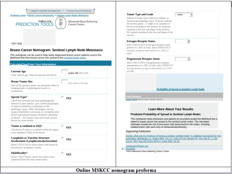
In view of this data and persistent comorbidities in post SLNB status it was suggested that SLNB can be avoided in low tumor burden patients.[19] Hence breast services at Memorial Sloan Kettering Cancer Centre; MSKCC developed nomogram to accurately predict probability of sentinel lymph node metastasis in breast cancer including isolated tumor cells. This nomogram was formulated based on multivariate analysis of database of 3786 patient’s following variables – age, tumor size, location, tumor type, LVI, multifocality, grade of tumor, ER status and PR status. It was validated in same study in about 1545 patients. This user-friendly prediction model is available on the Memorial Sloan-Kettering Cancer Centre (New York, NY) Web site (nomogram) based on a large data set to assist in predicting the presence of SLN metastasis. In today’s web oriented era patients want precise answers for their questions; they like to be informed precisely about their disease and chance of having positive SLN preoperatively. MSKCC nomogram is an effort to help patients in decision making regarding surgery and further treatment.
While SLN biopsy is an extremely safe and precise procedure with low morbidity, there are some patients, typically elderly these morbidities might be of great concern. In these patients if they have low burden disease, it has been suggested that they may be spared of SLNB as they pose low risk of axillary lymph node involvement.[19] It was thought that in this small patient population, the nomogram would help to weigh the risks and benefits of SLN biopsy. This nomogram was thought to be precise in selecting those patients at very low risk for SLN involvement who could be spared a SLN biopsy Alternatively, it may help surgeon reconsider an initial decision to spare a patient SLN biopsy in the event that a higher than expected risk is identified.
This nomogram has been validated by two other studies on larger basis. The current study is an effort to apply nomogram in Indian scenario. The feasibility of nomogram to predict probability of spread to sentinel lymph nodes in patients with early breast cancer will be studied by this study.
MATERIALS AND METHODS:
The current Prospective and retrospective was carried out from February 2013 to February 2016. All patients of early breast cancer with clinically node negative status were included in study. Patient’s clinical, histopathological [core biopsy] and radiological parameters were collected prior to surgery. All the patients were screened preoperatively clinically to mark tumor site, size and rule out positive axillary lymph nodes.
All patients underwent preoperative sono - mammography to know exact size, site, multi focality, multi centricity of tumor as well as axillary lymphnode status. All patients underwent core biopsy for diagnosis.
Patient’s tumor type, grade, estrogen receptor status[ER status], progesterone receptor status [PR status], Her 2 neu status, LVI were checked preoperatively. Probability of SLN metastasis was calculated for every patients by using MSKCC nomogram [which was in percentages] and was recorded.
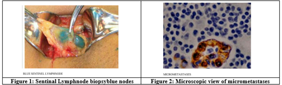
All patients underwent SLNB by dual tracer technique. Intraoperative frozen section analysis of sentinel lymph nodes was done for every patient and status of SLNB was noted. Post operatively final histopathology report was also checked to know status of SLN metastasis.
For retrospective data collection; all patients of early breast cancer with their true cut/ core biopsy report, mammography findings and clinical findings were traced from MRD collection. For the patients who underwent SLNB by dual tracer technique, axillary sampling frozen section report and final histopathology report were collected to know sentinel lymph node status.
Actual final histpath reports data of all 112 patients was compared to calculated predictability of positive SLNB by MSKCC nomogram for all 112 patients.
RESULTS:
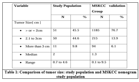
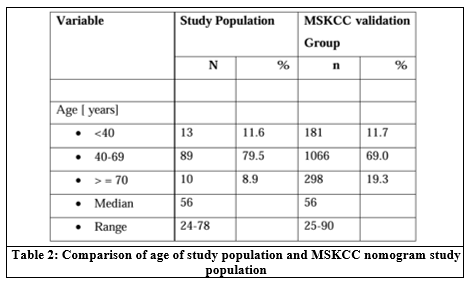
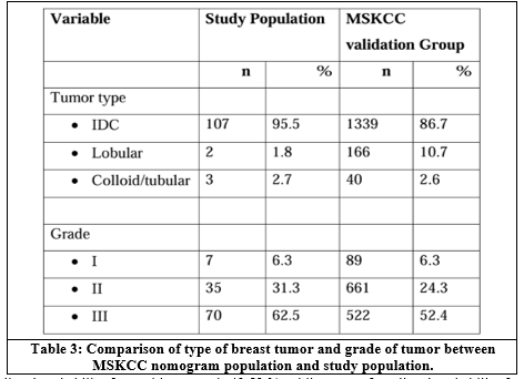
There were 13 patients who were below 40 yrs of age, 79 patients were in age group of 40 to 69 yrs and 10 patients were of 70yrs and above age. The mean age of study group was 54.81 yrs and median was 56 yrs. Range 24 to 78 yrs. Total 51 patients had tumor size of less than 2 cm, 2cm to 3cm tumor size was noted in 50 patients, and 11 patients had tumor size of more than 3 cm. The mean tumor size of population was 2.12 cm and median size was 2cm. Range of tumor size in study group was 0.7 to 4.6 cm. There were 55 cases with tumor of UOQ origin [49.1%], 27 from UIQ, 11from LIQ and 12 cases with tumor of LOQ. In 7 patients tumor was centrally located. Total 16 [14.3%] patients had multifocal breast tumor in study group than validation group [n = 390, 25.2%]. Total 107 [95.5%] patients reported IDC out of which grade 3 tumor was seen in 70 [62.5%], grade 2 in 35 [31.3%] and grade 1 in 7 patients [6.3%].
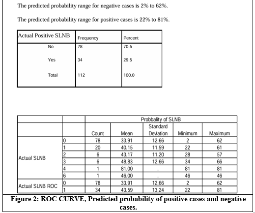
Two patients were diagnosed with lobular carcinoma, 2 with mucinous and 1 with tubular carcinoma from study population. Only two patients biopsy showed positive LVI status which was 2 % compared to22% of MSKCC validation population. Out of 112 patients 80 [71.4%] were Er positive, 75 patients [67%] were Pr positive, 15 cases tested Her positive ,89 Her negative, 8 Her equivocal. There were 23 [20.5%] triple negative cases, 89 patients were non TNBC. There were only 10 patients who were only Her positive. The study population is younger. The study population had larger tumors at presentation than western population; tumors seen were higher grade. On analysing it is seen that there were 34 cases of positive SLNB and 78 cases of negative SLNB.
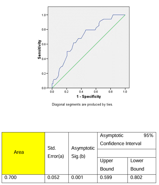
The mean of predicted probability for positive cases is 43.59 %, while mean of predicted probability fro negative cases is 33.92%. The predicted probability range for negative cases is 2% to 62%. The predicted probability range for positive cases is 22% to 81%. The area under the curve is 0.7 [p= 0.001] with 95% CI 0.599 to 0.802. This test showed that for positive SLNB cases the its predictive capability of MSKCC nomogram is more accurate than for negative cases.
The cut off point for maximum sensitivity and specificity for ROC curve of this analysis is 30.5%. The coordinates of ROC have shown that below and above this cut off point either the sensitivity or specificity decreases.
In original published paper of nomogram the categorization based on molecular subtypes of breast cancers was not done. In this study this categorization is done subsequent analysis was tried with help of Pearson correlation for molecular subtypes of breast cancer.
The guidelines to interpret Pearson Correlation coefficient are as follows:
Table 4
|
Strength of association |
Coefficient r |
|
|
|
Positive |
Negative |
|
Small |
0.1 to 0.3 |
-0.1 to -0.3 |
|
Medium |
0.3 to 0.5 |
-0.3 to -0.5 |
|
Large / strong |
0.5 to 1.0 |
-0.5 to -1.0 |
The hormone sensitive tumors showed medium strength correlation between actual positive SLNB and probability of positive SLNB. Pearson correlation coefficient is 0.317 [p= 0.004]. The correlation between Actual Positive SLNB and predicated probability of positive SLNB is weak for Her 2 neu positive patients [Pearson coefficient – 0.200]. This correlation is insignificant by Chi square test [p = 0.582].
There were 23 patients who were of triple negative pathology [20.5%]. The analysis showed negative correlation in all TNBC patients between probability of positive SLNB and actual positive SLNB Pearson correlation -0.010 [P =0.964].
DISCUSSION:
The first randomised control trial to substantiate evidence for modified radical mastectomy was NSABP B04 by Fisher B, Jeong JH, Anderson S, et al. The NSABP B-04 trial randomized patients with clinically negative nodes to one of three treatment regimens: radical mastectomy, total mastectomy with irradiation of the ipsilateral nodes, or total mastectomy alone with delayed axillary dissection. Patients with clinically positive nodes were randomized to radical mastectomy or total mastectomy with irradiation of the chest wall and regional lymphatics. About 62 % of patients from radical mastectomy underwent complete axillary node dissection which was not indicated; but added morbidity to them. About 18% patient from delayed node dissection and radiation group each developed metastatic disease in axilla in long term follow up. In spite of this overall survival and disease free survival in that trial was same in all three arms for node positive and negative disease at 25 years of follow-up. Patients with had delayed axillary dissection had equivalent survival. The results of this trial led to the conclusion that the mode and time of treatment of axillary nodes do not alter disease-free survival or overall survival. This trial also proved that total mastectomy or modified mastectomy is comparable to radical surgery in all aspects as well as it is less morbid.[20]
Milan 1, 2, 3 trials and NSABP6 confirmed role of radiation in breast conserving surgery.[21-25] One of the earliest randomized trials examining the use of SLNB was reported by Veronesi and colleagues in 2003.[5,12] They randomized 516 patients with breast cancer with tumors less than 2 cm in diameter to receive an SLNB followed by routine ALND or SLNB followed by an ALND only if the SLN contained metastases. The SLNs were identified in 98.5% of patients with a sensitivity of 91.2%, false negative rate was 8.8%. After 10 years of follow-up, no difference was observed between the groups for local axillary recurrence.
Another largest trial comparing SLNB with ALND in clinically node negative axilla was NSABPB 32.[6,7,11] Overall survival, disease-free survival, and regional control were statistically equivalent between groups. In addition, patients in the SLNB arm had a lower risk of morbidity compared with those in the standard ALND arm. In NSABP B32 trial it was seen that in patient who underwent SLNB and ALND , some patients had non sentinel lymph node that were negative on H and E staining but positive for micrometastasis on IHC staining. However these patients OS and DFS was same as other patients.
The study was undertaken to compare FNR [false negative rate] and cost of LAS at TMH Mumbai INDIA with SLNB in a consecutive series patients. The study was performed between March 2004 and December 2011 in 478 women with clinically node negative axilla. LAS and SLNB were performed in the same patient leading to comparison of each procedure with the gold standard (ALND).[8]
ALMANAC trial shows absolute risk of developing lymphedema 12 months after SLNB was 5% versus 13%(relative risk , 0.37; 95% CI, 0.23–0.60) in those who underwent an ALND. Similarly, the absolute risk of experiencing sensory loss at 12 months after SLNB was 11% compared with 31% of patients who had ALND (relative risk, 0.37; 95% CI, 0.27–0.50)8.
The ACOSOG Z0010 study[26] was designed to examine the incidence and prognostic significance of micro metastases in lymph nodes and bone marrow of women with early stage breast cancer. Patients with clinical T1 or T2, node-negative early-stage disease underwent lumpectomy, bone marrow aspiration, and SLNB. Those with negative sentinel nodes received no ALND or axillary treatment, while those with positive sentinel nodes or in whom the sentinel nodes were not identified underwent ALND. All patients then underwent breast irradiation and received systemic adjuvant therapy. ACOSOG Z0010 shows that micrometastasis detected by IHC does not affect OS if the patient is undergoing standard surgical excision along with adjuvant therapy. Adjuvant therapy taken in form of radiotherapy, chemotherapy and hormonal therapy take care of micrometastasis.
The American Joint Committee on Cancer (AJCC) defines macro metastases as foci of tumor cells greater than 2.0 mm, micro metastases are those foci measuring between 0.2 and 2.0 mm (pN1mi), and isolated tumor cells (pN0i+) foci measuring 0.2 mm or less. Micro metastases are found using deeper cross-sections and or immunohistochemistry (IHC).
An analysis of the Surveillance, Epidemiology, and End Results (SEER) data from 1998 to 2004 showed that 16% of SLN-positive patients did not undergo completion ALND. This proportion rose to 38% in those with micrometastasis. There were no differences in the incidence of axillary recurrence or in overall survival (OS) between patients who underwent ALND compared with those who did not.[27]
This raised the question of possible reduced need of ALND even in positive SLNB patients. This is because most patients of early breast cancer undergo breast conserving surgery along with radiation [with tangent field generally covering low axilla], systemic adjuvant therapy and hormonal therapy.
The ACOSOG Z0011 trial[10] presented by Dr. Giuliano and colleagues sought to examine whether SLND alone achieves similar locoregional control and survival rates compared to ALND in patients with positive SLNs who were undergoing lumpectomy with whole-breast radiation therapy. It enrolled clinically node-negative patients with T1 or T2 tumors who were treated with breast conservation surgery (BCS) and adjuvant radiotherapy who were found to have 1 or 2 positive SLN on standard pathologic examination with haematoxylin and eosin (H&E) staining(n = 891). They were randomized to receive ALND (n = 445) or no further axillary surgery (n = 446). Those with matted nodes or ≥ 3 positive sentinel nodes were excluded and all patients received breast-conserving therapy and whole-breast radiation therapy, as well as systemic adjuvant therapy as needed. No significant differences were seen in locoregional recurrence-free survival (P = .24), DFS (82% vs. 84%; P = .14), or OS (92% vs. 92.5%; P = 0.25) between ALND and SLNB, respectively.
Viviana Galimberti, MD, Bernard F. Cole, PhD, in their IBCSG 23-01 trial[28] for patients with early breast cancer tried to determine whether No ALND is non inferior to ALND. Eligibility was limited to patients with clinically-palpable axillary lymph node(s) and a primary tumour ≤ 5 cm who, after sentinel node biopsy, had one or more micrometastasis (≤ 2 mm) sentinel lymph nodes with no extracapsular extension. The primary endpoint disease-free survival (DFS) and OS was same.
These two trials i.e. ACOSOG Z 0011 and IBCSG 23-01 trial suggested and showed that in select patients with low axillary disease SLNB can substitute ALND even if SLNB is positive, provided that patients have favourable breast tumor size and grade, no extracapsular spread, receive radiotherapy and systemic adjuvant therapy.
Calculation of risk of positive lymph node metastasis was necessity not only in patients with favourable tumors but also for other patients, more so to counsel them as in era of multimedia and internet patients are well versed with disease.
The thought to calculate risk of positive SLNB for patients led to many studies. These studies identified variables as predictors of lymph node metastasis.[15-19] Tumor size, LVI [lympho vascular invasion], patient’s age, histological type of disease, multifocality, HER 2neu status tumor grade were known factors for lymph node metastasis. However, it is difficult to apply these data which is usually expressed as odds ratio to calculate the probability of sentinel lymph node (SLN) metastasis for a specific patient.
José Luiz B. Bevilacqua, Michael W. Kattan et al[29] developed a user-friendly prediction model (nomogram) based on a large data set to assist in predicting the presence of SLN metastasis. The main advantage of this nomogram is that it gives risk in percentages than odds ratio which is easy to explain to patients. This nomogram was developed with keeping the focus on consumer and patient use of information technologies eg, Internet, multimedia home-care informatics, computerized educational programs for medical information. This nomogram was an effort towards tailoring this information appropriately to individuals’ cancers, literacy, and culture-specific needs. It was an effort towards precise answers of patients’ questions. Clinical and pathologic features of 3,786 sequential SLN biopsy procedures were assessed with multivariable logistic regression to predict the presence of SLN metastasis in breast cancer. A nomogram was created from the logistic regression model. In multivariate analysis, variables that were associated with SLN metastasis in breast cancer were tumor type, LVI, tumor size, grade, tumor location, age, multifocality, and ER and PR status (P = .05 for all variables). There was no difference in frequency of SLN metastases between ductal and lobular tumor types (P= 0.25).This study identified that upper inner quadrant tumors are less likely to metastasize to axilla. ER and PR receptor status emerged as independent prediction factors in this study compared to all other studies. The frequency of SLN metastases in ER/PR-positive tumors were higher than those for ER/PR-negative tumors, corresponding to a small, but significant, increase of 2.0% for ER and 4.6% for PR.
A computerized version of the nomogram was developed and is available on the Memorial Sloan-Kettering Cancer Center (New York, NY) Web site. The model was subsequently applied to 1,545 sequential SLN biopsies. The nomogram was validated by accruing 1545 sequential SLNB. The nomogram analysis shows that area under the receiver operating characteristic curve of 0.754. This indicates that the nomogram is accurate in discriminating probable positive SLNB in every patient.
The rationale behind this nomogram was that in elderly patients with significant comorbidities; if consideration is to be given to avoid SLN biopsy because of comorbid conditions the nomogram could help to weigh the risks and benefits of SLN biopsy. Sentinel lymph node biopsy is an extremely safe procedure with low morbidity. This application was thought to be a potential help to physicians to select precisely those patients at very low risk for SLN involvement who could be spared an SLN biopsy. Alternatively, it might encourage a physician to reconsider an initial decision to spare the patient SLN biopsy in the event that a higher than expected risk is identified.
This study was validated at other centres as well M. Klar, MD, M. Foeldi, MD et al in their first validation study in German population found that collective tumor size, histology, lympho vascular infiltration, multifocality, Her-2-neu positivity, and nuclear grade correlated with the probability of SLN metastasis. The ROC of the validated nomogram in their breast cancer population revealed a value of 0.78 compared with 0.75 in the original publication.[30]
In a Dutch population, R.F.D. van la Parra a, C.M.T.P. Francissen et al[31] assessed the MSKCC nomogram to predict sentinel node metastases of breast cancer. They accrued 770 patients who underwent successful SLNB. Positive SLN were detected in 222 patients. They found that the nomogram estimated risk well, especially in the majority of patients at relatively low risk (<40%) for SLN metastases. Model discrimination was reasonable. The area under the ROC curve for the predictive nomogram was 0.67 (95% confidence interval 0.63–0.72) in Dutch population as compared to 0.75 in the original population.
As per all references over years ALND has been replaced by SLNB in clinically and radiologically negative axilla breast cancer. Initially even if one single SLNB was positive it was mandated to do ALND. However with new chemotherapy and targeted therapy lesions along with radiation therapy can take care of micrometastases. Patient with upto 3 positive SLNB can be spared of ALND specially in elderly and patient with Breast conserving surgery who eventually will require radiation. MSKCC nomogram tried to identified patient probability of SLNB positivity with reasonable accuracy even in validation study.
The present study was an effort to try MSKCC nomogram application in Indian scenario. The feasibility of nomogram to predict probability of spread to sentinel lymph nodes in patients with early breast cancer will be studied by this study.
CONCLUSION:
The study population is younger than MSKCC nomogram validation population. The study population had larger tumors at presentation than western population; tumors seen were higher grade. The cut-off point of predicted probability which has maximum sensitivity and specificity 30.5% for present study. The predicted probability range for negative cases is 2% to 62%. The predicted probability range for positive cases is 22% to 81%.
But the area under the curve is 0.7 [p= 0.001] with 95% CI 0.599 to 0.802. That means it may predict fairly about positive and negative SLN cases in present study. The MSKCC nomogram failed to predict probability of SLNB positivity in TNBC Indian patients. However it could predict SLNB positivity prior to surgery fairly well in hormone sensitive tumors.
In Indian population considering younger age, higher tumor size and higher grade of presentation may be reason of decreased negative predictive capability of MSKCC nomogram. Hence it cant be applied in Indian scenario to completely omit SLNB as per original MSKCC nomogram study.
In view of this nomogram can be applied for Indian scenario where logistics of SLNB is decreased in second and third tier cities to segregate patients requiring complete axillary dissection. Also there is always an option of second surgery as well as radiation to axilla.
REFERENCES:
[1] Halsted WS. The results of operations for the cure of cancer of the breast performed at the Johns Hopkins Hospital from June 1889 to January 1894. Annals of Surgery 1894;20(5):497–555.
[2] Meyer W. An improved method of the radical operation for carcinoma of the breast. New York Medical Record 1894;46(24):746-9.
[3] Halsted WS. Developments in skin grafting operation for cancer of the breast. Journal of the American Medical Association 1913;60(6):416-8.
[4] Veronesi U, Paganelli G, Viale G, et al. A randomized comparison of sentinel node biopsy with routine axillary dissection in breast cancer. N Engl J Med 2003;349(6):546.
[5] Veronesi U, Viale G, Paganelli G, et al. Sentinel lymph node biopsy in breast cancer: ten-year results of a randomized controlled study. Ann Surg 2010;251(4):595–600.
[6] Krag DN, Anderson SJ, Julian TB, et al. Technical outcomes of sentinel-lymph node resection and conventional axillary-lymph-node dissection in patients with clinically node-negative breast cancer: results from the NSABP B-32 randomised phase III trial. Lancet Oncol 2007;8(10):881-8.
[7] Krag D, Anderson S, Julian T, et al. Sentinel-lymph-node resection compared with conventional axillary-lymph-node dissection in clinically node-negative patients with breast cancer: overall survival findings from the NSABP B-32 randomised phase 3 trial. Lancet 2010;11(10):927-33.
[8] Mansel RE, Fallowfield L, Kissin M, et al. Randomized multicenter trial of sentinel lymph node biopsy versus standard axillary treatment in operable breast cancer: the ALMANAC trial. J Natl Cancer Inst 2006;98(9):599-609.
[9] Kell M, Burke JM, Barry M, et al. Outcome of axillary staging in early breast cancer: a meta-analysis. Breast Cancer Res Treat 2010;120(2):441-7.
[10] Giuliano AE, McCall LM, Beitsch PD, et al. ACOSOG Z0011: A randomized trial of axillary node dissection in women with clinical T1-2 N0 M0 breast cancer who have a positive sentinel node. J Clin Oncol 2010;28(15 suppl):69s.
[11] Ahikaga T, Krag D, Land S, et al. Morbidity results from the NSABP B-32 trial comparing sentinel lymph node dissection versus axillary dissection. J Surg Oncol 2010;102(2):111-8.
[12] Purushotham A, Upponi S, Klevesath MB, et al. Morbidity after sentinel lymph node biopsy in primary breast cancer: results from a randomized controlled trial. J Clin Oncol 2005;23(19):4312-21.
[13] Del Bianco P, Zavagno G, Burelli P, et al. Morbidity comparison of sentinel lymph node biopsy versus axillary lymph node dissection in breast cancer patients: results of the sentinella-GIVOM Italian randomised clinical trial. Eur J Surg Oncol 2008;34(5):508-13.
[14] Gill G. Sentinel-lymph-node-based management or routine axillary clearance? One year outcomes of sentinel node biopsy versus axillary clearance (SNAC): a randomized controlled surgical trial. Ann Surg Oncol 2008;16(2):266-75.
[15] Anan K, Mitsuyama S, Tamae K, et al. Axillary lymph node metastases in patients with small carcinomas of the breast: is accurate prediction possible? Eur J Surg 2000;166(8):610-5.
[16] Factors predicting the sentinel node metastases in T1 breast cancer tumor: an analysis of 1416 cases. Eur J Surg Oncol 2009;35(12):1245-9.
[17] Chua B, Ung O, Taylor R, et al. Frequency and predictors of axillary lymph node metastases in invasive breast cancer. ANZ J Surg 2001;71(12):723-8.
[18] Ramjeesingh R, Quan ML, Gardner S, et al. Prediction of involvement of sentinel and nonsentinel lymph nodes in a Canadian population with breast cancer. Can J Surg 2009;52(1):23-30.
[19] Yip CH, Taib NA, Tan GH, et al. Predictors of axillary lymph node metastases in breast cancer: is there a role for minimal axillary surgery? World J Surg 2009;33(1):54-7.
[20] Fisher B, Jeong JH, Anderson S, et al. Twenty-five-year follow-up of a randomized trial comparing radical mastectomy, total mastectomy, and total mastectomy followed by irradiation. N Engl J Med 2002;347(8):567-75.
[21] Fisher B, Anderson S, Bryant J, et al. Twenty-year follow-up of a randomized trial comparing total mastectomy, lumpectomy, and lumpectomy plus irradiation for the treatment of invasive breast cancer. N Engl J Med 2002;347(16):1233-41.
[22] Veronesi U, Cascinelli N, Mariani L, et al. Twenty-year follow-up of a randomized study comparing breast-conserving surgery with radical mastectomy for early breast cancer. N Engl J Med 2002;347(16):1227-32.
[23] Veronesi U, Volterrani F, Luini A et al. Quadrantectomy vs. lumpectomy for small size breast cancer. Eur J Cancer 1990;26(6):671-3.
[24] Veronesi U, Luini A, Del Vecchio M, et al. Radiotherapy after breast-preserving surgery in women with localized cancer of the breast. N Engl J Med 1993;328(22):1587-91.
[25] Veronesi U, Stafyla V, Luini A, et al. Breast cancer: from “maximum tolerable” to “minimum effective” treatment. Frontiers in Oncology 2012;2:125.
[26] Cote R, Giuliano E, Hawes D, et al. ACOSOG Z0010: A multicenter prognostic study of sentinel node (SN) and bone marrow (BM) micrometastases in women with clinical T1/T2 N0 M0 breast cancer. J Clin Oncol 2010; 28(15 suppl):69s.
[27] Yi M, Giordano S, Meric-Bernstam F, et al. Trends in and outcomes from sentinel lymph node biopsy (SLNB) alone vs. SLNB with axillary lymph node dissection for node-positive breast cancer patients: experience from the SEER database. Ann Surg Oncol 2010;17(Suppl 3):343-51.
[28] Galimberti V, Cole BF, Zurrida S, et al. IBCSG 23-01 randomised controlled trial comparing axillary dissection versus no axillary dissection in patients with sentinel node micro metastases. Lancet Oncol 2013;14(4):297-305.
[29] Bevilacqua JL, Kattan MW, Fey JV, et al. Doctor, What are my chances of having a positive sentinel node? A validated nomogram for risk estimation. J Clin Oncol 2007;25(24):3670-9.
[30] Klar M, Foeldi M, Markert S, et al. Good Prediction of the Likelihood for Sentinel Lymph Node Metastasis by Using the MSKCC Nomogram in a German Breast Cancer Population. Ann Surg Oncol 2009;16(5):1136-42.
[31] Van la Parra RF, Francissen CM, Peer PG, et al. Assessment of the Memorial SloanKettering Cancer Center nomogram to predict sentinel lymph node metastases in a Dutch breast cancer population. European Journal of Cancer 2013;49(3):564-71.