A Cross-Sectional Study of Cutaneous Manifestations in Patients with Diabetic Foot Ulcers Attending a Tertiary Care Hospital
- M Prasanna Kumari , Assistant Professor, Department of Dermatology, Venereology and Leprosy, Government Medical College, Vikarabad, Telangana, India.
- Goutham Krishna Reddy M , Assistant Professor, Department of General Surgery, Government Medical College, Vikarabad, Telangana, India.
- Shiva Kumar R , Assistant Professor, Department of General Surgery, Government Medical College, Vikarabad, Telangana, India.
Article Information:
Abstract:
Background: Diabetic foot ulcer is a serious complication of diabetes and is frequently accompanied by cutaneous changes that reflect neuropathy, vascular compromise, poor glycaemic control, and secondary infection. Careful documentation of these manifestations can support earlier recognition of high-risk feet and strengthen preventive care in tertiary settings. Objectives: To describe the spectrum of cutaneous manifestations in patients with diabetic foot ulcers and to evaluate their distribution in relation to clinical profile, ulcer characteristics, and glycaemic control. Methods: This hospital-based cross-sectional study was conducted at Government Medical College, Vikarabad, Telangana, from January 2025 to December 2025. One hundred adult patients with diabetic foot ulcers were enrolled. Demographic details, duration of diabetes, HbA1c status, comorbidities, ulcer site, duration, Wagner grade, and associated cutaneous findings were recorded through clinical examination and case-sheet review. Data were summarized using mean, frequency, and percentage. Results: The mean age of participants was 58.7 +/- 11.4 years, and 64% were men. Diabetes duration exceeded 10 years in 52% and poor glycaemic control was present in 62%. Toes were the commonest ulcer site (38%), while Wagner grade 2 ulcers predominated (42%). Xerosis was the leading cutaneous manifestation (72%), followed by fissures (48%), callosity (44%), hyperpigmentation (38%), and nail changes (36%). Interdigital fungal infection and tinea pedis were noted in 28% and 24%, respectively. Multiple cutaneous findings were frequent, with three manifestations in 32% and four or more in 28%. Selected manifestations were more concentrated among patients with HbA1c >8%. Conclusion: Cutaneous abnormalities were highly prevalent among patients with diabetic foot ulcers, particularly xerosis, fissuring, callosity, fungal infection, and nail dystrophy. These findings clustered in patients with longstanding diabetes, neuropathy, and poor glycaemic control, underscoring the need for integrated dermatologic and diabetic foot assessment in tertiary care practice.
Keywords:
Article :
INTRODUCTION:
Diabetes mellitus is a major chronic metabolic disorder with multisystem consequences, and the foot remains one of the most vulnerable target sites because of the combined effects of neuropathy, peripheral arterial disease, biomechanical stress, impaired immunity, and infection [1-3]. Diabetic foot disease imposes a substantial clinical and economic burden across the world and continues to be a leading cause of hospitalization, non-traumatic lower limb amputation, prolonged disability, and recurrent morbidity among people living with diabetes [1-3]. Global epidemiological evidence indicates that diabetic foot ulceration is common and is encountered more often in men, in older age groups, and in patients with longer duration of diabetes and associated vascular risk factors [2,7].
The cutaneous manifestations seen around a diabetic foot ulcer are not merely secondary surface findings; they often represent visible markers of deeper metabolic, neurologic, and vascular disturbance [4-6]. Xerosis, fissuring, callosity, nail dystrophy, hyperpigmentation, edema, fungal infection, and trophic skin changes can emerge because of autonomic denervation, barrier dysfunction, altered keratinization, repeated mechanical pressure, and reduced perfusion [4,5]. These changes compromise the integrity of the skin, facilitate microbial entry, increase the risk of ulcer progression, and reduce the capacity of the foot to withstand minor trauma [4,5].
Clinical reviews of diabetic foot disease consistently emphasize that neuropathy, plantar pressure, callus formation, infection, and peripheral arterial insufficiency act in concert rather than in isolation [5,8]. In daily practice, however, the dermatologic aspects of the diabetic foot are often under-documented when compared with ulcer depth, infection severity, or surgical outcome. Yet, recognition of associated cutaneous abnormalities has practical relevance because these findings can guide skin care, pressure off-loading, footwear counselling, fungal treatment, and intensified glucose control [3,8]. Their documentation is particularly relevant in tertiary care hospitals, where patients frequently present with longstanding disease, recurrent ulcers, and multiple coexisting risk factors.
Despite the clinical importance of these manifestations, local hospital-based descriptive data from Indian tertiary care settings remain limited. A systematic characterization of the skin changes accompanying diabetic foot ulcers can help refine bedside assessment and improve preventive strategies for recurrence and progression. Therefore, the present study was undertaken to describe the sociodemographic and clinical profile of patients with diabetic foot ulcers, to document the frequency and pattern of associated cutaneous manifestations, and to examine their distribution in relation to ulcer characteristics and glycaemic control.
METHODOLOGY:
Study design and setting.
This hospital-based cross-sectional study was carried out at Government Medical College, Vikarabad, Telangana, India, over a 12-month period from January 2025 to December 2025. The study was designed to describe the cutaneous manifestations associated with diabetic foot ulcers among adult patients attending a tertiary care institution. Patients were evaluated in the relevant outpatient and inpatient services where diabetic foot cases were routinely managed. The study followed standard clinical principles used in diabetic foot assessment, including ulcer grading and focused evaluation for infection, neuropathy, and vascular insufficiency [8-11].
Study population.
Adults aged 18 years and above with established diabetes mellitus and a clinically diagnosed diabetic foot ulcer were considered eligible for inclusion. Both men and women were enrolled. A diabetic foot ulcer was considered as a break in the skin involving the foot in a patient with diabetes, with or without evidence of infection or surrounding trophic change, consistent with accepted clinical descriptions in the diabetic foot literature [3,8]. Patients with traumatic ulcers unrelated to diabetes, venous ulcers, vasculitic ulcers, malignancy-associated ulcers, pressure injuries unrelated to diabetic pathology, and those unwilling to participate were excluded. Cases in whom detailed dermatologic examination of the foot was not feasible were also excluded.
Sample size and sampling.
A total of 100 patients fulfilling the inclusion criteria were included in the final analysis. Consecutive sampling was used, and all eligible patients presenting during the study period were enrolled until the target sample size was achieved. Because the primary objective was descriptive, the sample was intended to provide a practical clinical profile of the pattern of skin changes observed in diabetic foot ulcer patients attending the institution.
Data collection procedure.
After obtaining informed consent, each participant underwent structured history taking, review of diabetic status, and detailed clinical examination. Information recorded included age, sex, duration of diabetes, glycaemic control based on HbA1c category, smoking history, hypertension, peripheral neuropathy, and peripheral vascular disease. Ulcer-related variables included anatomical site, duration, clinical evidence of discharge, foul smell, surrounding cellulitis, and Wagner grade, which was used for descriptive ulcer stratification [8]. A focused dermatologic examination of both feet and lower limbs was performed to document xerosis, fissures, callosity, hyperpigmentation, nail changes, interdigital fungal infection, tinea pedis, edema, shiny atrophic skin, loss of hair, bullae, and eczematous changes. The clinical interpretation of these manifestations was informed by existing dermatologic and diabetic foot literature [4,5,12-14].
Statistical analysis.
Data were entered into a spreadsheet and analyzed using standard descriptive statistical methods. Continuous variables were summarized as mean with standard deviation, whereas categorical variables were expressed as frequency and percentage. Cross-tabulation was used to describe the distribution of selected cutaneous manifestations according to glycaemic control categories. The results are presented in tabular form with accompanying narrative interpretation.
Ethical considerations.
The study was conducted after approval from the Institutional Ethics Committee of Government Medical College, Vikarabad. Written informed consent was obtained from all participants before inclusion. Confidentiality of patient information was maintained throughout data collection, analysis, and manuscript preparation. Clinical care was not altered by participation in the study.
RESULTS:
A total of 100 patients with diabetic foot ulcers were included in this cross-sectional study. The results were analyzed with respect to demographic profile, diabetes-related characteristics, ulcer features, and associated cutaneous manifestations. The sociodemographic and clinical profile of the study population is summarized in Table 1.
Table 1. Sociodemographic and clinical characteristics of study participants (n = 100)
|
Variable |
Number |
Percentage (%) |
|
Age group (years) |
||
|
31-40 |
8 |
8.0 |
|
41-50 |
18 |
18.0 |
|
51-60 |
36 |
36.0 |
|
61-70 |
28 |
28.0 |
|
>70 |
10 |
10.0 |
|
Sex |
||
|
Male |
64 |
64.0 |
|
Female |
36 |
36.0 |
|
Duration of diabetes |
||
|
<5 years |
14 |
14.0 |
|
5-10 years |
34 |
34.0 |
|
>10 years |
52 |
52.0 |
|
Glycaemic control |
||
|
HbA1c <=8% |
38 |
38.0 |
|
HbA1c >8% |
62 |
62.0 |
|
Associated comorbidities |
||
|
Hypertension |
48 |
48.0 |
|
Peripheral neuropathy |
58 |
58.0 |
|
Peripheral vascular disease |
26 |
26.0 |
|
Smoking history |
32 |
32.0 |
As shown in Table 1, the majority of patients were in the age group of 51-60 years (36%), followed by 61-70 years (28%). The mean age of the study population was 58.7 +/- 11.4 years. Men constituted 64% of the participants, indicating a male predominance. More than half of the patients had diabetes for more than 10 years (52%), and poor glycaemic control with HbA1c >8% was observed in 62% of cases. Peripheral neuropathy was present in 58% of patients, while 48% had coexisting hypertension. The distribution of ulcer-related characteristics is presented in Table 2.
Table 2. Characteristics of diabetic foot ulcers (n = 100)
|
Variable |
Number |
Percentage (%) |
|
Site of ulcer |
||
|
Plantar surface |
34 |
34.0 |
|
Dorsum of foot |
12 |
12.0 |
|
Toes |
38 |
38.0 |
|
Heel |
16 |
16.0 |
|
Duration of ulcer |
||
|
<1 month |
22 |
22.0 |
|
1-3 months |
46 |
46.0 |
|
>3 months |
32 |
32.0 |
|
Wagner grade |
||
|
Grade 1 |
18 |
18.0 |
|
Grade 2 |
42 |
42.0 |
|
Grade 3 |
26 |
26.0 |
|
Grade 4 |
14 |
14.0 |
|
Ulcer discharge |
54 |
54.0 |
|
Foul smell |
37 |
37.0 |
|
Surrounding cellulitis |
41 |
41.0 |
Table 2 shows that the toes were the most common site of ulceration, accounting for 38% of cases, followed by the plantar surface in 34%. Nearly half of the ulcers had a duration of 1-3 months (46%). Wagner grade 2 ulcers were the most frequently observed lesions (42%), followed by grade 3 ulcers in 26%. Clinical signs suggestive of infection were common, with discharge seen in 54%, surrounding cellulitis in 41%, and foul smell in 37% of patients. The frequency of associated cutaneous manifestations is detailed in Table 3.
Table 3. Frequency of cutaneous manifestations associated with diabetic foot ulcers (n = 100)
|
Cutaneous manifestation |
Number |
Percentage (%) |
|
Xerosis (dry skin) |
72 |
72.0 |
|
Fissures/cracks |
48 |
48.0 |
|
Callosity |
44 |
44.0 |
|
Hyperpigmentation |
38 |
38.0 |
|
Nail changes (dystrophy/discoloration) |
36 |
36.0 |
|
Interdigital fungal infection |
28 |
28.0 |
|
Tinea pedis |
24 |
24.0 |
|
Edema |
33 |
33.0 |
|
Shiny atrophic skin |
30 |
30.0 |
|
Loss of hair over lower limb |
27 |
27.0 |
|
Bullae/blisters |
12 |
12.0 |
|
Dermatitis/eczematous changes |
10 |
10.0 |
As presented in Table 3, xerosis was the most common cutaneous finding, present in 72% of patients. Fissures were noted in 48% and callosity in 44%, indicating a frequent combination of skin dryness and pressure-related hyperkeratosis. Hyperpigmentation was seen in 38%, while nail changes were documented in 36% of cases. Interdigital fungal infection and tinea pedis were observed in 28% and 24% of patients, respectively. Features suggestive of trophic or vascular compromise, such as shiny atrophic skin and loss of hair over the lower limb, were noted in 30% and 27% of participants. The number of cutaneous manifestations seen per patient is shown in Table 4.
Table 4. Number of cutaneous manifestations per patient (n = 100)
|
Number of manifestations |
Number |
Percentage (%) |
|
One manifestation |
14 |
14.0 |
|
Two manifestations |
26 |
26.0 |
|
Three manifestations |
32 |
32.0 |
|
Four or more manifestations |
28 |
28.0 |
Table 4 demonstrates that most patients had multiple cutaneous findings. Three manifestations were present in 32% of patients, while 28% had four or more manifestations. Only 14% of participants had a single cutaneous manifestation, indicating that diabetic foot ulcers were usually accompanied by a cluster of skin and appendageal abnormalities rather than by an isolated lesion.The distribution of selected cutaneous manifestations according to glycaemic control is shown in Table 5.
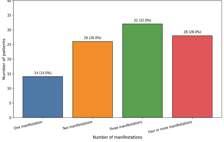
Figure 1: Number of cutaneous manifestations per patient
As evident from Table 5, selected cutaneous manifestations were more frequently observed among patients with poor glycaemic control. Of the 72 patients with xerosis, 51 had HbA1c >8%. Likewise, fissures were more common in poorly controlled diabetes, being present in 35 of 62 such patients. Fungal infections and nail changes also showed a greater concentration among patients with elevated HbA1c levels. Overall, the findings indicate that xerosis, fissuring, callosity, fungal involvement, and nail dystrophy formed the predominant dermatologic profile among patients with diabetic foot ulcers in this tertiary care cohort
Table 5. Distribution of selected cutaneous manifestations according to glycaemic control (n = 100)
|
Cutaneous manifestation |
HbA1c <=8% (n = 38) |
HbA1c >8% (n = 62) |
Total |
|
Xerosis |
21 |
51 |
72 |
|
Fissures/cracks |
13 |
35 |
48 |
|
Callosity |
14 |
30 |
44 |
|
Interdigital fungal infection |
7 |
21 |
28 |
|
Nail changes |
10 |
26 |
36 |
.
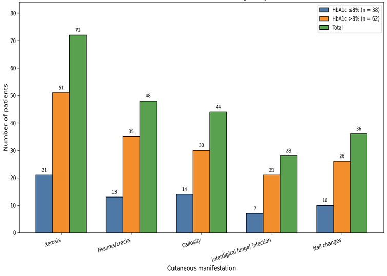
Figure 2: Distribution of selected cutaneous manifestations according to glycaemic control
DISCUSSION:
The present study provides a descriptive account of the cutaneous spectrum associated with diabetic foot ulcers in a tertiary care hospital. Most participants were older adults, and men constituted nearly two thirds of the study population. This profile is consistent with the broader epidemiology of diabetic foot ulceration described in systematic reviews, which report greater burden among males, older patients, and those with longer duration of diabetes [7]. The high proportion of patients with diabetes exceeding 10 years and HbA1c >8% in the present series reinforces the established relationship between chronic hyperglycaemia, cumulative tissue injury, and ulcer risk [7].
Peripheral neuropathy was present in more than half of the patients, and toes and plantar surfaces were the most frequently involved sites. These findings are pathophysiologically coherent. Neuropathy reduces protective sensation, alters foot biomechanics, and promotes repeated unrecognized trauma, while autonomic dysfunction contributes to dry skin and fissuring [8]. In classic and contemporary diabetic foot literature, neuropathy, deformity, callus formation, plantar pressure, and trauma are repeatedly identified as central pathways leading to ulceration [8]. The predominance of Wagner grade 2 ulcers in this cohort suggests that many patients presented after extension beyond superficial skin involvement but before the most advanced destructive stages became universal. The substantial proportion with discharge, foul smell, and cellulitis also highlights the persistent overlap between chronic ulceration and infection in tertiary care practice [9].
Among the dermatologic findings, xerosis emerged as the most frequent manifestation, followed by fissures and callosity. This pattern is strongly supported by dermatologic reviews describing xerosis and hyperkeratotic changes as common visible expressions of barrier dysfunction, autonomic denervation, and repetitive mechanical stress in diabetes [13,14]. Xerotic and cracked skin is clinically important because it weakens the cutaneous barrier and creates portals of entry for microbial colonization and infection. Callosity deserves particular attention because it reflects sustained focal pressure and is recognized as an important precursor of ulcer development and recurrence. The frequent coexistence of multiple skin findings in the present study indicates that diabetic foot ulcer patients often have cumulative rather than isolated dermatologic damage.
Fungal infection and nail changes were also notable in this series. Interdigital fungal infection was present in 28% and tinea pedis in 24%, while nail dystrophy or discoloration occurred in more than one third of patients. These observations are in agreement with previous work showing that fungal infections of the feet and nails are common in diabetes and are more frequent in individuals with poor glycaemic control and peripheral vascular compromise [12]. In practical terms, untreated interdigital maceration, tinea pedis, and onychodystrophy can aggravate skin breakdown, encourage secondary bacterial infection, and complicate foot hygiene. Similarly, shiny atrophic skin, edema, and loss of hair over the lower limb in the present study point toward a vascular component that deserves active evaluation, because peripheral arterial disease is closely linked with impaired healing and amputation risk [10,11].
Taken together, the findings support a broader clinical approach in which diabetic foot evaluation extends beyond ulcer size and depth to include systematic assessment of the surrounding skin, nails, and trophic changes. Early identification of xerosis, fissuring, callosity, fungal infection, and trophic vascular signs can inform skin hydration measures, callus care, antifungal treatment, pressure redistribution, vascular work-up, and intensified metabolic control. In tertiary hospitals, where patients often present late and with multiple overlapping risk factors, such integrated assessment has direct relevance for preventing progression, recurrence, and avoidable limb loss.
Limitations
This study was conducted in a single tertiary care hospital and included only 100 patients, which restricts external validity. The cross-sectional design captured associations at one time point and did not establish temporal sequence. Microbiological confirmation of fungal infections, vascular imaging, and formal neuropathy scoring were not uniformly incorporated. Community cases and milder lesions managed outside the hospital setting were not represented.
CONCLUSION:
Cutaneous manifestations were highly prevalent among patients with diabetic foot ulcers in this tertiary care hospital, with xerosis, fissures, callosity, hyperpigmentation, nail dystrophy, and fungal infection forming the dominant clinical pattern. These abnormalities were commonly accompanied by longstanding diabetes, poor glycaemic control, peripheral neuropathy, and signs of infection or trophic compromise. The clustering of multiple skin findings in a large proportion of patients shows that diabetic foot ulceration is rarely an isolated wound problem. Routine diabetic foot assessment should therefore include systematic examination of the surrounding skin and nails, as this can strengthen risk stratification, support earlier intervention, and improve comprehensive limb-preserving care.
REFERENCES:
1. Boulton AJM, Vileikyte L, Ragnarson-Tennvall G, Apelqvist J. The global burden of diabetic foot disease. Lancet. 2005;366(9498):1719-1724.
2. Zhang P, Lu J, Jing Y, Tang S, Zhu D, Bi Y. Global epidemiology of diabetic foot ulceration: a systematic review and meta-analysis. Ann Med. 2017;49(2):106-116.
3. Armstrong DG, Tan T-W, Boulton AJM, Bus SA. Diabetic Foot Ulcers: A Review. JAMA. 2023;330(1):62-75.
4. Lima AL, Illing T, Schliemann S, Elsner P. Cutaneous Manifestations of Diabetes Mellitus: A Review. Am J Clin Dermatol. 2017;18(4):541-553.
5. Troskot N, Duvancic T, Kolic M. Diabetic foot syndrome--dermatological point of view. Acta Clin Croat. 2013;52(1):99-106.
6. Kim J. The pathophysiology of diabetic foot: a narrative review. J Yeungnam Med Sci. 2023;40(4):328-334.
7. Tang WH, Zhao YN, Cheng ZX, Xu JX, Zhang Y, Liu XM. Risk factors for diabetic foot ulcers: a systematic review and meta-analysis. Vascular. 2024;32(3):661-669.
8. Frykberg RG, Armstrong DG, Giurini J, Edwards A, Kravette M, Kravitz S, et al. Diabetic foot disorders: a clinical practice guideline. American College of Foot and Ankle Surgeons. J Foot Ankle Surg. 2000;39(5 Suppl):S1-S60.
9. Lipsky BA, Senneville E, Abbas ZG, Aragon-Sanchez J, Diggle M, Embil JM, et al. Guidelines on the diagnosis and treatment of foot infection in persons with diabetes (IWGDF 2019 update). Diabetes Metab Res Rev. 2020;36 Suppl 1:e3280.
10. Hinchliffe RJ, Forsythe RO, Apelqvist J, Boyko EJ, Fitridge R, Hong JP, et al. Guidelines on diagnosis, prognosis, and management of peripheral artery disease in patients with foot ulcers and diabetes (IWGDF 2019 update). Diabetes Metab Res Rev. 2020;36 Suppl 1:e3276.
11. Brownrigg JRW, Schaper NC, Hinchliffe RJ. Diagnosis and assessment of peripheral arterial disease in the diabetic foot. Diabet Med. 2015;32(6):738-747.
12. Akkus G, Evran M, Gungor D, Karakas M, Sert M, Tetiker T. Tinea pedis and onychomycosis frequency in diabetes mellitus patients and diabetic foot ulcers. A cross sectional - observational study. Pak J Med Sci. 2016;32(4):891-895.
13. Parker J, Scharfbillig R, Jones S. Moisturisers for the treatment of foot xerosis: a systematic review. J Foot Ankle Res. 2017;10:9.
14. Lechner A, Akdeniz M, Tomova-Simitchieva T, Bobbert T, Moga A, Lachmann N, et al. Comparing skin characteristics and molecular markers of xerotic foot skin between diabetic and non-diabetic subjects: An exploratory study. J Tissue Viability. 2019;28(4):200-209.