Anaesthetic Management of Abdominal Hysterectomy for Adnexal Mass in Elderly Lady with Corrected CHD and Co-Existing CAD - A Case Report
- Sirpikha V , Postgraduate in Anesthesia, Department of Anaesthesia, Dhanalakshmi Srinivasan Medical College and Hospital, Siruvachur, Perambalur, Tamil Nadu, India.
- Swetha Reddy Shetty , Postgraduate in Anesthesia, Department of Anaesthesia, Dhanalakshmi Srinivasan Medical College and Hospital, Siruvachur, Perambalur, Tamil Nadu, India.
- Geetha J. , Professor, Department of Anaesthesia, Dhanalakshmi Srinivasan Medical College and Hospital, Siruvachur, Perambalur, Tamil Nadu, India.
Article Information:
Abstract:
Health awareness and access have improved longevity along with rising trend of geriatric population presenting with complex and contrasting comorbidities. Atrial septal defects constitute 5-10% of congenital heart diseases. Although they survive to adulthood an unrepaired ASD leads to long term exposure to chronic right heart volume overload causing atrial arrythmias, pulmonary vascular disease and right heart failure. These are often the precipitating factors necessitating ASD closure in adulthood. ASD with poor reduce left ventricular compliance enhances left to right shunt while reduced right ventricular compliance diminishes and may also reverse the shunt, age and post ASD closure have their own sequalae. This case is presented to direct a clear understanding of hemodynamics in a patient with both raised right and left atrial pressure and reduced threshold for Tachyarrythmias.
Keywords:
Article :
CASE REPORT:
History
65 yrs old thin lady presented with vague abdominal pain and diagnosed as adnexal mass in post-menopausal state. Closure of atrial septal defect was done with pericardial patch 15 yrs ago at the age of 50. Known to have had systemic hypertension since 1 month and on AMLONG twice a day. She has not been on any antiplatelets or anticoagulants medications. History of palpitations on and of was present but no syncopal attacks.
Clinical Features
This elderly lady was conscious, oriented, anxious, talkative with a clinical Frailty score of 5 had a pulse rate of 68/min and blood pressure off 110/70mmhg mercury. BMI of 27.1, mid sternal scar+ with wires felt subcutaneously. Chest clear, CVS nil abnormality detected on auscultating.
ECG: Sinus Rhythm heart rate 72 per minute regular
X-Ray Chest: Emphysematous chest, sternal wires+ tubular heart +
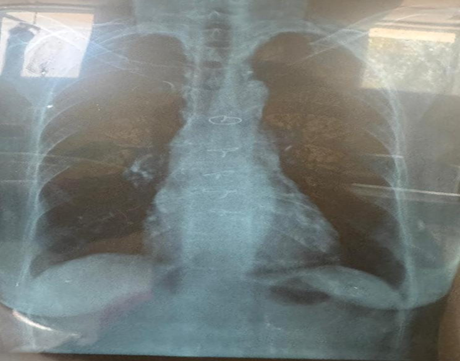
Figure 1
Table 1
|
Hb |
13gms |
Platelets |
28lakhs/cumm |
Creatinine |
0.6mg/dl |
|
BT |
2min45sec |
Sugar |
102mg/dl |
Sodium |
137mmol/ltr |
|
CT |
4min30sec |
urea |
17mg/dl |
potassium |
4.0mmol/ltr |
Table 2
|
Before ASD closure |
After ASD closure |
Pre-operative |
|
Large ostium secundum ASD |
Pericardial ASD patch+ |
ASD patch closure, no residual shunt |
|
L R shunt |
Cardiomegaly |
bicuspid aortic valve |
|
Normal LV function |
Dilated pulmonary artery |
CAD+LAD dilated, hypokinetic basal and inferior wall |
|
Bicuspid aortic valve |
Bicuspid aortic valve |
EF=55% MR+ No PAH+ AML Prolapse |
|
Normal coronaries |
Atherosclerotic aorta |
|
ECHO Reports
Problems and Risks
· Age related
· bicuspid aortic valve
· dilated LA systolic LV dysfunction
· ASD closure done 25yrs ago
· PAH
· Poor respiratory reserve
· CAD, CCF, CVA
· IE
· Acute AR/LV FAILURE
· Pre-op IHD/ arrhythmia
· High risk consent
Goals of Anaesthesia
· Aim of adequate tissue oxygenation and cardiac output.
· Prevent rise in pulmonary pressures- as LA is already enlarged.
· Avoid-laparoscopy and hypercarbia as Trendelenberg, rise in intra-abdominal pressure both could increase airway pressure that is detrimental to left atrial preload, left atrium is already enlarged and left ventricle has regional wall motion abnormalities.
· Avoid tachycardia and hypertension ass sympathetic stimulation could increase myocardial oxygen demand and reduce oxygen supply which could precipitate a coronary event namely myocardial ischemia.
· Maintain normovolemia replacing blood for blood, prevent crystalloid overload and ensure adequate urine output of 0.5-1ml/kg/hour.
· Adequate pain relief to prevent sympathetic stimulation and activation of renin angiotensin mechanism.
Optimization
· Poor respiratory reserve evidenced by sabarasez breath holding test (9secs) improved by deep breathing exercises, incentive spirometry, and desensitization and humidification of respiratory tract by Budecort nebulization every eight hours.
· Cilnidipine on which the patient is already on to be continued.
· Metoprolol 25 XL once daily to be started.
· Benzodiazepines namely alprazolam 0.5mg every night for calming down.
· Planning for anticoagulation in the post-operative to prevent left atria clot and thromboembolism. Baseline coagulation profile was available for comparision.
· Clinically frail lady with evidence of moderate degenerative changed in the vertebral bodies and hemodynamic management made regional analgesia questionable in this patient.
Plan of Anesthesia
Two good IV accesses, oxygenation and the benzodiazepine in the pre-operative area following positive counselling and reassurance.
General Anaesthesia
Monitors: standard monitors with continuous ST segment monitoring of all leads along with QT interval monitoring, capnography.
· Monitoring of hourly urine output.
Induction
Fentanyl 2mcg/kg, Propofol 2mg/kg and Vecuronium 0.1mg/kg, lignocaine 2mg/kg, preloaded nitroglycerine and dopamine were ready to face hemodynamic changes.
Intubation
7mm endotracheal intubation using 4 size Mccoy blade for minimal manoevering.
Maintenance
IPPV/ TV-400ml/ RR-12/ I: E= 1:2/ PEEP 4.
Monitors
Airway pressure was maintained around 14cmH2O, ST <0.5mm and QT around 420 milliseconds. Blood loss calculated as 400ml ovarian mass was excised along with uterus and bilateral adnexae.
Extubation was smooth following reversal of residual neuromuscular blockade with neostigmine 25mg and glycopyrrolate 0.6mg. Post-operative period oxygenation by mask for 4 hours, heparin 5000 units subcutaneously after 6 hours was started. Immediate post-op ECG showed no fresh ST-T changes and heart rate was 72/min with sinus rhythm and BP was 110/70mmhg as patient was sensitive to fentanyl she was found comfortable with tramadol 5omg IV and paracetamol 1g IV every 8 hours.
Post-Operative Event
By the end of first POD patient had an argument with her son and her heart rate increased to 120/min as sinus tachycardia with rhythm becoming supraventricular and stabilized as atrial fibrillation at 182/min. patient was conscious, oriented with no chest pain or dyspnea, troponins were negative.
The heart rate was stabilized with oral metoprolol 25mg, oral verapamil and Amiodarone bolus 150mg followed by infusion of Amiodarone 1mg/minute for 6 hours and 0.5m/min for 18hrs.
The patient was well, stable and shifted to ward on 4th post-operative day and discharged on 7th post-operative day and discharged on 7th day with stabilized atrial fibrillation and oral antiplatelet metoprolol and verapamil for her cardiac status.
This case is presented to emphasize how post-operative monitors is important and the fact that it can be more eventful than the intra-operative period.
DISCUSSION:
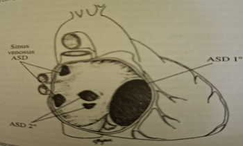
Figure 2
Discussion of this case shall be on understanding the complex pathophysiology and hemodynamics in this case. First is adulthood ASD presentation, surgery for AD followed by coronary artery disease, regional wall motion abnormalities in echocardiogram, left atrial dilation, post-menopausal ovarian cyst, arrhythmias in the post-operative period and some common drugs in the event of arrythmias namely Lignocaine, Verapamil, Amiodarone and Beta blockers and other suggested drugs in refractory cases.
Adulthood ASD
Atrial communications may go unrecognised into adulthood due to subtle clinical symptoms and manifestations. ASD is the most common form of congenital heart disease found among adults. Our patient has bicuspid aortic valve and anterior mitral leaflet prolapse as well. An unrepaired ASD reduces overall life expectancy of the individual due to long-term exposure to chronic right heart volume overload. Subsequently atrial arrhythmias, pulmonary vascular disease and right heart failure could ensue.[1] Four types of atrial communication exist: ostium primum, ostium secundum, sinus venosus and coronary sinus defects. Ostium secundum defects are the only true atrial septal defects; all others are atrial level shunts as they are not surrounded by true atrial septal tissue.[2] In any type of atrial level shunt, the degree Left to right atrial shunting depends on the size of the defect and relative diastolic filling properties of the two ventricles.
Our patient had an enlarged left atrium and increase in left atrial pressure and coronary artery disease reducing the compliance of left ventricle. Consequently, the left to right shunt would have increased. Significant pulmonary hypertension at the age of 50 necessitated ASD closure and the sequelae of effects of age and stress-related coronary artery disease followed.
Surgical closure of an ASD improves the functional status and exercise capacity in symptomatic patients and improves survival rates. It also improves or eliminates congestive heart failure, especially when patients are operated at a younger age, namely less than 25 years. However, surgical closure of ASDs in adult life does not prevent atrial fibrillation, flutter or stroke, especially when patients are Operated after 40 years, a left to right shunt is considered significant when the ratio of pulmonary to systemic blood flow or shunt fraction Qp /Qs is more than 1.5 : 1.0.
Mechanisms of Cardiac Arrhythmias
Electrical signalling in the heart involves passage of ions through ion channels. The sodium, potassium, calcium and chloride ions are the major charge carriers and their movement across the cell membrane through channel pores creates a flow of current that generates excitation and signals in cardiac myocytes. Opening of ion channels allows selected ions to flow passively down the electrochemical activity gradient at a very high rate, more than 10⁶ ions per second. The high transfer rates and restriction to downhill fluxes not coupled to hydrolysis of energy-rich phosphates distinguish ion channel mechanisms from pumps and exchangers.[3] The AV node and His bundle region are innervated by a rich supply of cholinergic and adrenergic fibers, with densities exceeding those found in ventricular myocardium. The vagus nerve modulates cardiac sympathetic activity at preganglionic and postganglionic sites by regulating the amount of epinephrine released and by inhibiting cyclic AMP–induced phosphorylation of cardiac proteins including ion channels and calcium pumps. Tonic vagal stimulation can produce a greater absolute reduction in the sinus rate in the presence of tonic background sympathetic stimulation, a phenomenon termed accentuated antagonism. In contrast, changes in AV conduction during concomitant sympathetic and vagal stimulation are the algebraic sum of individual effects of tonic vagal and sympathetic stimulation alone.
Cardiac responses to brief vagal bursts begin after a short latency and dissipate quickly, whereas cardiac responses to sympathetic stimulation commence and dissipate slowly. The rapid onset and offset of responses to vagal stimulation allow dynamic beat-to-beat vagal modulation of heart rate and AV conduction, whereas the slow temporal response to sympathetic stimulation precludes any beat-to-beat regulation by sympathetic activity.
Sympathetic stimulation increases and reverses the spatial gradients of ventricular repolarization, as the direction of repolarization shifts from apex-to-base in sinus rhythm to base-to-apex. Nonuniform distribution of sympathetic nerve contributes to nonuniform electrophysiologic effects. The ventricular content of norepinephrine is greater at the base than at the apex of the heart. Thus, both direct and reflex sympathetic stimulation increase regional differences in cardiac repolarization. Afferent vagal activity appears to be higher in the posterior ventricular myocardium. This may account for vasoactive vagomimetic effects of inferior wall myocardial infarction. The dispersion of repolarization is significantly enhanced in ischemic cardiomyopathy.[4]
Mechanisms of Arrhythmogenesis Include
a) Disorders of impulse formation
b) Abnormal automaticity
c) Triggered activity
d) Delayed afterdepolarizations
e) Intracellular Ca++ ions overload handling
f) Early afterdepolarization
g) Parasystole.
Disorders of impulse formation include re-entry phenomena, spatio-temporal organization and focal discharge (AF).
Bicuspid Aortic Valve
The case presented in this report operated for closure of secundum atrial septal defect also had bicuspid aortic valve. Congenital bicuspid aortic valve is present in 1–2% of the population. 70–80% of patients have fusion of right and left coronary cusps. Patients have fusion of right and left coronary cusps. Bicuspid aortic valve may be associated with aortopathy with dilatation of ascending aorta and accelerated degeneration of aortic media.[5] The risk of aortic dissection is 5 to 9 times higher in patients with bicuspid aortic valve than general population.[6] A bicuspid aortic valve is often associated with mitral valve prolapse as our patient.
Pharmacological Basis of Management
The frail elderly lady with a closed atrial septum secundum defect with bicuspid aortic valve, anterior mitral valve prolapse had to undergo the definitely indicated surgery due to the presence of cystic adnexal mass. In this section two salient drugs for managing perioperative complications shall be discussed, namely Amiodarone and Verapamil.
Amiodarone is a class III anti-arrhythmic agent. It is an iodinated benzofuran derivative
Electrophysiological Actions
The drug prolongs the action potential duration and refractoriness of all cardiac fibers without affecting the resting membrane potentiall.[7] Amiodarone and its metabolite desmethyl amiodarone prolong APD of ventricular muscle but shorten APD of Purkinje fibers. It reduces sinus and junctional discharge rates and prolongs AV conduction time. It depresses conduction more at faster rates than slower rates.
Amiodarone noncompetitively inhibits or antagonizes alpha and beta receptors and blocks conversion of thyroxine to triiodothyronine (T4 → T3). The sinus rate is slowed by 20–30% and prolongs QT interval and effective refractory period, PR interval and AV nodal conduction time are also prolonged. Amiodarone inhibits sodium channels, potassium channels, calcium channels and is also anti-adrenergic.
Hemodynamic Effects
Amiodarone is a peripheral and coronary vasodilator, reduces heart rate, systemic vascular resistance, left ventricular contractile force and left ventricular dP/dt when administered by intravenous route. Oral doses do not suppress left ventricular ejection fraction even in patients with reduced EF. The negative inotropic action is due to its antiadrenergic properties. Hence it should be given cautiously in patients with marginal cardiac compensation.
Pharmacokinetics
Amiodarone is slowly, variable and incompletely absorbed with a systemic bioavailability of 25% to 65% plasma concentrations peak 3 to 6 hours after single oral dose.

Figure 3
Structure of Amiadarone
Elemental Formula: C₂₅H₂₉I₂NO₃
Amiodarone’s structure is similar to that of thyroxine (T₄). This allows interaction with thyroid hormone receptors, causing side effects.
Desethylamiodarone is the major metabolite following extensive hepatic metabolism. The concentration in myocardium is 10–50 times that found in plasma. Plasma clearance is low and renal clearance is negligible. Amiodarone and desethylamiodarone are not dialysable. The drug has a large volume of distribution (60 L/kg). It is 96% protein bound, and 10–50% crosses the placenta and is also secreted into breast milk.
The onset of action after intravenous administration occurs within 1 to 2 hours, and after oral dose the onset of action may require 2 to 3 days or even 1 to 2 weeks. Plasma concentration is 0.5 mg/ml. For each 100 mg/day, the elimination half-life of amiodarone is multiphasic with an initial 50% reduction of plasma concentration in 3 to 10 days after cessation of drug ingestion, followed by a terminal half-life of 26–107 days. Therapeutic serum concentrations range from 0.5 to 1.5 mg/ml.
In emergency situations, amiodarone is administered intravenously at 15 mg/minute for 10 minutes followed by infusion of 1 mg/min for 6 hours and 0.5 mg/min for 18 hours.
Indications
• Wide spectrum of supraventricular and ventricular tachyarrhythmias
• AV nodal, AV reentry, junctional tachycardia.
• Atrial flutter and fibrillation.
• VT and VF associated with coronary artery disease and hypertrophic obstructive cardiomyopathy.
• Improves survival in post-MI tachyarrhythmias.
• If given prior to open heart surgery, it reduces the incidence of postoperative AF.
• It is superior to class I antiarrhythmics in maintaining sinus rhythm in recurrent AF.
Adverse Effects
A. Non-Cardiac
Long term oral administration for 5 years causes pulmonary and gastrointestinal complications in 15% of patients leading to stoppage of drug. Most adverse effects are reversible.
Non-cardiac adverse effects include
Pulmonary toxicity may be due to hypersensitivity reaction or widespread phospholipidosis. Pulmonary toxicity[8] often manifests as dyspnea, non-productive cough and crepitations on auscultation, and hypoxia. Abnormal gallium scan and reduced carbon monoxide diffusion capacity are observed.
Elderly age and drug doses more than 300 mg/day are the risk factors. Periodic assessment helps to detect toxic symptoms early. Elevated liver enzymes, when more than three times normal, could lead to fatal cirrhosis.[9] Neurologic dysfunction, blue flush discoloration of skin due to photosensitivity.
Hyperthyroidism in 1–2% cases and hypothyroidism in 2–4%. Peripheral conversion of T4 to T3 is inhibited leading to increase in T4, reverse T3 and TSH and mild reduction in T3. Thyroid function must be monitored every 3 months. Corneal microdeposits occur in 100% of cases when the drug is taken for 6 months.
B. Cardiac Side Effects
• Symptomatic bradycardia – 2%
• Worsening of tachyarrhythmias and Torsades de pointes – 1–2%
• Congestive heart failure – 2%
Drug Interactions
Concomitant use of warfarin, digoxin should necessitate ½ to ⅓ reduction in dosage. Beta blockers and calcium channel blockers are synergistic.
Use of amiodarone in pregnancy is controversial and not indicated in lactation. Dronedarone is the next generation of amiodarone with better sodium channel blocking effects.
Verapamil
Verapamil belongs to class IV anti-arrythmic rarely calcium channel antagonists. It is a synthetic papevareine derivative that blocks that blocks the slow calcium channel and reduce Ical in cardiac muscle
Electrophysiological Actions
Verapamil potentially blocks the sodium ion channel. The clinically used raecemic mixture has local anesthetic effects through a d-isomer which does a weak blockade while the l isomer blocks the slow inward current caused by calcium. Verapamil reduces the plateau height of action potential and Purkinje fibre action potential by blocking Ical in all cardiac fibres. It suppresses electrical activity in the normal sinus and AV nodes. It depresses the slope of diastolic potential and prolongs conduction time and refractory periods of the AV node. the AV node blocking effects are more apparent of faster rates of stimulation. Verapamil slows the activation of the slow channel and delays its recovery from inactivation.
Verapamil does not affect calcium activated ATPase, nor does it block beta receptors but it may block alpha receptors and potentiate vagal effects on AV node. Verapamil prolongs conduction time through the AV node AH interval) and lengthens AV nodal anterograde and retrograde refractory periods without affecting p wave, QRS duration and HV interval.
The sinus rate does not change significantly as verapamil causes peripheral vasodilatation, transient hypotension and reflex sympathetic stimulation which mitigates any direct slowing effect on SA node. Verapamil does not exerts significant direct effect on atrial or ventricular refractiveness or the antero or retrograde properties of accessory pathways.
Verapamil
Verapamil decreases platelet adhesiveness and reduces extent of myocardial ischemia.
Hemodynamic Effects
Verapamil inhibits vascular smooth muscle contraction and causes marked coronary and peripheral vasodilation. Verapamil reduces myocardial oxygen demand while decreasing coronary vascular resistance. Peak effects in hemodynamics occur from 3 to 5 minutes on completion of injection. Systemic resistance, mean arterial pressure and left ventricular dp/dt, the LVEDP increases. Heart rate, cardiac index and mean pulmonary arterial pressure do not change.
Pharmacokinetics
Measurable prolongation of AV nodal conduction time occurs in 30 minutes and lasts for 4 to 6 hours. After intravenous administration AV nodal conduction delay occurs within 1 to 2 minutes and AH interval prolongation is detectable for 6 hours. Verapamil undergoes substantial first pass metabolism in liver leading to a bioavailability of 20–30%. Elimination half-life is 3 to 8 hours and 70% is excreted by kidneys. Norverapamil is the major metabolite that contributes to electrophysiologic actions. Serum protein binding is up to 90%.
Dosage and Administration
Cardiac rhythm and blood pressure is monitored while 10 mg is infused over 1 to 2 minutes. The dose can be repeated 30 minutes later. The clinical effect can be maintained by a continuous infusion at a rate of 0.005 mg/kg/min. The oral dose is 240 to 480 mg/day in divided doses.
Indications
Sustained AV node reentry or orthodromic AV reciprocating tachycardia associated with an accessory pathway. The drugs of choice in this situation after vagal manoeuvres will be first adenosine and then verapamil. Verapamil terminates 60–90% of the tachycardic episodes. Must be tried before digoxin, pacing or electrical cardioversion. Decreases ventricular response over AV node in recent onset atrial fibrillation or flutter.
Adverse Reactions
• Hypotension
• Bradycardia
• The effects are pronounced in children and in patients already on beta blockers.
Contraindications
• Advanced congestive heart failure
• Second degree AV block
• Complete heart block
• Cardiogenic shock
Pharmacologic rationale in this patient was this patient on Metoprolol sustained release tablet 25 mg in the pre-operative period, careful monitoring and balancing of drugs to maintain hemodynamics and Prevention of sympathetic stimulation in the intraoperative period maintained a sinus rhythm and stable heart rate.
A post-operative emotional family outburst precipitated tachyarrhythmia which progressed from sinus tachycardia through supraventricular tachycardia to atrial fibrillation. Patient was not in congestive failure and non-STEMI event was ruled out by Troponin T and I. Rate control was initiated with amiodarone and oral beta blocker followed by amiodarone infusion and oral verapamil did the final trick of treatment.
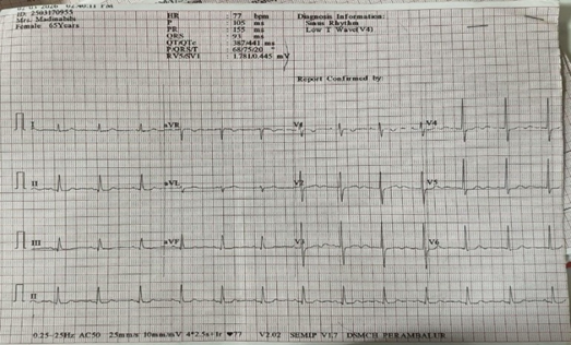
Figure 4: Pre-Op ECG
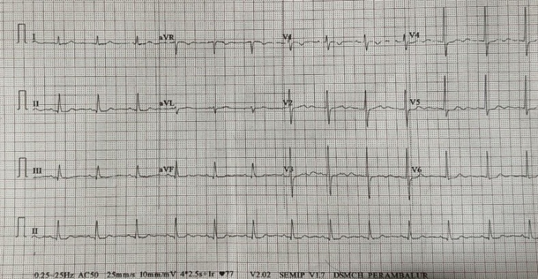
Figure 5: On shifting to Post OP Ward From OT
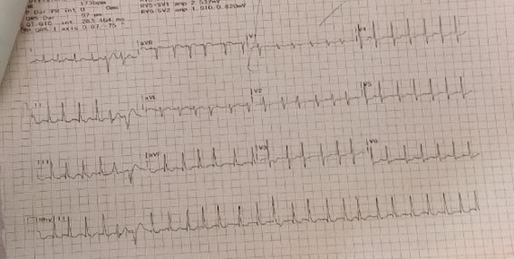
Figure 6: Postop Day One HR 182/Min
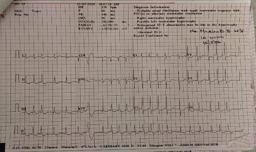
Figure 7: Bolus Amiadarone Followed by Infusion Started, Atrial Fibrillation with VPCS
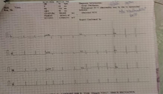
Figure 8: Fourth POD ECG after Verapamil
CONCLUSION:
It is not uncommon to detect secundum atrial defect during routine evaluation in an adult for elective surgery. A history Closure of an atrial septal defect at the age of 50 years often suggests a large shunt with pulmonary hypertension. Aging, frailty, atherosclerotic coronary artery disease with left ventricular dysfunction may all be anticipated as an evolution of cardiovascular disease with progression of age. NYHA classification for dyspnea and cardiac symptomatology such as angina or palpitations may not indicate any triggers as there is restricted physical activity and biochemistry is normal. They may not be able to perform treadmill exercise test either due to arthritis or their pathology as addressed in this case at the age of 65 years. High degree of suspicion for cardiac complications should be triggered by an enlarged left atrium and ventricular regional wall motion abnormalities. Incidence of atrial fibrillation and coronary events may even occur after the first postoperative day .This case is presented for the successful outcome through vigilant monitoring, detection of arrhythmias and aggressive pharmacotherapy in an elderly lady post ASD closure and enlarged left atrium with coronary artery disease. Successful outcome through vigilant monitoring, detection of arrhythmias and aggressive pharmacotherapy in an elderly lady post ASD closure and enlarged left atrium with coronary artery disease
REFERENCES:
1. Krasuski RA. Atrial septal defect and patent foramen ovale. Chap- 29. In: Griffin BP, Topol EJ, Nair D, et al, eds. Manual of cardiovascular medicine. 4th edn. Wolters Kluwer 2012:516-8.
2. Webb GD, Smallhorn JF, et al. Congenital heart disease in adult and pediatric patients. Chap- 75, Part IX. In: Zipes DP, ed. Braunwald’s heart disease. 11th edn. Elsevier 2019: p 1535.
3. Tomaselli GF et al. Mechanisms of cardiac arrhythmias. Chap- 34. In: Zipes DP, ed. Braunwald’s heart disease. 11th edn. Elsevier 2019: p. 619.
4. Vaseghi M, Lux RL, Mahajan A, et al. Sympathetic stimulation increases dispersion of repolarization in humans with myocardial infarction. American Journal of Physiology-Heart and Circulatory Physiology 2012;302(9):H1838-46.
5. Verma S, Siu SC. Aortic dilatation in patients with bicuspid aortic valve. New England Journal of Medicine 2014;370:1920-9.
6. Bonow RO. Bicuspid valves and dilated aortas: a critical review of ACC/AHA practice guidelines. American Journal of Cardiology 2008;102:111-4.
7. Miller J, Zipes DP, et al. Therapy for cardiac arrhythmias. Chap- 36. In: Zipes DP, ed. Braunwald’s heart disease. Elsevier 2019:684-3.
8. Mankikian J, Favelle O, Guillou A, et al. Initial characteristics and outcome of amiodarone pulmonary toxicity. Respiratory Medicine 2014;108(4):638-46.
Hussain N, Bhattacharyya A, Prueksaritanond S. Amiodarone‐induced cirrhosis of liver: what predicts mortality? International Scholarly Research Notices 2013;2013(1):617943.