Estimation of vitamin D, insulin resistance, and uric acid levels in psoriasis patients.
- Shaik Yaseen Chand Basha , Assistant Professor, Department of Biochemistry, Government Siddhartha Medical College, Vijayawada, Andhra Pradesh, India.
- M.Sarala , Assistant Professor, Department of Biochemistry, Government Siddhartha Medical College, Vijayawada, Andhra Pradesh, India.
- Kanumuru Balu Mahendran3, , Assistant Professor, Department of Biochemistry, Government Siddhartha Medical College, Vijayawada, Andhra Pradesh, India.
- Shaik Kausar Tabassum , MBBS 2nd Year Student, Government Siddhartha Medical College, Vijayawada, Andhra Pradesh, India.
Article Information:
Abstract:
Background: Psoriasis is a chronic immune-mediated inflammatory disorder increasingly linked with metabolic disturbances. Altered vitamin D homeostasis, insulin resistance, and elevated serum uric acid have each been implicated in systemic inflammation and cardiometabolic risk in psoriasis. Objectives: To estimate serum vitamin D, insulin resistance, and serum uric acid levels in patients with psoriasis and to examine their distribution across clinical severity categories. Methods: This cross-sectional study was conducted in the Department of Dermatology at Government Siddhartha Medical College, Vijayawada, Andhra Pradesh, from 17-August-2025 to 16-February-2026. One hundred consecutive adult patients with clinically diagnosed psoriasis were enrolled. Clinical details, Psoriasis Area and Severity Index score, serum vitamin D, fasting blood glucose, fasting insulin, HOMA-IR, and serum uric acid were recorded and analyzed using descriptive statistics. Results: The mean age of participants was 42.8 ± 11.6 years, and 62% were male. Plaque psoriasis was the commonest subtype [78%], while 42% had moderate disease. Vitamin D deficiency was present in 54% of patients and insufficiency in 28%, with a mean serum vitamin D level of 21.4 ± 8.7 ng/mL. Insulin resistance was identified in 60% of patients, with a mean HOMA-IR value of 4.1 ± 1.7. Elevated serum uric acid was noted in 42%, and the mean uric acid level was 6.3 ± 1.5 mg/dL. Vitamin D deficiency, insulin resistance, and elevated uric acid were progressively more frequent with increasing psoriasis severity. Conclusion: Psoriasis patients in this cohort demonstrated a high burden of hypovitaminosis D, insulin resistance, and hyperuricemia. These abnormalities were more pronounced in moderate and severe disease, supporting the concept that psoriasis is associated with broader metabolic dysregulation.
Keywords:
Article :
INTRODUCTION:
Psoriasis is a chronic, relapsing, immune-mediated inflammatory disorder characterized by epidermal hyperproliferation, aberrant keratinocyte differentiation, and dysregulated interactions between innate and adaptive immune pathways [1]. Although traditionally considered a skin-limited disease, psoriasis is now recognized as a systemic inflammatory condition with important metabolic and cardiovascular associations [2]. The inflammatory cascade involving dendritic cells, T helper 17 cells, tumor necrosis factor-alpha, and interleukin-23 contributes not only to cutaneous plaque formation but also to broader metabolic dysfunction. As a result, psoriasis has increasingly been linked with obesity, dyslipidemia, insulin resistance, diabetes mellitus, hypertension, and non-alcoholic fatty liver disease [2,3].
Among the biomarkers of current interest, vitamin D has received particular attention because of its immunomodulatory and antiproliferative effects on keratinocytes and T-cell function [3,4]. Vitamin D analogues have long been used in topical psoriasis therapy, and several observational studies have shown lower circulating 25-hydroxy vitamin D levels in patients with psoriasis compared with controls [4,5]. Meta-analytic evidence also suggests that hypovitaminosis D is more frequent in psoriasis, although the strength of association with clinical severity varies across populations [5,6]. Reduced sun exposure due to covered lesions, chronic inflammation, dietary factors, obesity, and altered vitamin D metabolism have all been proposed as contributors [3,4].
Insulin resistance represents another major metabolic abnormality increasingly recognized in psoriasis. Chronic systemic inflammation can disrupt insulin signaling through cytokine-mediated pathways, while shared risk factors such as central adiposity further amplify this association [2,7]. Earlier clinical studies have documented higher fasting insulin levels and increased HOMA-IR values in psoriasis patients [8,9]. More recent longitudinal evidence has suggested that insulin resistance can even precede incident psoriasis, indicating a bidirectional relationship between inflammatory and metabolic processes [10]. From a clinical perspective, detection of insulin resistance in psoriasis is relevant because it may identify patients at increased risk of type 2 diabetes and cardiovascular disease.
Serum uric acid has also emerged as a meaningful biochemical marker in psoriasis. Accelerated epidermal turnover in psoriasis increases nucleic acid catabolism, thereby contributing to uric acid generation. In parallel, obesity, insulin resistance, and metabolic syndrome can impair urate handling and sustain hyperuricemia [2,11]. Previous studies have reported higher serum uric acid concentrations and a greater prevalence of hyperuricemia among psoriasis patients, particularly in those with extensive or severe disease [11,14]. Elevated uric acid is clinically important because it is associated with gout, endothelial dysfunction, and adverse cardiometabolic outcomes.
Given the increasing recognition of psoriasis as a multisystem inflammatory disease, evaluation of metabolic biomarkers alongside clinical severity assumes practical relevance in routine dermatologic care. However, Indian data examining vitamin D status, insulin resistance, and uric acid together in a single psoriasis cohort remain limited. The present study was therefore undertaken to estimate serum vitamin D levels, insulin resistance, and serum uric acid levels in patients with psoriasis attending a tertiary care teaching hospital. The specific objectives were to describe the demographic and clinical profile of patients with psoriasis, assess serum vitamin D status, determine the burden of insulin resistance and elevated serum uric acid, and examine their distribution across psoriasis severity categories assessed using the Psoriasis Area and Severity Index.
METHODOLOGY:
Study design and setting.
This was a hospital-based cross-sectional study conducted in the Department of Dermatology, Government Siddhartha Medical College, Vijayawada, Andhra Pradesh, over a six-month period from 17-August-2025 to 16-February-2026.
Study population.
Adult patients aged 18 years and above with a clinical diagnosis of psoriasis attending the dermatology outpatient or inpatient services during the study period were considered eligible. Both newly diagnosed and previously known cases were included provided they fulfilled the study criteria and consented to participate.
Inclusion and exclusion criteria.
Patients with clinically diagnosed psoriasis of any major subtype were included. Individuals with psoriatic arthritis on long-term urate-lowering therapy, chronic kidney disease, chronic liver disease, known endocrine disorders affecting vitamin D metabolism, pregnancy, malignancy, or current use of systemic vitamin D supplementation were excluded. Patients receiving drugs known to markedly alter uric acid or insulin metabolism were also excluded to reduce biochemical confounding.
Sample size and sampling technique.
A total of 100 patients were enrolled by consecutive sampling. This sample size was considered adequate for descriptive estimation of the selected biochemical parameters and for subgroup analysis according to psoriasis severity within the study duration.
Clinical evaluation.
After obtaining written informed consent, a detailed clinical history was recorded, including age, sex, duration of disease, and clinical subtype of psoriasis. A complete dermatological examination was performed in all participants. Disease severity was assessed using the Psoriasis Area and Severity Index [8]. For descriptive interpretation, PASI categories were grouped as mild, moderate, and severe based on composite clinical severity used in the department.
Biochemical assessment.
Venous blood samples were collected after an overnight fast. Serum vitamin D was measured as serum 25-hydroxy vitamin D and categorized as deficient when <20 ng/mL, insufficient when 20-29 ng/mL, and sufficient when >=30 ng/mL [3-5,12]. Fasting blood glucose and fasting serum insulin were measured using standard laboratory methods. Insulin resistance was estimated using the homeostasis model assessment formula HOMA-IR = fasting insulin x fasting glucose / 405, in accordance with established methodology [6,7]. For the purpose of this study, HOMA-IR >2.5 was considered indicative of insulin resistance. Serum uric acid was estimated by standard enzymatic methods and categorized as normal or elevated according to laboratory reference values.
Data management and statistical analysis.
All data were entered into a structured proforma and transferred to a spreadsheet for analysis. Continuous variables were summarized as mean ± standard deviation, while categorical variables were presented as number and percentage. The distribution of vitamin D deficiency, insulin resistance, and elevated uric acid across PASI severity categories was examined descriptively. The findings are presented in tabular form to facilitate interpretation of the metabolic profile of the study population.
Ethical considerations.
The study was conducted after approval from the Institutional Ethics Committee of Government Siddhartha Medical College, Vijayawada(IECSMCGGH/2025/AP/197 dated 2nd August 2025). Written informed consent was obtained from all participants before enrolment. Confidentiality of participant data was preserved throughout data collection, analysis, and manuscript preparation.
RESULTS:
A total of 100 patients diagnosed with psoriasis were included in this cross-sectional study. The findings were analyzed with respect to demographic profile, clinical characteristics, serum vitamin D levels, insulin resistance, and serum uric acid levels.
The age of the patients ranged from 19 to 68 years, with the highest proportion belonging to the 41-50 years age group [30%]. The mean age of the study population was 42.8 ± 11.6 years. Male patients constituted 62% of the sample, showing a male predominance. The majority of patients had disease duration of 1-5 years [46%]. Plaque psoriasis was the most common clinical type, observed in 78% of patients. Based on PASI score, 42% had moderate disease, followed by 34% with mild disease and 24% with severe disease [Table 1].
Table 1. Demographic and clinical characteristics of study participants [n = 100]
|
Variable |
Number |
Percentage [%] |
|
Age group [years] |
|
|
|
18-30 |
18 |
18.0 |
|
31-40 |
26 |
26.0 |
|
41-50 |
30 |
30.0 |
|
51-60 |
16 |
16.0 |
|
>60 |
10 |
10.0 |
|
Sex |
|
|
|
Male |
62 |
62.0 |
|
Female |
38 |
38.0 |
|
Duration of psoriasis |
|
|
|
<1 year |
22 |
22.0 |
|
1-5 years |
46 |
46.0 |
|
>5 years |
32 |
32.0 |
|
Type of psoriasis |
|
|
|
Plaque psoriasis |
78 |
78.0 |
|
Guttate psoriasis |
8 |
8.0 |
|
Palmoplantar psoriasis |
9 |
9.0 |
|
Pustular psoriasis |
5 |
5.0 |
|
Severity by PASI score |
|
|
|
Mild |
34 |
34.0 |
|
Moderate |
42 |
42.0 |
|
Severe |
24 |
24.0 |
The mean serum vitamin D level among psoriasis patients was 21.4 ± 8.7 ng/mL. Vitamin D deficiency was the most common biochemical abnormality and was present in 54% of patients, while another 28% had insufficient levels. Only 18% of participants had sufficient vitamin D levels. The mean fasting blood glucose level was 104.6 ± 18.2 mg/dL, the mean fasting serum insulin level was 16.8 ± 6.4 µIU/mL, and the mean HOMA-IR value was 4.1 ± 1.7. Insulin resistance was identified in 60% of patients. The mean serum uric acid level was 6.3 ± 1.5 mg/dL, and elevated values were observed in 42% of the cohort [Table 2].
Table 2. Biochemical categories among psoriasis patients [n = 100]
|
Biochemical variable |
Category |
Number |
Percentage [%] |
|
Vitamin D status |
Deficient [<20 ng/mL] |
54 |
54.0 |
|
|
Insufficient [20-29 ng/mL] |
28 |
28.0 |
|
|
Sufficient [>=30 ng/mL] |
18 |
18.0 |
|
Insulin resistance |
Present |
60 |
60.0 |
|
|
Absent |
40 |
40.0 |
|
Serum uric acid status |
Elevated |
42 |
42.0 |
|
|
Normal |
58 |
58.0 |
Vitamin D deficiency was more frequent among patients with greater disease severity. Among patients with mild psoriasis, 35.3% had vitamin D deficiency, compared with 57.1% of those with moderate psoriasis and 75.0% of those with severe psoriasis. This pattern indicates an inverse relationship between serum vitamin D levels and psoriasis severity [Table 3].
Table 3. Association of vitamin D deficiency with psoriasis severity [n = 100]
|
PASI severity |
Vitamin D deficient |
Not deficient |
Total |
|
Mild |
12 |
22 |
34 |
|
Moderate |
24 |
18 |
42 |
|
Severe |
18 |
6 |
24 |
|
Total |
54 |
46 |
100 |
Insulin resistance and elevated serum uric acid were more commonly observed in patients with moderate-to-severe psoriasis. Insulin resistance was present in 41.2% of mild cases, 66.7% of moderate cases, and 75.0% of severe cases. Elevated uric acid levels were observed in 23.5% of mild cases, 42.9% of moderate cases, and 66.7% of severe cases. These findings suggest that metabolic abnormalities were more pronounced with increasing clinical severity [Table 4].
Table 4. Association of insulin resistance and elevated uric acid with psoriasis severity [n = 100]
|
PASI severity |
Insulin resistance present |
Elevated uric acid |
Total |
|
Mild |
14 |
8 |
34 |
|
Moderate |
28 |
18 |
42 |
|
Severe |
18 |
16 |
24 |
|
Total |
60 |
42 |
100 |
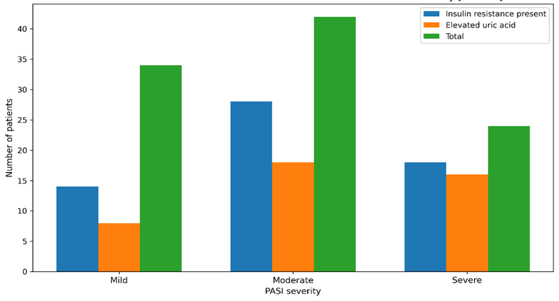
Figure1: Association of insulin resistance and elevated uric acid with psoriasis severity
Overall, the present study demonstrated that vitamin D deficiency, insulin resistance, and elevated serum uric acid levels were common among patients with psoriasis. The frequency of these abnormalities increased with greater disease severity, supporting the metabolic and systemic dimension of psoriatic disease.
DISCUSSION:
This study evaluated three clinically relevant metabolic markers in psoriasis and demonstrated a high burden of hypovitaminosis D, insulin resistance, and elevated serum uric acid among affected patients. The predominance of plaque psoriasis and the greater proportion of moderate disease in the present series are in keeping with the usual clinical spectrum described in tertiary care dermatology practice. More importantly, the biochemical findings support the contemporary view that psoriasis is not merely a cutaneous disorder but a systemic inflammatory disease with measurable metabolic correlates [8].
The mean serum vitamin D level in our cohort was low, and more than half of the patients had frank vitamin D deficiency. This observation is consistent with previous reviews and meta-analyses showing a higher frequency of reduced serum 25-hydroxy vitamin D among psoriasis patients than in healthy controls [9]. Pokharel et al. reported significantly lower vitamin D levels in psoriatic cases and an inverse association with disease severity, while Patil et al. also documented a substantial prevalence of deficiency in Indian psoriasis patients [10]. In the present study, vitamin D deficiency increased progressively from mild to severe psoriasis, reinforcing the clinical relevance of this association. Mechanistically, low vitamin D status can influence keratinocyte proliferation, differentiation, and immune regulation, thereby providing a biologically plausible link with disease activity [3,4].
Insulin resistance was identified in 60% of participants, and the mean HOMA-IR value was elevated. Earlier studies by Pereira et al. and Boehncke et al. similarly demonstrated increased insulin resistance in psoriasis, indicating that glucose homeostasis is frequently altered in these patients [9,10]. The observation that insulin resistance was more common in moderate and severe disease in our study also aligns with the broader concept that inflammatory burden and metabolic dysfunction intensify together [13]. Longitudinal evidence from the Women's Health Initiative further suggests that insulin resistance is not only a consequence but can also precede psoriasis, highlighting a bidirectional inflammatory-metabolic axis [11]. These findings support the usefulness of fasting glucose, fasting insulin, and HOMA-IR-based screening in routine evaluation of psoriasis patients, especially those with more extensive disease [12].
Elevated serum uric acid was present in 42% of our patients, with prevalence rising sharply in severe psoriasis. This pattern corresponds with previously published evidence showing higher serum uric acid and hyperuricemia in psoriasis compared with controls [14]. Gui et al. observed significantly higher uric acid concentrations and a greater prevalence of hyperuricemia in psoriatic patients, while Gisondi et al. emphasized hyperuricemia as a common feature in chronic plaque psoriasis [14]. Increased epidermal cell turnover, purine catabolism, obesity, and insulin resistance likely contribute together to uric acid elevation in psoriasis. Because uric acid is also linked with endothelial dysfunction and metabolic syndrome, its estimation can provide additional insight into systemic risk profiling [2,14].
The combined interpretation of these findings suggests that psoriasis severity parallels deterioration in metabolic status. In practical terms, patients with moderate and severe psoriasis appear to represent a subgroup with greater biochemical vulnerability. For tertiary care centers, this has implications for integrated assessment, early counseling, and interdisciplinary follow-up. The study therefore adds institution-level evidence from coastal Andhra Pradesh to the growing literature supporting routine metabolic evaluation in psoriasis. Future analytical studies with controls and longitudinal follow-up would help clarify causality, define local cutoffs with greater precision, and determine whether correction of vitamin D deficiency, reduction of insulin resistance, or control of hyperuricemia influences disease course and long-term cardiovascular risk.
Limitations
This study was conducted at a single tertiary care center with a cross-sectional design, which limits causal interpretation. The absence of a healthy control group restricted comparative analysis. Seasonal variation in vitamin D status, dietary intake, sun exposure, body mass index stratification, and detailed treatment history were not analyzed separately. The biochemical associations were therefore descriptive and institution-specific rather than population-based.
CONCLUSION:
The present study demonstrates that patients with psoriasis carry a substantial burden of metabolic abnormalities in addition to cutaneous disease. Vitamin D deficiency and insufficiency were highly prevalent, insulin resistance was common, and a considerable proportion had elevated serum uric acid levels. These abnormalities were increasingly frequent in patients with moderate and severe psoriasis, indicating a close relationship between disease severity and metabolic derangement. Routine clinical evaluation of psoriasis should therefore extend beyond lesion assessment to include relevant biochemical screening. Early identification of these abnormalities can support comprehensive risk stratification, timely counseling, and more holistic long-term management in dermatology practice.
REFERENCES:
1. Rendon A, Schakel K. Psoriasis pathogenesis and treatment. Int J Mol Sci. 2019;20(6):1475.
2. Gisondi P, Del Giglio M, Girolomoni G. Psoriasis and the metabolic syndrome. Clin Dermatol. 2018;36(1):21-28.
3. Barrea L, Savanelli MC, Di Somma C, Napolitano M, Megna M, Colao A, et al. Vitamin D and its role in psoriasis: An overview of the dermatologist and nutritionist. Rev Endocr Metab Disord. 2017;18(2):195-205.
4. Soleymani T, Hung T, Soung J. The role of vitamin D in psoriasis: a review. Int J Dermatol. 2015;54(4):383-392.
5. Formisano E, Proietti E, Borgarelli C, Pisciotta L. Psoriasis and vitamin D: a systematic review and meta-analysis. Nutrients. 2023;15(15):3387.
6. Matthews DR, Hosker JP, Rudenski AS, Naylor BA, Treacher DF, Turner RC. Homeostasis model assessment: insulin resistance and beta-cell function from fasting plasma glucose and insulin concentrations in man. Diabetologia. 1985;28(7):412-419.
7. Wallace TM, Levy JC, Matthews DR. Use and abuse of HOMA modeling. Diabetes Care. 2004;27(6):1487-1495.
8. Langley RG, Ellis CN. Evaluating psoriasis with Psoriasis Area and Severity Index, Psoriasis Global Assessment, and Lattice System Physician's Global Assessment. J Am Acad Dermatol. 2004;51(4):563-569.
9. Pereira RR, Amladi S, Varthakavi P. A study of the prevalence of diabetes, insulin resistance, lipid abnormalities, and cardiovascular risk factors in patients with chronic plaque psoriasis. Indian J Dermatol. 2011;56(5):520-526.
10. Boehncke S, Thaci D, Beschmann H, Ludwig RJ, Ackermann H, Badenhoop K, et al. Psoriasis patients show signs of insulin resistance. Br J Dermatol. 2007;157(6):1249-1251.
11. Chan AA, Li H, Li W, Pan K, Yee JK, Chlebowski RT, et al. Association between baseline insulin resistance and psoriasis incidence: the Women's Health Initiative. Arch Dermatol Res. 2022;314(9):869-880.
12. Patil A, Deo K, Kalyan D, Deora MS, Sharma Y, Deoghare S. Association of vitamin D deficiency with psoriasis and metabolic syndrome: A case-control study in Indian patients. Indian J Dermatol. 2022;67(1):92.
13. Pokharel R, Agrawal S, Pandey P, Lamsal M. Assessment of vitamin D level in patients with psoriasis and its correlation with disease severity: A case-control study. Psoriasis (Auckl). 2022;12:251-258.
14. Gui XY, Jin HZ, Wang ZJ, Xu TD. Serum uric acid levels and hyperuricemia in patients with psoriasis: a hospital-based cross-sectional study. An Bras Dermatol. 2018;93(5):761-763.