ASSOCIATION BETWEEN IONIZED CALCIUM LEVELS AND SEVERITY OF DENGUE FEVER
- M. Abishek. , Post graduate student, final year, Department of General Medicine, Vinayaka Missions Medical College, Karaikal. India.
- Babu Rajendran. , MD, Professor, Department of General Medicine, Vinayaka Missions Medical College, Karaikal, India
- V. Sakthivel N. , MD, HOD & Professor, Department of General Medicine, Vinayaka Missions Medical College Karaikal, India.
Article Information:
Abstract:
Background: Dengue fever is a rapidly expanding mosquito-borne viral disease in tropical and subtropical regions. The clinical spectrum varies from mild febrile illness to severe dengue characterized by plasma leakage, hemorrhage, and organ dysfunction. Early identification of biochemical markers associated with disease severity is essential for improving clinical outcomes. Objective: To evaluate the association between ionized calcium levels and the severity of dengue fever. Methods: A hospital-based observational study was conducted in the Department of General Medicine at Vinayaka Missions Medical College and Hospital, Karaikal, India, among 50 adult patients with laboratory-confirmed dengue infection from July 2025 to December 2025. Demographic data, clinical manifestations, and laboratory parameters including ionized calcium levels were recorded. Patients were classified according to WHO 2009 dengue severity criteria. Statistical analysis included descriptive statistics, independent Student’s t-test, and Pearson correlation analysis. Results: A total of 50 participants were included, with the majority aged <30 years (46%) and a slight male predominance (56%). Fever was present in all cases (100%), while other symptoms such as vomiting (18%), abdominal pain (4%), and bleeding manifestations (4%) were less frequent. Most participants had dengue without warning signs (73%), followed by dengue with warning signs (20%) and severe dengue (7%). Mean ionized calcium levels showed significant variation over time, decreasing from Day 0 to Day 4 and increasing by Day 7. Repeated measures ANOVA demonstrated a statistically significant difference across time points (F = 61.16, p < 0.001), with all pairwise comparisons being significant (p < 0.001). Correlation analysis revealed no significant association between ionized calcium and platelet count at any time point. No significant correlation was observed with serum albumin at Day 0 and Day 4; however, a moderate negative correlation was noted at Day 7 (r = -0.35, p = 0.01). A weak positive correlation between ionized calcium and hematocrit was observed at Day 0 (r = 0.28, p = 0.04), but not at later time points.Serum albumin levels did not differ significantly across dengue severity categories (p = 0.89). Conclusion: Ionized calcium levels were significantly associated with dengue severity. Monitoring calcium levels may help identify patients at risk of severe disease and complications.
Keywords:
Article :
INTRODUCTION:
Dengue fever is one of the most significant mosquito-borne viral infections worldwide and poses a major public health challenge in tropical and subtropical regions. The disease is caused by four serotypes of the dengue virus (DENV-1 to DENV-4), which are transmitted primarily by Aedes aegypti mosquitoes. The global incidence of dengue has increased dramatically in recent decades due to urbanization, climate change, and increased international travel.¹
It is estimated that approximately 390 million dengue infections occur annually worldwide, with nearly 96 million individuals developing clinically apparent disease.² The burden of dengue is particularly high in South and Southeast Asia, Latin America, and parts of Africa, where outbreaks frequently occur.³
Clinically, dengue infection presents with a wide spectrum ranging from asymptomatic infection to severe disease. Common symptoms include fever, headache, retro-orbital pain, myalgia, arthralgia, and rash.⁴ Some patients may develop severe complications such as plasma leakage, severe bleeding, and organ impairment.⁵
The World Health Organization (WHO) revised the classification of dengue in 2009 to improve early recognition of severe disease. According to this classification, dengue is categorized into dengue without warning signs, dengue with warning signs, and severe dengue.⁶ Early identification of patients at risk of severe disease is essential for timely intervention and improved outcomes.⁷
The pathogenesis of severe dengue involves complex interactions between viral replication, host immune response, and endothelial dysfunction. Increased vascular permeability and plasma leakage play a crucial role in the progression of severe disease.⁸
Hematological abnormalities such as thrombocytopenia and hemoconcentration are commonly observed in dengue infection and are widely used as indicators of disease severity.⁹ However, several biochemical abnormalities have also been reported, including disturbances in liver enzymes, electrolytes, and metabolic parameters.¹⁰
Electrolyte imbalance is frequently observed in viral infections and may reflect systemic inflammatory responses or organ dysfunction. Among these abnormalities, hypocalcemia has been increasingly reported in patients with dengue fever.¹¹
Calcium plays an essential role in numerous physiological processes including muscle contraction, vascular tone regulation, neuronal signaling, and blood coagulation.¹² Ionized calcium represents the biologically active fraction of total serum calcium and provides a more accurate assessment of calcium status in critically ill patients.¹³
Previous studies have suggested that decreased calcium levels may be associated with disease severity, hypotension, and bleeding manifestations in dengue infection.¹⁴ However, limited data are available regarding the relationship between ionized calcium levels and dengue severity.
Therefore, the present study was undertaken to evaluate the association between ionized calcium levels and the severity of dengue fever among hospitalized patients
MATERIALS AND METHODS:
Study Design
The present study was conducted as a prospective observational study aimed at evaluating the association between serum ionized calcium levels and the severity of dengue fever among hospitalized adult patients. The study was designed to assess biochemical changes occurring during dengue infection and to determine whether ionized calcium levels could serve as a potential marker of disease severity.
Study Setting
The study was carried out in the Department of General Medicine at Vinayaka Missions Medical College and Hospital, Karaikal, a tertiary care teaching hospital that caters to patients from both urban and rural populations. The institution functions as a referral center for infectious diseases including dengue fever, particularly during seasonal outbreaks.
Study Duration
The study was conducted over a six-month period from July 2025 to December 2025, corresponding to the peak seasonal incidence of dengue infection in the region.
Study Population
A total of 50 adult patients diagnosed with dengue fever were included in the study. All patients admitted to the Department of General Medicine during the study period who fulfilled the inclusion criteria were consecutively enrolled. Patients were categorized according to disease severity into:
• Dengue without warning signs
• Dengue with warning signs
• Severe dengue
as per World Health Organization (WHO) 2009 classification guidelines. Inclusion Criteria
Patients were included in the study if they met the following criteria:
• Age 18 years or above
• Laboratory-confirmed dengue infection based on NS1 antigen detection and/or IgM ELISA positivity
• Patients admitted for clinical management of dengue fever
• Patients who provided informed consent for participation in the study
Exclusion Criteria
Patients were excluded if they had medical conditions that could independently affect calcium metabolism. These included:
• Chronic kidney disease
• Parathyroid disorders
• Chronic liver disease
• Known metabolic bone disorders
• Patients receiving calcium supplementation or vitamin D therapy
• Patients with other systemic infections or chronic illnesses influencing serum calcium levels
Data Collection
After obtaining informed consent, detailed clinical and laboratory information was collected using a structured proforma.
Demographic Data
The following demographic details were recorded:
• Age
• Gender
• Area of residence
Clinical Evaluation
All patients underwent a thorough clinical examination at the time of admission. The following symptoms and signs were documented:
• Fever
• Vomiting
• Abdominal pain
• Bleeding manifestations
• Hypotension
• Signs of shock
• Other warning signs of dengue
Clinical records were reviewed throughout the hospital stay to identify any complications related to dengue infection.
Classification of Dengue Severity
Patients were classified according to the World Health Organization (WHO) 2009 dengue classification, which includes:
1. Dengue without warning signs
2. Dengue with warning signs
3. Severe dengue
The classification was based on clinical presentation, laboratory findings, and evidence of complications such as plasma leakage, bleeding, or organ involvement.
Laboratory Investigations
Blood samples were collected from all patients at the time of admission and processed in the hospital laboratory using standard protocols.
The following laboratory parameters were recorded:
• Serum ionized calcium levels
• Platelet count
• Hematocrit levels
• Other routine laboratory investigations relevant to dengue management
Measurement of Ionized Calcium
Serum ionized calcium levels were measured using the ion-selective electrode method, which provides an accurate estimation of biologically active calcium in blood.
Results were expressed in millimoles per liter (mmol/L).
Based on laboratory reference values, patients were categorized into:
• Normal ionized calcium levels
• Hypocalcemia Outcome Measures
The primary objective of the study was to evaluate the association between ionized calcium levels and dengue severity.
The following parameters were analyzed:
• Distribution of dengue severity categories
• Prevalence of hypocalcemia among dengue patients
• Mean ionized calcium levels in severe and non-severe dengue
• Association between ionized calcium levels and clinical manifestations such as bleeding, hypotension, and shock
• Relationship between ionized calcium levels and laboratory indicators including platelet count and hematocrit
Statistical Analysis
All collected data were entered and analyzed using Statistical Package for Social Sciences (SPSS) software.
Descriptive Statistics
Continuous variables such as ionized calcium levels, platelet count, and hematocrit were expressed as mean ± standard deviation (SD).
Categorical variables such as clinical features, dengue severity categories, and hypocalcemia prevalence were expressed as frequency and percentage.
Comparative Analysis
Repeated Measures Anova
Correlation Analysis
The relationship between ionized calcium levels and laboratory parameters such as platelet count and hematocrit was evaluated using the Pearson correlation coefficient (r).
Level of Significance
A p-value less than 0.05 was considered statistically significant for all statistical tests.
Ethical Considerations
The study protocol was reviewed and approved by the Institutional Ethics Committee of Vinayaka Missions Medical College and Hospital, Karaikal. Written informed consent was obtained from all participants prior to enrollment. Patient confidentiality was maintained throughout the study, and all data were used exclusively for research.
RESULTS:
The majority of participants (73%) were classified as having dengue without warning signs, indicating that most cases were mild in nature. A smaller proportion of participants (20%) had dengue with warning signs, suggesting the presence of clinical features that require closer monitoring and management. Severe dengue was observed in only 7% of participants, representing the least frequent category but indicating a subset of patients with serious complications. Overall, the findings demonstrate that most participants experienced.
mild dengue, while a smaller proportion progressed to more severe forms of the disease. The mean ionized calcium levels showed variation over time, with a value of 1.33 ± 0.06 mmol/L on Day 0, decreasing to 1.06 ± 0.61 mmol/L on Day 4, and subsequently increasing to 1.20 ± 0.08 mmol/L by Day 7. This trend indicates a transient decline followed by partial recovery. The mean platelet count was 1.33 ± 0.61 ×10³/µL, reflecting thrombocytopenia among the participants, which is a common hematological finding in dengue infection. The mean hematocrit level was 45.55 ± 7.33%, suggesting relative hemoconcentration in some cases. Liver function parameters were elevated, with mean SGOT and SGPT levels of
105.74 ± 43.94 IU/L and 97.34 ± 29.73 IU/L, respectively, indicating hepatic involvement among the study population. The mean serum albumin level was 3.26 ± 0.37 mg/dL, which may suggest mild hypoalbuminemia in some participants. Overall, the laboratory findings highlight characteristic features associated with dengue infection, including thrombocytopenia, elevated liver enzymes, and variations in ionized calcium levels.
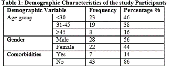
Table 1 presents the demographic profile of the study participants. A total of 50 individuals were included in the study.
With respect to age distribution, nearly half of the participants (46%) were aged below 30 years, followed by 38% in the 31–45 years age group. Participants aged above 45 years constituted the smallest proportion (16%), indicating that the study population was relatively younger.
In terms of gender, males represented a slightly higher proportion (56%) compared to females (44%), suggesting a marginal male predominance in the study sample.
Regarding comorbidities, the majority of participants (86%) did not report any existing comorbid conditions, while only 14% had comorbidities. This indicates that most participants were relatively healthy with respect to underlying health conditions.
Overall, the study population was predominantly younger, with a slight male predominance and a low prevalence of comorbidities.
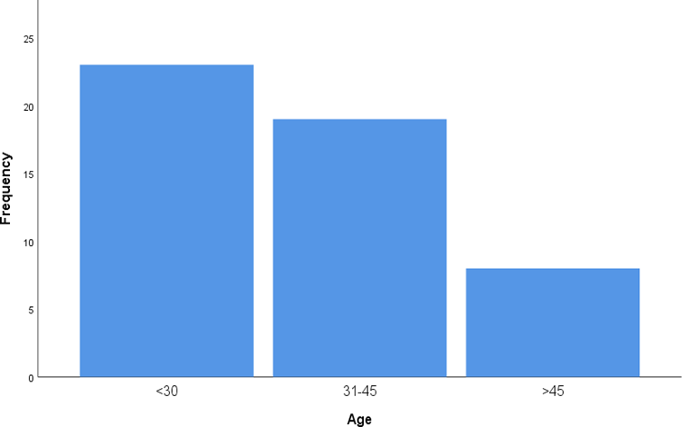
Graph1:Age Group
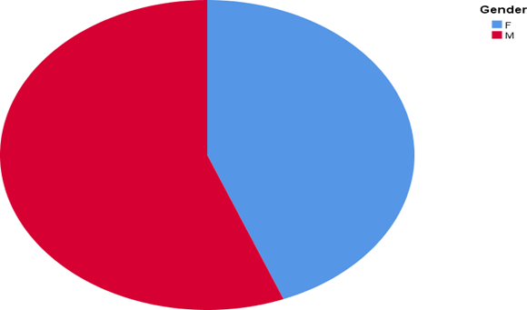
Graph 2:Gender
Table 2.Clinical Features of the study participants
|
Clinical features |
Frequency |
Percentage |
|
Fever |
50 |
100 |
|
Vomiting |
9 |
18 |
|
Abdominal pain |
2 |
4 |
|
Bleeding Manifestation |
2 |
4 |
Table 2 illustrates the clinical features observed among the study participants. All participants (100%) presented with fever, indicating that it was the most common symptom in the study population.
Other symptoms were reported less frequently. Vomiting 18% and abdominal pain were 4% in the study participants, suggesting that gastrointestinal manifestations were relatively uncommon. Bleeding manifestations were reported in 4% of participants, indicating a notable proportion of cases with more severe or complicated clinical presentation.
Overall, fever was the predominant clinical feature, while other symptoms such as vomiting, abdominal pain, and bleeding manifestations were less commonly observed among the participants
Table 3.Dengue severity of the study participants
|
Severity category |
Frequency |
Percentage (%) |
|
Dengue without warning signs |
37 |
73 |
|
Dengue with warning signs |
10 |
20 |
|
Severe dengue |
3 |
7 |
Table 3 presents the distribution of dengue severity among the study participants. The majority of participants (73%) were classified as having dengue without warning signs, indicating that most cases were mild in nature.
A smaller proportion of participants (20%) had dengue with warning signs, suggesting the presence of clinical features that require closer monitoring and management. Severe dengue was observed in only 7% of participants, representing the least frequent category but indicating a subset of patients with serious complications.
Overall, the findings demonstrate that most participants experienced mild dengue, while a smaller proportion progressed to more severe forms of the disease.
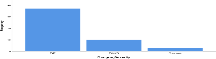
Graph 3: Dengue Severity
Table 4: Baseline Laboratory Parameters
|
Parameter |
Mean ± SD |
|
|
Ionized calcium (mmol/L) |
Day 0 |
1.33±0.06 |
|
Day 4 |
1.06±0.61 |
|
|
Day 7 |
1.20±0.08 |
|
|
Platelet count (×10³/µL) |
1.33±0.61 |
|
|
Hematocrit (%) |
45.55±7.33 |
|
|
SGOT |
105.74 ± 43.94 |
|
|
SGPT |
97.34± 29.73 |
|
|
Albumin mg/dl |
3.26 ± 0.37 |
|
Table 4 summarizes the baseline laboratory parameters of the study participants. The mean ionized calcium levels showed variation over time, with a value of 1.33 ± 0.06 mmol/L on Day 0, decreasing to 1.06 ± 0.61 mmol/L on Day 4, and subsequently increasing to 1.20 ±
0.08 mmol/L by Day 7. This trend indicates a transient decline followed by partial recovery.
The mean platelet count was 1.33 ± 0.61 ×10³/µL, reflecting thrombocytopenia among the participants, which is a common hematological finding in dengue infection. The mean hematocrit level was 45.55 ± 7.33%, suggesting relative hemoconcentration in some cases.
Liver function parameters were elevated, with mean SGOT and SGPT levels of 105.74 ±
43.94 IU/L and 97.34 ± 29.73 IU/L, respectively, indicating hepatic involvement among the study population.
The mean serum albumin level was 3.26 ± 0.37 mg/dL, which may suggest mild hypoalbuminemia in some participants.
Overall, the laboratory findings highlight characteristic features associated with dengue infection, including thrombocytopenia, elevated liver enzymes, and variations in ionized calcium levels.
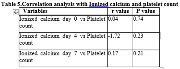
Table 5 presents the correlation between ionized calcium levels at different time points and platelet count among the study participants.
At Day 0, ionized calcium demonstrated a very weak positive correlation with platelet count (r = 0.04), which was not statistically significant (p = 0.74). Similarly, on Day 4, although a negative correlation was observed (r = -1.72), this association was not statistically significant (p = 0.23). By Day 7, a weak positive correlation was again noted (r = 0.17), which also failed to reach statistical significance (p = 0.21).
Overall, the findings indicate that there is no statistically significant correlation between ionized calcium levels and platelet count at any of the measured time points. The observed correlations are weak and inconsistent in direction, suggesting that ionized calcium may not be a reliable predictor of platelet count in this study population.
These results imply that changes in ionized calcium levels occur independently of platelet dynamics in dengue patients, and further studies with larger sample sizes may be required to explore any potential association.
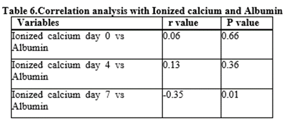
Table 6 presents the correlation between ionized calcium levels at different time points and serum albumin among the study participants.
At Day 0, ionized calcium showed a very weak positive correlation with albumin levels (r= 0.06), which was not statistically significant (p = 0.66). Similarly, on Day 4, a weak positive correlation was observed (r = 0.13), but this association also did not reach statistical significance (p = 0.36).
In contrast, at Day 7, a moderate negative correlation was identified between ionized calcium and albumin levels (r = -0.35), which was statistically significant (p = 0.01). This suggests that, at this time point, lower albumin levels were associated with relatively higher ionized calcium levels, or vice versa.
Overall, the results indicate that there is no meaningful association between ionized calcium and albumin levels during the early phase of illness (Day 0 and Day 4). However, by Day 7, a significant inverse relationship emerges, which may reflect physiological changes during the recovery or late phase of the disease.
These findings suggest that the relationship between ionized calcium and albumin may be dynamic over the course of illness, potentially influenced by factors such as changes in protein binding, fluid shifts, or disease progression. Further studies are warranted to better understand the clinical significance of this association.
Table7: Correlation analysis between Ionized calcium with Hematocrit
|
Variables |
r value |
P value |
|
Ionized calcium day 0 vs Albumin |
0.28 |
0.04 |
|
Ionized calcium day 4 vs Albumin |
0.11 |
0.4 |
|
Ionized calcium day 7 vs Albumin |
0.06 |
0.66 |
Table 7 presents the correlation between ionized calcium levels at different time points and hematocrit among the study participants.
At Day 0, ionized calcium demonstrated a weak but statistically significant positive correlation with hematocrit (r = 0.28, p = 0.04). This suggests that higher ionized calcium levels were modestly associated with higher hematocrit values during the early phase of illness.
However, at Day 4, the correlation between ionized calcium and hematocrit was weak and not statistically significant (r = 0.11, p = 0.40). Similarly, by Day 7, the association remained very weak and non-significant (r = 0.06, p = 0.66).
Overall, these findings indicate that a statistically significant relationship between ionized calcium and hematocrit is present only at the initial stage (Day 0), but this association diminishes over time and is not observed in later stages of the disease. The early positive. correlation may reflect concurrent physiological changes such as hemoconcentration and electrolyte variation during the acute phase of illness.
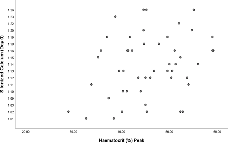
Graph 4: Relationship between serum ionized calcium (day 0)and peak haematocrit levels
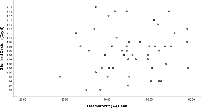
Graph 5: Relationship between serum ionized calcium (day 4)and peak haematocrit levels.
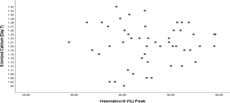
Graph 6: Relationship between serum ionized calcium (day 7)and peak haematocrit levels.
Table 8.Changes in Ionized Calcium Levels Across Time (Repeated Measures ANOVA,
(n = 50)
|
Time Point |
Mean ± SD |
F value |
p-value |
Pairwise Comparison (p-value) |
|
Day 0 |
1.13 ± 0.07 |
|
|
Day 0 vs Day 4: <0.001* |
|
Day 4 |
1.06 ± 0.06 |
61.16 |
<0.001* |
Day 0 vs Day 7: <0.001* |
|
Day 7 |
1.20 ± 0.08 |
|
|
Day 4 vs Day 7: <0.001* |
Table 8 presents the changes in ionized calcium levels across different time points using repeated measures ANOVA. The analysis demonstrated a statistically significant difference in mean ionized calcium levels over time (F= 61.16, p < 0.001), indicating that ionized calcium levels varied significantly across the study period. The mean ionized calcium level decreased from 1.13 ± 0.07 mmol/L on Day 0 to 1.06 ± 0.06 mmol/L on Day 4, followed by a notable increase to 1.20 ± 0.08 mmol/L by Day 7.
Pairwise comparisons revealed that all differences between time points were statistically significant. There was a significant decline in ionized calcium levels from Day 0 to Day 4 (p < 0.001), followed by a significant increase from Day 4 to Day 7 (p < 0.001). Additionally, the difference between Day 0 and Day 7 was also statistically significant (p< 0.001), indicating an overall increase in ionized calcium levels by the end of the observation period.
These findings suggest a dynamic pattern of ionized calcium changes during the course of illness, characterized by an initial decline followed by a significant recovery. This trend may reflect underlying pathophysiological processes during different phases of the disease.
Table 9: Serum Albumin Levels Across Dengue Severity (One-Way ANOVA)
|
Variable |
DF |
DWS |
Severe |
F value |
P value |
|
Mean ± SD |
Mean ± SD |
Mean ± SD |
|||
|
Albumin mg/dl |
3.27 ± 0.30 |
3.21 ± 0.60 |
3.30 ± 0.52 |
0.11 |
0.89 |
Table 9 compares serum albumin levels across different categories of dengue severity using one-way ANOVA, where DF represents dengue without warning signs, DWS represents dengue with warning signs , and the third group corresponds to severe dengue.
The mean serum albumin levels were similar across all three groups, with values of 3.27 ±0.30 mg/dL in the DF group, 3.21 ± 0.60 mg/dL in the DWS group, and 3.30 ± 0.52 mg/dL in the severe dengue group.
The one-way ANOVA analysis demonstrated no statistically significant difference in serum albumin levels among the groups (F= 0.11, p = 0.89). This indicates that serum albumin levels remain relatively consistent irrespective of disease severity.
DISCUSSION:
The study highlights a significant and dynamic alteration in ionized calcium levels during the clinical course of dengue fever. Our findings revealed a "U-shaped" trend, where ionized calcium significantly declined from Day 0 (1.13 ± 0.07 mmol/L) to a nadir on Day 4 (1.06 ± 0.06 mmol/L), before rebounding by Day 7 (1.20 ± 0.08 mmol/L). This transient hypocalcemia during the acute phase (Day 4) is consistent with the findings of Manjunath et al. (2019) and Kodilkar et al. (2023), who both observed that lower ionized calcium levels are a hallmark of the symptomatic phase of dengue infection.
The mechanism behind this decline is multifaceted. It has been suggested that the influx of calcium into host cells is obligatory for the cytotoxic activity of the dengue virus, as noted by Dahanayaka et al. (2017). Furthermore, the significant variation across time points in our study (F = 61.16, p < 0.001) underscores the rapid metabolic shifts occurring during the transition from the febrile to the critical phase. A point of divergence in our study was the relationship between calcium and platelet counts. While Sreenivas et al. (2019) reported a significant association where lower calcium levels correlated with worsening thrombocytopenia (p = 0.019), our data showed very weak and non-significant correlations at Day 0 (r = 0.04), Day 4 (r = -1.72), and Day 7 (r = 0.17). This suggests that in our cohort, ionized calcium levels and platelet dynamics may fluctuate independently, or that the recovery of calcium levels by Day 7 precedes the full recovery of the platelet count.
In evaluating plasma leakage markers, we observed a significant positive correlation between ionized calcium and hematocrit at Day 0 (r = 0.28, p = 0.04). This aligns with the theory that electrolyte disturbances are most pronounced during the early phase of hemoconcentration. However, unlike the study by Pramiladevi et al. (2023), which found serum calcium to be a strong predictor of disease severity, our analysis of serum albumin across severity groups (DF, DWS, and Severe) showed no significant difference (p = 0.89). This indicates that, in this specific study population, albumin may not be a sensitive marker for differentiating severity.
Notably, a significant inverse relationship between ionized calcium and albumin emerged at Day 7 (r = -0.35, p = 0.01). While Kavitha et al. (2020) emphasized the importance of albumin-corrected calcium, our finding of a late-stage correlation suggests that as patients enter the recovery phase, the redistribution of fluids and proteins significantly alters the free ionized fraction of calcium. This dynamic relationship suggests that a single baseline measurement is insufficient to capture the electrolyte imbalances in dengue.
CONCLUSION:
The findings demonstrated that ionized calcium levels undergo significant variation during the course of illness, with a decline observed by Day 4 followed by a significant recovery by Day 7. This dynamic pattern suggests that ionized calcium is influenced by the evolving pathophysiological changes in dengue, possibly related to fluid shifts and metabolic alterations.
Correlation analyses revealed no significant association between ionized calcium and platelet count at any time point, indicating that ionized calcium does not reflect thrombocytopenia in dengue. Similarly, no meaningful relationship was observed between ionized calcium and serum albumin during the early phase of illness; however, a significant negative correlation was noted at Day 7, suggesting a possible late-phase interaction influenced by changes in protein binding or recovery processes.
A weak but statistically significant positive correlation between ionized calcium and hematocrit was observed at Day 0, which was not sustained at later time points. This transient association may reflect early hemoconcentration and electrolyte changes during the acute phase of the disease.
Despite the known role of albumin as a marker of plasma leakage, the study did not demonstrate any significant difference in serum albumin levels across dengue severity categories. This suggests that albumin may have limited utility as a standalone marker for disease severity in this study population.
REFERENCES:
1. Thomas RT, Sanghamithra P. A cross sectional study on the relationship between serum calcium level and severity of dengue fever. Int J Health Sci. 2022;6(S9):3865-3873. doi: 10.53730/ijhs.v6nS9.13495.
2. Desai B, Dhangar V, Chaudhary M. A cross-sectional study of association between serum calcium levels and severity of dengue virus infection in a tertiary care hospital in Western Province of India. Int J Sci Res. 2024 Nov;13(11):27-29. doi: 10.36106/ijsr.
3. Mahajan A, Shetty IN. A Study on Correlation between Serum Ionized Calcium and Severity of Dengue Infection. IOSR J Dent Med Sci. 2019;18(3):44-52.
4. Marin-Lopez A, Jiang J, Wang Y, Cao Y, MacNeil T, Hastings AK, et al. Aedes aegypti SNAP and a calcium transporter ATPase influence dengue virus dissemination. PLoS Negl Trop Dis. 2021 Jun 11;15(6):e0009442
5. Kesavan S, Sadhasivam P. An association between serum calcium level and severity of dengue virus infection in Govt. Mohan Kumaramangalam Medical College Hospital, Salem. Int J Med Sci Curr Res. 2022 Nov-Dec;5(6):843-850.
6. Reddy T, Reddy V, Lakshmaiah V, Garlapati R. Hypocalcemia as a prognostic marker for severity in patients with dengue fever. Global Journal for Research Analysis. 2017 Nov;6(11):334-336.
7. Constantine GR, Rajapakse S, Ranasinghe P, Parththipan B, Wijewickrama A, Jayawardana P. Hypocalcemia is associated with disease severity in patients with dengue. J Infect Dev Ctries. 2014 Sep 12;8(9):1205-1209.
8. Joshi A, Sahu MC, Arun G. Prevalence, severity and clinical impact of hypocalcemia in dengue fever patients. Int J Mosq Res. 2024;11(3):102-107.
9. Adikari M, Perera C, Fernando M, Loeb M, Premawansa S, de Silva D, et al. Prevalence of hypocalcemia and its potential value as a biochemical marker in patients with severe dengue infection. J Trop Dis. 2015;4(2):188. doi: 10.4172/2329-891X.1000188.
10. Remya SR, Kem AK, Sharma R. Role of serum calcium levels as a biomarker for dengue severity and prognosis: insights from a cross-sectional analytic study. Natl J Med Res. 2024;14(2):56-60
11. Deshwal R, Sharma A. Serum Calcium Levels as a Marker of Dengue Severity: A Clinical Observational Study. J Assoc Physicians India. 2025;73(12):48-50.
12. Manjunath VG, Balla S, Kumar KJ. Serum ionic calcium levels and hypocalcemia in dengue fever in children and its correlation with its severity: case control study. Int J Contemp Pediatr. 2019 May;6(3):1289-93.
13. Deva JA, Premkumar G, Aarthi J, Sarkar G. Study of Serum Calcium Levels as Early Prognostic Indicator and Its Association with Severity and Outcome of Dengue Fever. J Chem Health Risks. 2025;15(4):1390-1396.
14. Rohidas WS. The prevalence of hypocalcemia in children with dengue infection. Int J Pharm Biol Sci Arch. 2015;3(1):47-50.
15. Chatterjee R, Khanna P. Trend of Serum Calcium to Platelet Count: A Prognostic Indicator in Dengue. Quest J Med Dent Sci Res. 2021;8(2):52-57.
16. Suresh SR, Srikanth AK, Sriharsha K, Harshith CS. Utility of serum free calcium as a predictor of severity in dengue fever. J. Evolution Med. Dent. Sci. 2018 Sep 24;7(39):4277- 4281. doi: 10.14260/jemds/2018/954.
17. Jayachandra, Kavya ST, Pai SP, Balakrishna A. Utility of serum free calcium as a predictor of severity in dengue fever. Cukurova Med J. 2017;42(4):609-616.
18. Sheshan VS, Deepali, Shenoy GC, Sudheendra, Kavya ST. Analysis of Hypocalcemia in Dengue and Correlation of Serum Calcium Levels with Severity of Dengue Disease. Acad. J Med. 2021;4(1):74-77. doi: 10.47008/ajm.2021.4.1.14
19. Kodilkar J, Dhamanskar S, Dixit M, Mishra S, Singh J. Assessment of Association of Hypocalcemia with Severity of Disease in Patients with Dengue Fever in a Tertiary Care Hospital. Int J Pharm Clin Res. 2023;15(2):1361-1364.
20. Dosi RV, Bhatt PA, Pandya KJ. Association of elevated LDH and hypocalcemia in dengue fever and its severity and outcomes. IOSR J Dent Med Sci. 2023 Apr;22(4):30-4.
21. Pramiladevi R, Siddramappa S, Patil. A study on correlation of serum calcium with severity of dengue fever - a cross-sectional study. Glob J Res Anal. 2023 May;12(5):232- 234. doi: 10.36106/gjra.-
22. Dahanayaka NJ, Agampodi SB, Kodithuwakku Arachchi UP, Vithange SP, Rajapakse R, Ranathunga K, et al. Dengue fever and ionized calcium levels: significance of detecting hypocalcaemia to predict severity of dengue. Ceylon Med J. 2017;62(1):67-69. doi: 10.4038/cmj.v62i1.8438.