Anaesthesia for C-Section in Post-Polio Syndrome – A Case Report
- Naveenprasath G. , Postgraduate in Anesthesia, Department of Anaesthesia, Dhanalakshmi Srinivasan Medical College and Hospital, Siruvachur, Perambalur, Tamil Nadu, India.
- Pavithra J. , Postgraduate in Anesthesia, Department of Anaesthesia, Dhanalakshmi Srinivasan Medical College and Hospital, Siruvachur, Perambalur, Tamil Nadu, India.
- Geetha J. , Professor, Department of Anaesthesia, Dhanalakshmi Srinivasan Medical College and Hospital, Siruvachur, Perambalur, Tamil Nadu, India.
Article Information:
Abstract:
Aim: To highlight the long-term progressive musculoskeletal complications of childhood poliovirus infection and their clinical implications during pregnancy. Methods: A case study of a 28-year-old pregnant woman with a history of childhood paralytic poliomyelitis presenting with asymmetric flaccid paralysis and severe spinal deformity for safe confinement. Clinical features and functional limitations were assessed in the context of post-poliomyelitis sequelae. Results: The patient demonstrated significant residual paralysis predominantly involving the lower limbs, consistent with thoracolumbar grey matter involvement. Progressive weakening of compensatory mechanisms led to abnormal weight bearing, early degenerative changes in bones and joints, reduced physical activity, obesity, and osteoporosis. Disuse atrophy contributed to worsening limb disability and marked spinal deformity. These findings are consistent with post-poliomyelitis syndrome, characterized by gradual musculoskeletal deterioration over time. Conclusion: Childhood poliovirus infection can result in lifelong and progressively worsening deformities due to secondary musculoskeletal changes. This case underscores the importance of long-term monitoring and multidisciplinary management, particularly in special conditions such as pregnancy, to ensure optimal outcomes.
Keywords:
Article :
CASE REPORT:
History: 28 years old second gravida lady presented to pre anaesthetic clinic for elective C section. She had no systemic comorbidities due to obstetric or general reasons, she was afflicted by poliomyelitis at the age of 4 years with a residual right lower limb flaccid paralysis. She got married to a kind man with mild deformities and had a previous pregnancy delivering a term baby by elective C section under spinal anaesthesia. She remembers unpleasant awareness of pain and handling during the procedure two and a half years ago. Though there was no new post-operative sensory or motor deficit, she gives history of increasing deformity of spine and the stiffness of back with increased disuse atrophy of her right lower limb than during her previous pregnancy. No history of use of any medications apart from iron and folic acid.
Clinical Features
Conscious, oriented, cheerful, with positive attitude. Pulse 76 per minute, blood pressure 120/70 mm Hg and SpO2 99% in room air. Chest was clear and heart sounds normal.
Airway examination showed mouth opening more than 3 finger breadths, Mallampati class 2 and normal range of neck movements. Right lower limb was flaccid, dystrophic with fixed flexion deformity of knee joint, grossly tilted pelvis, distorted spine with thoracic scoliosis and lumbar lordosis. Lumbar paraspinal muscles were hard, stiff and rigidity over L3, L4, L5 region in the spinal and paraspinal regions. All baseline investigations were within normal limits. Imaging modalities were not done in view of pregnancy and inability to be positioned for radiological evaluation.
Anaesthetic Considerations
• Maternal and fetal safety.
• Maintenance of uteroplacental circulation at optimal limits.
• Prevention of pressure and manipulation apart from protection of the disused right lower limb and distorted spine.
• Beware of neuromuscular complications which might occur in coexisting spasticity and flaccidity of erector spinae and right lower limb muscles respectively.
• Watch out for difficult delivery of the baby due to distorted pelvis.
• Anticipation of adhesions due to the scar of previous C section and blood loss.
• Difficult dural puncture and accumulation or inadequate spread of the local anaesthetic due to the deep trough of the spine curvature.
• Presence of extra-junctional receptors of acetylcholine due to the progressive neuromuscular changes in this residual anterior horn cell paralytic form of disease during 24 years of dysuse on mobility.
• Unaware of any bulbar component of the type of afflicted polio in childhood, suspicion of intercostal and general respiratory effort to cough out secretions and open out collapsed alveoli due to diaphragmatic splinting of pregnancy and approximate BMI of more than 30.
Optimisation
The limitations of good optimisation are inability to do a perfect pulmonary function test to evaluate and improve as well as any imaging modalities for chest and spine, both due to term pregnancy.
Deep breathing exercises, Budecort nebulisation to humidify and desensitize the airway were adopted. Reassurance, counselling and consent for any plan of anaesthesia and complications were discussed.
Plan
The risk-benefit ratio between the pros and cons of regional and general culminated towards a safe and careful general anaesthesia. Patient’s refusal for regional technique was an added factor.
Premedication
Antacid and anti-reflux prophylaxis with ranitidine 50 mg and metoclopramide 4 mg were given.
Preparation
Two 18G intravenous access, two laryngoscopes, two suction, two anaesthetist, difficult airway cart, drug and monitor check along with check of anaesthesia work station.
Monitors
Pulse oximeter, electrocardiogram, capnogram, non-invasive blood pressure, urine output.
Induction
Pre-oxygenation, abdomen painted and draped by obstetrician, rapid sequence induction, Sellick’s manoeuvre offered by assistant. Thiopentone sodium 300 mg, succinylcholine 100 mg, glycopyrrolate 0.2 mg. 7.00 mm cuffed endotracheal tube used to intubate trachea, cuff inflated and secured. 50% O2 / N2O and isoflurane 0.6% / IPPV/incision/delivery time, two minutes, well baby delivered. Atracurium 25 mg for NDMR, pentazocine 30 mg following delivery, oxytocin 20 units intravenous infusion as uterotonic, abdomen closed in layers after hemostasis. Residual neuromuscular blockade was reversed after TOF 3/4 with neostigmine 2.5 mg and glycopyrrolate 0.6 mg.
Extubation was smooth, recovery complete, and patient was shifted to post-operative ward with analgesic of tramadol 100 mg with promethazine12.5 mg. Hemodynamics were stable in the perioperative period.
DISCUSSION:
Childhood poliomyelitis often presents in adulthood with residual asymmetric flaccid paralysis and these patients attend surgical clinics for various elective procedures. The neuroskeletal muscular derangement should be considered as a progressive phenomena as a part of post-polio syndrome.
This discussion will include an elaboration, evolution and progression of poliomyelitis, pathophysiology and residual effects, description and challenges in modes of anaesthesia.
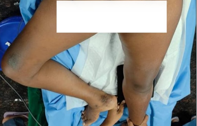
Figure 1: Post-Polio Contracture
History
The first report of the ancient disease of poliomyelitis dates back to 14th century BC at Egypt. Epidemic outbreaks in Stockholm and Vermont in 1887 and 1894 respectively progressed to more virulent cycles of epidemics peaking in mid-20th century.[1] Salk and Sabin vaccines, parenteral and oral, invented in 1950s improved scenario along with tank ventilation supporting bulbar polio. Global eradication programme on polio was instituted in 1988 with extended immunisation programme. Now reporting presence of the enteroviral infection only in Pakistan and Afghanistan, inadequate vaccination coverage and poor sanitation facilities being the main causes of failure to eradicate the disease.[2]
Aetiology
Poliovirus, a human enterovirus belongs to the family Picornaviridae with 3 subtypes, polio 1, 2 and 3. The virus is composed of a single-stranded positive sense RNA genome with a protein capsid.
The route of transmission is from gastrointestinal, respiratory entry followed by hematogenous spread to central nervous system and spreads along axons of peripheral nerves.[3] Depending on the virus type and location of invasion, aseptic meningitis, bulbar and paralytic disorders develop. Destruction of alpha motor neurons in the anterior horn cells of the spinal cord, cranial nerve nuclei of medulla, pons and midbrain finds inflammatory infiltration initially and glial cell scarring later.
In the recovery phase from acute polio, degeneration of reversibly damaged neurons, collateral axonal sprouting and development of other reparative and compensatory processes can result in slow improvement in paralysis.[4] Human axons may be able to reinnervate as much as 5 times their original muscle fibre territory. In chronic cases denervated muscles atrophy and cause functional disorders of limbs, trunk, respiratory muscles and diaphragm. Thus deformities develop progressively due to muscular imbalances or dysfunctions of growing skeleton as in children. This results in joint contractures, kyphosis, scoliosis, foot deformities or limb length discrepancies. Chronic overloading results in unstable joints, degenerative changes and pain.
Post-Polio Syndrome
Poliomyelitis has been classified variously as acute, chronic, paralytic and nonparalytic, spinal, bulbospinal and bulbar types. Morbidity following paralytic polio results from biomechanical alterations due to muscle weakness, imbalance of muscle power, deformities and complications related polio surgeries. Pain, easy fatiguability, limited function, reduced quality of life are common and especially precipitated by stressful events in life such as pregnancy or trauma. Lack of limb function leads to osteoporosis and eventual fractures. Approximately 25–40% of patients with previous paralytic polio develop post-polio syndrome in their lifetime. The most common theory behind the pathogenic mechanisms of PPS involves degeneration of the enlarged motor units formed following initial infection. Longitudinal research demonstrate a parallel between a decreasing size and number of active motor units and gradual strength decline in PPS. Overuse of remaining motor units, stress-induced aging of motor units, detercorating neuromuscular junctions, weight gain and deconditioning, PPS accelerates age-related sarcopenia and results in annual loss of up to 8% of muscle strength.[5] PPS is a clinical diagnosis and neither EMG nor muscle biopsy is able to differentiate patients with or without PPS. The severity and speed of progression are variable and are influenced by factors such as severity of acute paralysis, age of onset and socioeconomic status.[6]
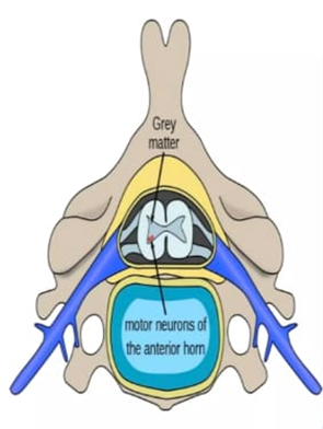
Figure 2: Spinal Polio Diagram
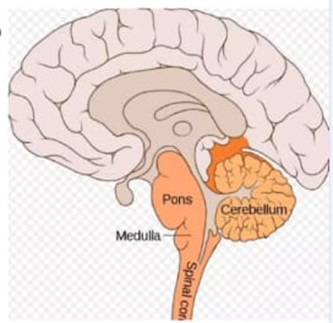
Figure 3: Bulbar Polio Diagram
Partial failure of subarachnoid block for previous C-section in this patient was an important reason for patient refusal for a lumbar puncture. This section explains the patient factors for the failed subarachnoid block, focusing on drug spread in the deformed spine. Inadequate intrathecal spread often occurs due to kyphosis, scoliosis, septations of ligaments within the theca that could act as barriers to spread, spinal stenosis and abnormal cysts or saccular dilatations with subarachnoid space.[6] The volume variability of lumbar CSF is another reason affecting the spread. Alfred F. Barker (1907) described all the conditions needed for a successful spinal analgesia: to enter the lumbar dural sac effectually with the point of the needle and to discharge through this, all the contemplated dose of the drug directly and freely into the cerebrospinal fluid, below the termination of the cord.
A patchy block is often due to insufficient drug delivered due to anatomical abnormality.[7] Neuromuscular disorder associated scoliosis may be classified as neuropathic and myopathic causes. Neuropathic causes include poliomyelitis, cerebral palsy and syringomyelia. Thus the subarachnoid space too becomes anatomically distorted due to lateral, rotational and sagittal changes in the vertebral column. The epidural and subarachnoid spaces often deviate towards convexity of spinal curve. The abnormal curvature can lead to reduced CSF flow. Inserting the spinal needle towards the convex side of the scoliosis curve is often more successful as the interlaminar space is typically wider there. Patients with early onset or complex scoliosis have a higher chance of associated intradural abnormalities that may affect the subarachnoid space. Asymmetry in bilateral spread of local anaesthetic in kyphoscoliosis often results in patchy or unilateral sensory block.[8] A modified paramedian approach directing the needle towards convexity of the curve may be beneficial.[9]
About two thirds of patients with paralytic polio have residual sequelae, the post-polio syndrome is thought to be due to progressive dysfunction and loss of motor neurons that compensated for the neurons lost during the original infection and not to persistent or reactivated poliovirus infection.[10]
Recent recommendations for poliovirus vaccination of adults towards global eradication of polio have been formulated in the United States in the year 2000 - an all inactivated poliovirus vaccine (IPV) for all adults at high risk of contracting the virus such as travel to endemic regions namely Pakistan, Afghanistan, health care workers and public authorities.[11]
Poliovirus is shed from some immunocompromised patients for more than 25 years, live attenuated vaccine or oral polio drops may cause vaccine derived poliovirus to circulate and cause disease and wild type of virus may be present in research laboratories; these facts pose concerns about discontinuing vaccination.
Oral Polio vaccine and Inactivated Polio vaccine induce antibodies that persist for 5 years. Both induce IgG and IgA antibodies. Adults who are unvaccinated or vaccination status unknown or had a primary series of oral polio vaccine are recommended to receive three doses of inactivated polio vaccine if they belong to the high risk group of reinfection.
Antivirals and monoclonal antibodies are in development to reduce or terminate shedding of poliovirus by long-term virus excreters. Pocapavir has been shown to reduce shedding of OPV type I in a clinical trial but further studies are awaited.
CONCLUSION:
We are in an era of intensified global eradication programme against polio virus in the form of extended immunisation schedules. Paralytic polio leaves deformities which in the course of life time causes various adaptations and deterioration of altered muscular imbalances, altered tone in different groups of muscles, sequelae of denervation, osteoporosis and obesity due to reduced physical activity. Thanks to awareness among people and employment opportunities for the motivated physically challenged group they live through a near normal socio economic domestic life.
The residual paralysis of polio when presenting for general or obstetric or orthopedic surgeries should not be considered or ignored as an isolated entity but rather as a post-polio syndrome. This article is presented to highlight proper evaluation of neuromusculoskeletal system as a whole along with chest physiotherapy in every case of residual polio presenting for anaesthesia and surgery, remembering the fact that it is only the active viral infection that has halted but not the neuromuscular skeletal deformities which are gradually progressive
REFERENCES:
1. Mehndiratta MM, Mehndiratta P, Pande R. Poliomyelitis: historical facts, epidemiology, and current challenges in eradication. The Neurohospitalist 2014;4(4):223-9.
2. Polio Global Eradication Initiative. https://polioeradication.org/polio this week/ accessed 20/Aug/2025
3. Murphy OC, Messacar K, Benson L, et al. Acute flaccid myelitis: cause, diagnosis, and management. The Lancet 2021;397(10271):334-46.
4. LoJK, Robinson LR. Post polio syndrome and late effects of poliomyelitis Part 1: pathogenesis, biomechanical considerations, diagnosis and investigations. Muscle Nerve 2018;58(6):751-9.
5. Lo JK, Robinson LR. Post‐polio syndrome and the late effects of poliomyelitis: Part 2. Treatment, management, and prognosis. Muscle & Nerve 2018;58(6):760-9.
6. Pokharel A. Study of failed spinal anesthesia undergoing caesarean section and its management. Postgraduate. Medical Journal of NAMS 2011;11(2):11-5.
7. Girard T, Savoldelli GL. Failed spinal anesthesia for Cesarean delivery: prevention, identification and management. Curr Opin Anaesthesiol 2024;37(3):207-12.
8. Misra S, Shukla A, Rao KG. Subarachnoid block in kyphoscoliosis: a reliable technique? Medical Journal of Dr. DY Patil Vidyapeeth 2016;9(6):761-4.
9. Huang J. Paramedian approach for neuroaxial anesthesia in parturients with scoliosis. Anesth Analg 2010;111(3):821-2.
10. Cohen JI. Enterovirus, parechovirus and reovirus infections. Chap- 210. In: Longo D, Fauci AS, Kasper DL, eds, et al. Harrison’s principles of internal medicine. 22nd edn McGraw Hill 2025: p. 1634.
11. Recommendations for polio virus vaccinator in adults. https/www.cdc.gov/vaccines/vpid/polio/hcp/recommendations./html.