Perioperative Management of Rupture Uterus – A Case Report
- Dinesh K. , Postgraduate in Anesthesia, Department of Anaesthesia, Dhanalakshmi Srinivasan Medical College and Hospital, Siruvachur, Perambalur, Tamil Nadu, India.
- Sirpika V. , Postgraduate in Anesthesia, Department of Anaesthesia, Dhanalakshmi Srinivasan Medical College and Hospital, Siruvachur, Perambalur, Tamil Nadu, India.
- Geetha J. , Professor, Department of Anaesthesia, Dhanalakshmi Srinivasan Medical College and Hospital, Siruvachur, Perambalur, Tamil Nadu, India.
Article Information:
Abstract:
Aim: To present and analyze a case of uterine rupture along a previous caesarean scar in a second-trimester pregnancy following trauma, and to highlight associated risks and management strategies, Objectives : To understand the role of fibroblastic proliferation in scar formation and its mechanical implications, To evaluate the risk of uterine rupture in women with previous caesarean sections, To assess how trauma and increased mechanical stress contribute to scar rupture during pregnancy , To discuss maternal morbidity in relation to multiple prior caesarean deliveries, To review the clinical presentation and management of uterine rupture in such cases. Method: A case-based observational approach was used. The subject was a 41-year-old pregnant woman in her second trimester with a history of two previous caesarean sections. Clinical history, including trauma (fall from height), presentation, diagnostic findings, and management strategies were documented and analyzed in the context of existing literature. Result: The patient presented with uterine rupture along the previous caesarean scar following trauma. The condition was associated with increased mechanical stress on the weakened scar tissue. The case demonstrated that prior multiple caesarean sections and external trauma significantly increase the risk of uterine rupture. Prompt diagnosis and intervention were critical in managing maternal morbidity. Conclusion: Uterine scars from previous caesarean sections remain structurally vulnerable despite healing through fibroblastic proliferation. The risk of rupture increases with the number of prior caesarean deliveries and can be further exacerbated by trauma. Early recognition and timely management are essential to reduce maternal morbidity and improve outcomes in such high-risk pregnancies.
Keywords:
Article :
CASE REPORT:
History
A 41-year-old lady with a history of an accidental fall from a high stool 2 days ago has persistent lower abdominal pain. She was a gravida three with a gestational age of 18 weeks. She had two live children delivered by lower segment caesarean sections. The last childbirth was five years ago. No history of maternal comorbidities in the previous and present pregnancy. She was a known case of hypothyroidism on 125 mcg of thyroxine tablet.
Clinical Features
Obese lady with a weight of 80 kg, approximate BMI of 30, conscious, oriented, mildly tachypnoeic with pulse 88/min and blood pressure 110/70 mmHg, respiratory rate of 26 per minute with SpO₂ of 98% in RA. Urine output was 50 ml of clear urine over the last 4 hours.
Ultrasonogram showed extrauterine Pregnancy with fetal demise and gestational age of 19 to 20 weeks. MRI of the pelvis showed an empty uterine cavity and cervical canal with a deformed fetus and a surrounding amniotic sac seen in extra-uterine location between the uterus and bladder, without any myometrial lining. The posterior portion of the sac and placenta is seen attached to the lower uterine segment. Both ovaries could not be visualised distinctly, mild ascites with free fluid in the pouch of Douglas, and at the right iliac fossa was noted. Anterolateral displacement of bowel loops by an extrauterine sac is seen on the left side. Bowel loops are normal. The general impression shows features of a caesarean scar ectopic pregnancy with mild ascites. The salient points to be noted in the MRI findings towards the hemodynamic status of the patient are:
1. No hemoperitoneum
2. Amniotic sac is intact
3. Fetal demise and small gestational age (18 weeks)
4. No adhesions with the bladder due to the previous two C sections.
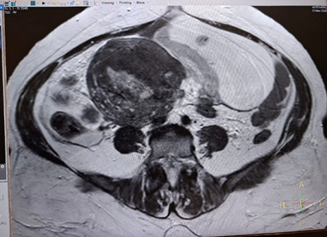
Figure 1: Pre-operative MRI showing a deformed fetus in an extrauterine location
Investigations
• Hb: 9.5 g%
• Platelets: 2 lakh/cumm
• PT/INR: 16.1 / 1.2
• APTT: 35.3 sec
• RBS (Random Blood Sugar): 121 mg%
• Blood group: O+
• Serology: Non-reactive
• ECG: Within normal limits
Problems and Risks
1. Extension of rupture along the scar on the uterus and shock
2. Hemorrhage due to rupture of dilated uterine vessels
3. Atony of the uterus
4. Disseminated intravascular coagulation (DIC)
5. Rupture of the amniotic sac and amniotic fluid embolism
6. Sepsis as the event is two days old
7. Obesity-related complications of airway, chest, and wound healing
8. Hypothyroidism – thyroid dysfunction
9. Respiratory distress syndrome
10. Thromboembolic phenomena
Goals of Anaesthesia
1. Hemodynamic stability
2. Replacement of blood and plasma
3. Beware of adhesions and injury to other organs in the vicinity, bowel, bladder and ureters
4. Maintain good tissue perfusion, oxygenation, and adequate urine output of more than 40 ml/hour
Surgical Plan
As the patient was forty-one years old and had already completed her family, the weak and already opened scar of the previous two C-sections and fetal demise following trauma, it was decided by the obstetricians to proceed immediately for hysterectomy after delivery of the fetus.
Conduct of Anaesthesia
Premedication
• Midazolam 2 mg as an anxiolytic
• Hydrocortisone 100 mg to combat stress and to act on the HPA axis
• Glycopyrrolate 0.2 mg as antisialagogue
Induction
Two 16G venflons were secured for intravenous access, and the patient was shifted from the pre-operative area by trolley with pulse oximeter monitoring to the operating table.
Monitors
• Pulse oximetry
• Five-lead ECG
• Non-invasive blood pressure
• Capnogram
• Airway pressure (Paw) in the anaesthesia workstation
• Urine output
Intubation
The patient was in sympathetic response with tachycardia and hypertension. Pulse: 100/min, BP: 154/96 mmHg. Hence patient was induced with:
• Fentanyl 100 mcg
• Propofol 120 mg
• Vecuronium 6 mg
Without fear of shock, fetal safety and uterine atony. She was intubated with a 7.0 mm cuffed endotracheal tube, secured, and general anaesthesia was maintained with:
• Oxygen (33%)
• Nitrous oxide
• IPPV
• Isoflurane ½ MAC
The fetus was delivered intact with the amniotic membrane along with placenta. No uterotonics were given, and a hysterectomy was done in the usual way. 2 units of packed cells and 1 unit of 6% hetastarch were given. Blood loss was estimated to be 750 ml, and tissue loss was about 650 grams. The intraoperative period was uneventful, and smooth extubation was achieved. The postoperative period was uneventful.
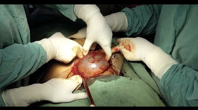
Figure 2: The amniotic sac is being delivered.
DISCUSSION:
Uterine rupture could cause serious morbidity and mortality for the mother and fetus.[1] A complete division of all three layers of the uterus namely the outer serosal layer or perimetrium, the middle smooth muscle layer or myometrium, and the inner endothelial layer or endometrium, is called rupture, while the discontinuity of the layers with intact serosa is termed uterine dehiscence. Previous caesarean sections are the foremost cause of uterine rupture of the gravid uterus.
The gravity of complications are proportional to the number of previous caesarean sections and the period of rupture in the third trimester. Uterine rupture can also occur in nonpregnant women when the uterus is exposed to trauma, infection or malignancy. In recent times, the trial of labour has often been attempted after caesarean delivery (TALOC), which throws more light on interest in uterine rupture. Uterine rupture can occur also in nonpregnant women when the uterus is exposed to trauma, infection or malignancy.[2] In the recent times trial of labour after caesarean delivery (TALOC) has been attempted which throws more light on interest in uterine rupture. Subsequent pregnancy on a previous C-section scar causes thinning of myometrium. Incidence of uterine rupture varies strongly depending on factors such as previous uterine surgery, delivery modes and healthcare access in different regions. The risk of uterine rupture during a trial of labour after C-section is 0.5%, while it is 0.02% in women who have an elective repeat C-section without labour.[3] Clinical symptoms associated with uterine rupture include acute abdominal pain, abnormal cardiotocography, vaginal bleeding, unstable hemodynamics, tachycardia, hypotension, altered sensorium, acute absence of contractions and dystocia.[4]
Risk factors for uterine Rupture include a previous uterine scar, excessive use of oxytocin or prostaglandins, short interdelivery interval, maternal age over 40, obesity, macrosomia, multiple pregnancy, abnormal placentation, fundal pressure in labour and obstructed labour.[5] Our patient had three of the above risk factors, namely previous two C-sections, obesity, maternal age of 41 years, and trauma. Several studies were conducted across the globe to assess whether there were significant differences in measurable prepartum and antepartum factors between maternal and neonatal high-risk ruptures and complete uterine rupture. Maternal high-risk rupture was characterised by blood loss exceeding one litre, necessitating transfusion. No significant association was identified between maternal high-risk rupture and complete uterine rupture. In contrast, neonatal high-risk rupture was defined by a need for NICU. Admission was significantly associated with differences in parity, gestational age, birth weight and number of prior C-sections compared to complete uterine rupture.[6]
In our patient with para 2 C-sections through Pfannenstiel incision and maternal age of 41, the accidental fall precipitated a silent rupture as the fetus was only at 18 weeks of gestational age. As the maximum peak of uterine blood flow is attained during 16–20 weeks of gestation, this patient had a blood loss, and as hysterectomy was planned, a near one litre blood loss was replaced.
Trauma in Pregnancy
Traumatic injuries in pregnancy are not uncommon, accounting for 6% to 7% of all traumatic injuries.[7] Whether the trauma is trivial or severe, pregnant trauma patients are important because traumatic injuries during pregnancy are associated with abortion, premature labor, rupture of membranes, maternal death and stillbirth.[8] The changes in maternal physiology and gestational age during trauma complicate an accurate evaluation of fetal outcomes.[9] In pregnant women, blunt trauma is the most common mechanism of injury.[10] Motor vehicle crashes are the leading cause of trauma in pregnant women, followed by falls.[11] In the first trimester of pregnancy, the fetus is protected by the pelvis. At 20 weeks of gestation, the uterus is palpable at the umbilical level. At 36 weeks of gestation, the uterus enlarges, and the uterine wall becomes thinner, making it more vulnerable to trauma.[12] In pregnant women, tachycardia and hypotension may not develop until significant blood loss has occurred, which may delay the diagnosis of shock.[13] Haemorrhage during pregnancy significantly reduces blood flow to the uterus. The mechanism of injury may be direct trauma to the abdomen or indirect trauma due to deceleration or shearing forces. Direct fetal trauma accounts for fewer than 1% of cases of severe abdominal trauma occur in pregnant women, but fetal deaths are reported in 3.7% to 17.5% of those cases.[14] The risk of fetal loss correlates with the severity of trauma and the maternal condition. An injury severity score of more than 9 depicts a higher risk.[9] Traumatic uterine rupture often allows fetal mortality to reach 100%.
CONCLUSION:
Trauma or fall in pregnancy, especially in women with previous multiple C-sections, forms a major contributory cause for uterine rupture. Advanced pregnancy, big baby, ruptured amniotic sac and uterine blood vessels present with early shock syndrome than a first or mid-trimester pregnancy, which depicts a gradual fall in BP and tachycardia. The surgical plan is the obstetrician’s decision considering her family history, obstetric history, and safety. Caring for pregnant trauma patients requires an organised and integrated approach with multidisciplinary involvement.
REFERENCES:
1. Gibbins KJ, Weber T, Holmgren EM, et al. Maternal and fetal morbidity associated with uterine rupture of unscarred uterus. Am J Obstet Gynecol 2015;213(3):382.e1–6.
2. Herrera FA, Hassanein AH, Bansal V. Atraumatic spontaneous rupture of the nongravid uterus. J Emerg Trauma Shock 2011;4(3):439.
3. Vaginal birth after caesarean birth. NICE. Available at: https://www.nice.org.uk/guidance/qs32/chapter/quality-statement-1 Accessed on: 16 Feb 2025
4. Savukyne E, Bykovaite-Stankeviciene R, Machtejeviene E, et al. Symptomatic uterine rupture: a fifteen year review. Medicina 2020;56(11):574.
5. Al-Zirqi I, Daltveit AK, Forsen L, et al. Risk factors for complete uterine rupture. Am J Obstet Gynecol 2017;216:165.e1-8.
6. Baek S, Froese V, Morgenstern B. Risk profiles and outcomes of uterine rupture: a retrospective and comparative single-center study of complete and partial ruptures. J Clin Med 2025;14(14):4987.
7. Shah KH, Simons RK, Holbrook T, et al. Trauma in pregnancy: maternal and fetal outcomes. Journal of Trauma and Acute Care Surgery 1998;45(1):83-6.
8. El Kady D, Gilbert WM, Anderson J, et al. Trauma during pregnancy: an analysis of maternal and fetal outcomes in a large population. Am J Obstet Gynecol 2004;190(6):1661-8.
9. Curet MJ, Schermer CR, Demarest GB, et al. Predictors of outcome in trauma during pregnancy. J Trauma 2000;49(1):18-24.
10. Petrone P, Jiménez-Morillas P, Axelrad A, et al. Traumatic injuries to the pregnant patient: a critical literature review. European Journal of Trauma and Emergency Surgery 2019;45(3):383-92.
11. Dunning K, LeMasters G, Bhattacharya A. A major public health issue: the high incidence of falls during pregnancy. Maternal and Child Health Journal 2010;14(5):720-5.
12. Weintraub AY, Lezon E, Mazor M. The pathophysiology of trauma in pregnancy: a review. J Matern Fetal Neonatal Med 2006;19(10):601-5.
13. La Rosa M, Loaiza S, Zambrano MA, et al. Trauma in pregnancy. Clin Obstet Gynecol 2020;63(2):447-54.
14. Weiss HB, Songer TJ, Fabio A. Fetal deaths related to maternal injury. JAMA 2001;286(15):1863-8.