PAROTID SIALOLITHIASIS.
- P. Balaji , MS, PhD, FRCS, FCLS, Director of General Surgery and Minimal Invasive Surgery, SRM Institute of Medical Science, Vadapalani, Chennai.
- Siddharth.D , MS, FRCS, Senior Consultant, General General Surgery and Minimal Invasive Surgery, SRM Institute of Medical Science, Vadapalani, Chennai.
- Vidya Lakshmi , MS, Consultant, General Surgery and Minimal Invasive Surgery, SRM Institute of Medical Science, Vadapalani, Chennai.
- Nitish R Jayaharan , Junior Resident, General Surgery and Minimal Invasive Surgery, SRM Institute of Medical Science, Vadapalani, Chennai.
Article Information:
Abstract:
Background: Sialolithiasis is a common benign disorder of the salivary glands, predominantly affecting the submandibular gland. Parotid sialolithiasis is uncommon and often poses diagnostic and therapeutic challenges, particularly when associated with persistent sialadenitis unresponsive to conventional management. Case Presentation: We report a 44-year-old male presenting with acute painful swelling of the left parotid gland exacerbated by meals. Imaging revealed multiple sialoliths within the parotid gland and Stensen’s duct, associated with ductal dilatation and glandular inflammation. Despite spontaneous expulsion of calculi following conservative management, the patient developed persistent bilateral parotid inflammation unresponsive to antibiotics and analgesics. Extensive infectious, neoplastic, and rheumatological evaluations were negative. Empirical corticosteroid therapy led to marked clinical improvement, suggesting an underlying autoimmune inflammatory process despite negative serological markers. Conclusion: This case highlights the importance of considering autoimmune etiologies such as seronegative Sjögren’s syndrome in patients with persistent parotid sialadenitis following resolution of ductal obstruction. Early recognition and timely initiation of corticosteroid therapy may prevent chronic glandular damage and improve outcomes.
Keywords:
Article :
INTRODUCTION:
Sialolithiasis or salivary gland stones is a benign condition which refers to the formation of calcified concretions or stones within the salivary glands or their excretory ducts and accounts for the most common cause of obstructive salivary gland disease.It is the most common cause of salivary gland swelling, with an incidence of 1 in 10,000–30,000.This leads to blockage of the normal salivary flow leading to painful swollen glands .Obstruction can in turn lead to sialadenitis, secondary infection, or rarely abscess formation.
It involves formation of hard mineral deposits more commonly of calcium phosphate (sialoliths) either within the duct or the gland parenchyma. The exact etiology is unclear however both anatomical and compositional factors have a role to play in the same. Dehydration, drugs (e.g diuretics) and smoking are some of the risk factors under study.Approximately 72–94% of cases involve the submandibular gland, while parotid gland involvement is rare, accounting for only 6–15% of cases.They are often asymptomatic unless obstructive. Obstruction of salivary flow leads to ductal ectasia, glandular enlargement, and predisposition to ascending infection, manifesting clinically as painful post-prandial swelling.
Parotid sialolithiasis typically affects adults between 30 and 60 years of age, with a male predominance.Salivary Calculi affecting the parotid gland are usually unilateral and predominantly located within Stensen’s duct rather than the gland parenchyma.
Although most cases respond to conservative or minimally invasive interventions, persistent or recurrent parotid sialadenitis despite stone removal should prompt evaluation for systemic inflammatory or autoimmune conditions. Sjögren’s syndrome, particularly its seronegative variant, has been increasingly recognized as an underdiagnosed contributor to chronic parotid pathology.
Sjögren’s syndrome (SS) is a chronic autoimmune exocrinopathy characterized by lymphocytic destruction of salivary and lacrimal glands, leading to xerostomia and xerophthalmia. Chronic salivary hypofunction in Sjögren’s predisposes to parotid duct obstruction and stone formation.
CASE PRESENTATION:
A 44-year-old male presented with a one-week history of painful swelling over the left parotid region, progressively worsening during mastication. There was no history of fever, xerostomia, xerophthalmia, weight loss, radiation exposure, or prior salivary gland disease.
Clinical Examination
A 5 × 4 cm firm, tender swelling was noted in the left parotid region, extending from the pre-auricular area to the angle of the mandible, with elevation of the ear lobule.
Intraoral examination revealed inflamed mucosa around the left Stensen’s duct orifice with absent salivary flow on gland massage.
No cervical lymphadenopathy or facial nerve dysfunction was observed.
Radiological Evaluation
Non-contrast CT scan of the neck demonstrated:
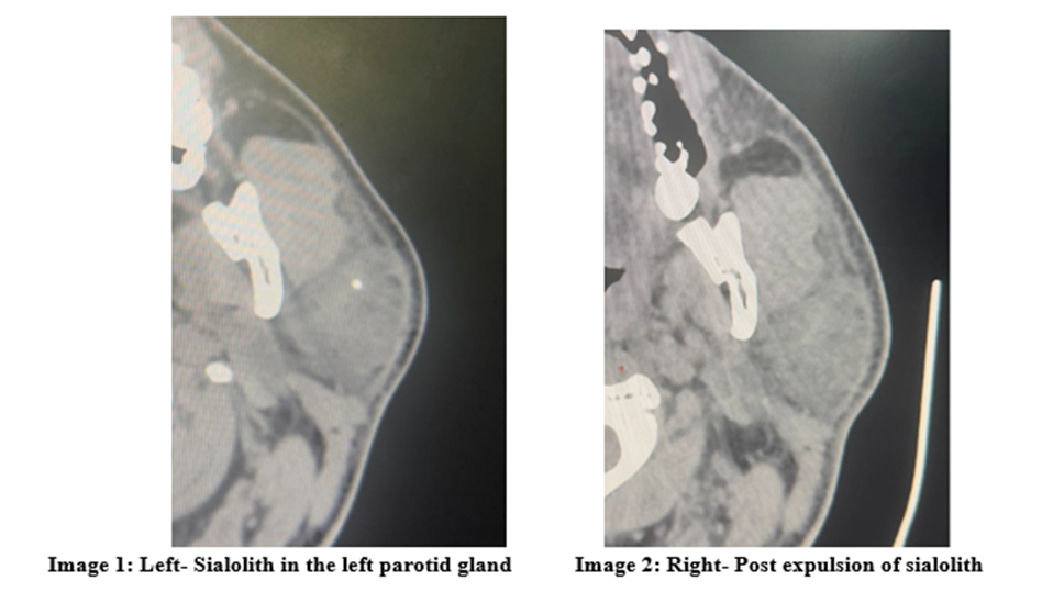
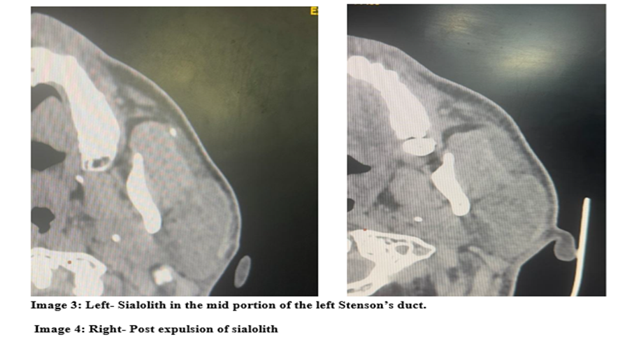
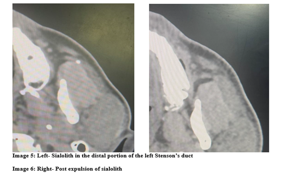
Three sialoliths: one intraparenchymal and two within the mid and distal portions of the left Stensen’s duct (measuring 3 mm and 3.6 mm).
Dilatation of the proximal duct.
Enlarged left parotid gland with heterogeneous attenuation and mild enhancement, consistent with acute sialadenitis.
Management and Clinical Course
The patient was initially managed conservatively with:
· Broad-spectrum antibiotics
· Analgesics
· Adequate hydration
· Sialogogues
· Chlorhexidine mouthwash
The sialoliths subsequently expelled spontaneously. However, pain and swelling persisted, and the patient developed new-onset right parotid swelling. Repeat CT imaging showed complete clearance of calculi but revealed bilateral parotid gland edema with extensive fat stranding and subcutaneous edema involving the lower jaw and submental regions.
Further Evaluation
Comprehensive investigations were performed to exclude alternative etiologies:
· ANA, anti-SSA/Ro, IgG4 levels, and ACE levels were all within normal limits.
· Findings were inconsistent with bacterial or viral sialadenitis, sarcoidosis, IgG4-related disease, or neoplasia.
· Given the refractory nature of symptoms and bilateral gland involvement, an autoimmune etiology—specifically seronegativeSjögren’s syndrome—was suspected.
Therapeutic Intervention
Systemic corticosteroid therapy was initiated, resulting in significant symptomatic improvement with marked reduction in pain and swelling. The patient was discharged on maintenance therapy including corticosteroid mouthwash and sialogogues and remains under regular follow-up.
DISCUSSION:
Sialolithiasis is more common in submandibular gland due to antigravity drainage, thick mucinous secretions and alkaline pH.Parotidsialolithiasis represents a rare but clinically significant cause of obstructive sialadenitis. It leads to pain and swelling in the parotid region usually pronounced during or after consumption of food .This condition can cause significant discomfort when left untreated and could lead to chronic infection.AlthoughSialolithiasis occurs mainly in the submandibular gland , patients with Sjogren syndrome more commonly present with Sialolithiasis of the parotid gland . These stones usually made of calcium phosphate and hydroxyl apatite can cause significant facial pain and swelling.Degree of obstruction can be assessed by milking the duct to see the flow of saliva.
Unlike submandibular calculi, parotid stones are typically smaller, less radiopaque, and more difficult to diagnose clinically. Non contrast CT imaging remains the diagnostic modality of choice due to its high sensitivity for detecting calcifications and assessing associated inflammatory changes.Other modalities include conventional radiotherapy, USG and Sialography. Sialography includes injecting dye into the duct which is contraindicated in acute inflammation and allergy to dye.
Treatment is typically conservative for sialoliths of less than 5mm which includes adequate hydration, Sialogogues, gland massage , NSAIDs and antibiotics if infection is present.Stones of moderate size less than 7mm can be managed with minimally invasive techniques like sialendoscopy with intracorporeal shock wave lithotripsy if required. Sialendoscopy has both diagnostic and therapeutic roles and helps in differentiating between obstructive inflammatory conditions and calculi ( more than 10mm). In case of parotid calculi associated with Sjogren syndrome management with corticosteroids is advised.
The persistence of sialadenitis despite stone expulsion in this patient underscores the multifactorial nature of parotid gland inflammation. Sjögren’s syndrome is increasingly recognized as a contributor to parotid sialolithiasis, with salivary stasis, altered glandular secretion, and chronic inflammation promoting calculus formation.
The typical initial symptoms of patients with Sjögren’s syndrome are dry eyes and mouth. However, sialadenitis caused by atypical inflammation or obstruction of Stensen’s duct, due to a sialolith, is occasionally the first presentation of Sjögren’s syndrome
Importantly, up to 17–20% of patients with Sjögren’s syndrome are seronegative for anti-SSA/Ro antibodies, making diagnosis challenging when relying solely on serology . In such cases, clinical judgment plays a pivotal role, and empirical corticosteroid therapy may be both diagnostic and therapeutic.
This case further highlights the role of conservative and minimally invasive approaches in managing small parotid calculi, while emphasizing the need for a broader diagnostic perspective when standard treatment fails.
CONCLUSION:
Persistent parotid sialadenitis following resolution of ductal obstruction should raise suspicion for an underlying autoimmune process, even in the absence of serological markers. SeronegativeSjögren’s syndrome remains an important diagnostic consideration. Early initiation of corticosteroid therapy in carefully selected patients can result in significant clinical improvement and prevent chronic glandular dysfunction.
This case is presented mainly to stress the importance of considering an autoimmune background like Sjogren syndrome in patients presenting with parotid sialadenitis with sialolithiasis which do not resolve in traditional conservative management and keep progressing.
In such cases, a thorough rheumatological blood work up need to be done and if ANA study turns positive, corticosteroids need to be initiated into the treatment plan. It should be borne in mind that inpatients presenting with persistent parotid sialadenitis despite expulsion of sialoliths and negative ANA, the possibility of autoimmune disorders like Sjogren syndrome should still be considered as 17.2% of cases have been found to have Sjogren syndrome despite absence of anti- SSA/ Ro antibodies. In such cases starting empirical corticosteroid therapy will offer clinical benefits as in the case presented above.
REFERENCES:
1. Jáuregui E, Kiringoda R, Ryan WR, Eisele DW, Chang JL. Chronic parotitis with multiple calcifications: Clinical and sialendoscopic findings. Laryngoscope. 2017 Jul;127(7):1565-1570. doi: 10.1002/lary.26386. Epub 2016 Nov 15. PMID: 27861944.
2. Lan J, Deng C, Huang H, Rao P, Chen Y, Shi Y, Chen J, Shi G, Liu Y, Chen S. Seronegative primary Sjögren's syndrome, a distinct subtype of primary Sjögren's syndrome in Chinese patients. BMC Rheumatol. 2024 Apr 16;8(1):15. doi: 10.1186/s41927-024-00384-9. PMID: 38627838; PMCID: PMC11020423.
3. AnduezaGuembe M, ChiesaEstomba CM, Saga Gutiérrez C, Thomas ArrizabalagaI,ÁbregoOlano M, VázquezQuintano M, AltunaMariezcurren X. Utility of sialendoscopyinthe management of juvenile recurrent parotitis. Retrospective study.ActaOtorrinolaringolEsp (Engl Ed). 2024 Sep-Oct;75(5):304-309. [PubMed: 39038536]
4. Bukhari AF, Bugshan AS, Papas A, Desai B, Farag AM. Conservative Management of Chronic SuppurativeParotitis in Patients with Sjögren Syndrome: A Case Series. Am J CaseRep. 2021 Mar 19;22:e929553. [PMC free article: PMC7988433] [PubMed: 33739960]
5. van der Lans RJL, Lohuis PJFM, van Gorp JMHH, Quak JJ. Surgical Treatment of ChronicParotitis.Int Arch Otorhinolaryngol. 2019 Jan;23(1):83-87. [PMC free article:PMC6331305] [PubMed: 30647789]
6. Denny MC, Fotino AD. The Heerfordt-Waldenström syndrome as an initial presentation of sarcoidosis.Proc (BaylUniv Med Cent). 2013 Oct;26(4):390-2. [PMC free article:PMC3777100] [PubMed: 24082416]
7. Fragoulis GE, Zampeli E, Moutsopoulos HM. IgG4-related sialadenitis and Sjögren'ssyndrome. Oral Dis. 2017 Mar;23(2):152-156. [PubMed: 27318181]
8. Brooks KG, Thompson DF. A review and assessment of drug-induced parotitis.AnnPharmacother. 2012 Dec;46(12):1688-99. [PubMed: 23249870]
9. Park KK, Tung RC, de Luzuriaga AR. Painful parotid hypertrophy with bulimia: a report of medical management. J Drugs Dermatol. 2009 Jun;8(6):577-9. [PubMed: 19537384]
10. Berta E, Angel G, Lagarde F, Fonlupt B, Noyelles L, Bettega G. Role of sialendoscopyinjuvenile recurrent parotitis (JRP). Eur Ann Otorhinolaryngol Head Neck Dis. 2017Dec;134(6):405-407. [PubMed: 28669808]
11. Wang S, Marchal F, Zou Z, Zhou J, Qi S. Classification and management of chronic sialadenitis of the parotid gland. J Oral Rehabil. 2009 Jan;36(1):2-8. [PubMed: 18976271]
12. Capaccio P, Torretta S, Pignataro L, Koch M. Salivary lithotripsy in the era of sialendoscopy. ActaOtorhinolaryngol Ital. 2017 Apr;37(2):113-121. [PMC free article:PMC5463518] [PubMed: 28516973]
13. Ichinose M, Matsushima T, Hataya H. Purulent Discharge from Stensen Duct in Neonatal Suppurative Parotitis. J Pediatr. 2022 Apr;243:230-231. [PubMed: 34952006]
14. Salehzadeh F, Molatefi R, Mardi A, Nahanmoghaddam N. Juvenile idiopathic recurrentparotitis (JIRP) treated with short course steroids, a case series study and one decade followup for potential autoimmune disorder. PediatrRheumatol Online J. 2024 Jan 04;22(1):8.[PMC free article: PMC10765850] [PubMed: 38178123]
15. Hammett JT, Walker C. Sialolithiasis. [Updated 2024 Sep 10]. In: StatPearls [Internet]. Treasure Island (FL): StatPearls Publishing; 2025 Jan-. Available from: https://www.ncbi.nlm.nih.gov/books/NBK549845/
16. Guzzo M, Locati LD, Prott FJ, Gatta G, McGurk M, Licitra L. Major and minor salivaryglandtumors. Crit Rev OncolHematol. 2010 May;74(2):134-48. [PubMed: 19939701]
17. Kao WK, Chole RA, Ogden MA. Evidence of a microbial etiology for sialoliths.Laryngoscope. 2020 Jan;130(1):69-74. [PubMed: 30861582]
18. Thakur J, Thakur A, Mohindroo N, Mohindroo S, Sharma D. Bilateral parotid tuberculosis.J Glob Infect Dis. 2011 Jul;3(3):296-9. [PMC free article: PMC3162820] [PubMed:21887065]
19. Li X, Yang J, Qu LY, Zheng DN, Xie XY, Liu DG, Yu GY. Diagnosis and Treatment of Radioactive Iodine-Induced Sialadenitis: A 10-Year Endoscopic Experience. Laryngoscope.2024 Nov;134(11):4506-4513. [PubMed: 38761158]
20. Thomson J P S and Hobsley M. Parotid duct calculus. Proceedings of the Royal Society of Medicine 1973, 66:352-3
21. Patey, D.H.The clinical features and diagnosis of parotid calculi. Practitioner 1966: 197: 67
22. Eddey H.H. Recurrent parotitis and associated calculus, cyst or tumour. Medical Journal of Australia 1966; 2:581
23. Ottaviani F, Capaccio P, Rivolta R, et al. Salivary gland stones US evaluation in shock wave lithotripsy. Radiology 1997; 204:437-41.
24. Moghe S, Pillai A, Thomas S, Nair PP. Parotid sialolithiasis. BMJ Case Rep. 2012 Dec 14;2012:bcr2012007480. doi: 10.1136/bcr-2012-007480. PMID: 23242089; PMCID: PMC4543829.
25. Konstantinidis I, Paschaloudi S, Triaridis S, Fyrmpas G, Sechlidis S, Constantinidis J. Bilateral multiple sialolithiasis of the parotid gland in a patient with Sjögren's syndrome. ActaOtorhinolaryngol Ital. 2007 Feb;27(1):41-4. PMID: 17601211; PMCID: PMC2640017.
26. Billings M, Dye BA, Iafolla T, Baer AN, Grisius M, Alevizos I. Significance and Implications of Patient-reported Xerostomia in Sjögren's Syndrome: Findings From the National Institutes of Health Cohort. EBioMedicine. 2016 Oct;12:270-279. doi: 10.1016/j.ebiom.2016.09.005. Epub 2016 Sep 9. PMID: 27639822; PMCID: PMC5078600